. 2019 Aug 21;23(1):1745.
doi: 10.4102/sajr.v23i1.1745. eCollection 2019.

Meralgia paresthetica: Now showing on 3T magnetic resonance neurography

Affiliations

Meralgia paresthetica: Now showing on 3T magnetic resonance neurography

Raihaan M Ally et al. SA J Radiol. .

Abstract

Meralgia paresthetica is a neuropathy of the lateral femoral cutaneous nerve. Traditionally, the diagnosis is based on classical symptoms and signs. In cases where there is a diagnostic dilemma, the role of magnetic resonance imaging has been to exclude other causes for the patient's presentation, as the small extraspinal peripheral nerves were not well visualised at imaging. The development of 3-Tesla magnetic resonance neurography, however, has made pathology of these nerves more conspicuous.

Keywords: Meralgia paresthetica; fluid-sensitive sequences; lateral femoral cutaneous nerve; magnetic resonance neurography; neuropathy.


Conflict of interest statement

The authors declare no conflict of interest regarding the write up of this article.

Figures
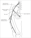
FIGURE 1
FIGURE 1
Diagram illustrating the anatomy of the lateral femoral cutaneous nerve.
FIGURE 2
FIGURE 2
3T proton density fat-saturated axial MR images at the level of the anterior superior iliac spine in a 54-year male patient presenting with a clinical suspicion of meralgia paresthetica on the left. Note the high signal and prominence of the lateral femoral cutaneous nerve on the left (long arrow) as compared to the right (dashed arrow) as it runs over the (a) iliacus muscle (short arrow) and (b) anterior superior iliac spine.
FIGURE 3
FIGURE 3
3T proton density fat-saturated axial MR image in the same patient described in Figure 2 demonstrates the persistent high signal of the lateral femoral cutaneous nerve in the subcutaneous tissue where it divides into anterior and lateral branches.

References

    1. Patijn J, Mekhail N, Hayek S, Lataster A, Van Cleef M, Van Zundert J. Meralgia paresthetica. Pain Pract. 2011;11(3):302–308. 10.1111/j.1533-2500.2011.00458.x - DOI - PubMed
    1. Chhabra A, Del Grande F, Soldatos T, et al. . Meralgia paresthetica: 3-Tesla magnetic resonance neurogaphy. Skeletal Radiol. 2013;42(6):808–808. 10.1007/s00256-012-1557-4 - DOI - PubMed
    1. Petchprapa CN, Rosenberg ZS, Sconfienza LM, Cavalcanti CFA, La Rocca Vieira R, Zember JS. MR imaging of entrapment neuropathies of the lower extremity. Part 1. The Pelvis and Hip. Radiographics 2010;30:983–1000. 10.1148/rg.304095135 - DOI - PubMed
    1. Grossman MG, Ducey SA, Nadler SS, Levy AS. Meralgia paresthetica: Diagnosis and treatment. J Am Acad Orthop Surg. 2001;9:336–344. - PubMed
    1. Muniz Neto FJ, Kihara Filho EN, Miranda FC, Rosemberg LA, Santos DCB, Taneja AK. Demystifying MM neurography of the lumbosacral plexus: From protocols to pathologies. Biomed Res Int. 2018;9608947:20 10.1155/2018/9608947 - DOI - PMC - PubMed

LinkOut - more resources