Review
. 2020 Jan 10;48(1):16-35.
doi: 10.1093/nar/gkz1101.

Multiple roles of DNA2 nuclease/helicase in DNA metabolism, genome stability and human diseases

Affiliations
Review

Multiple roles of DNA2 nuclease/helicase in DNA metabolism, genome stability and human diseases

Li Zheng et al. Nucleic Acids Res. .

Abstract

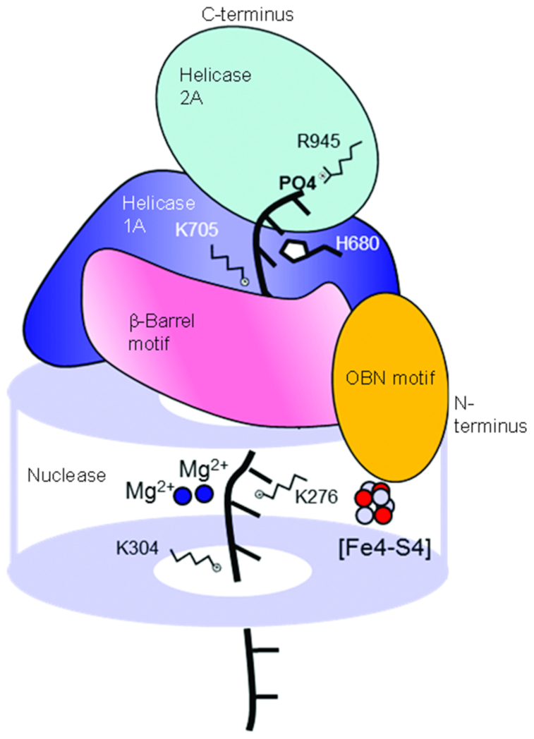
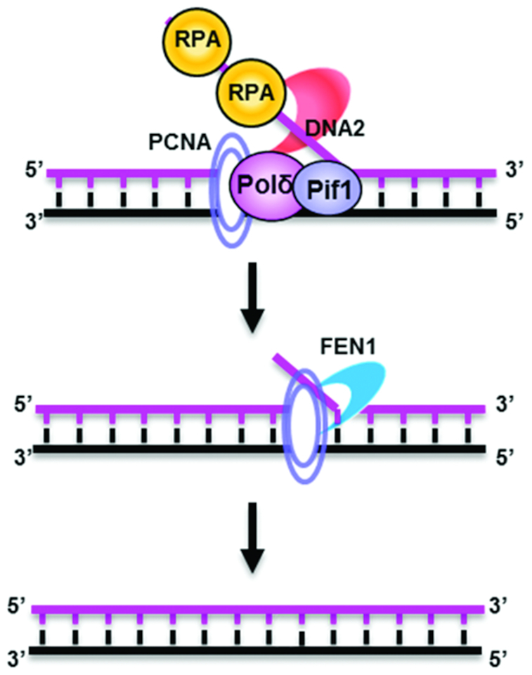
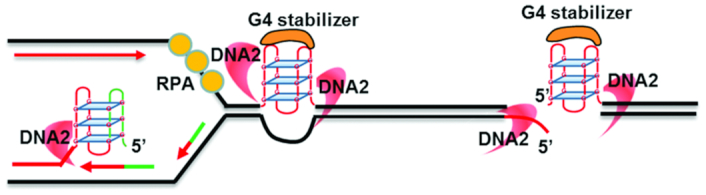
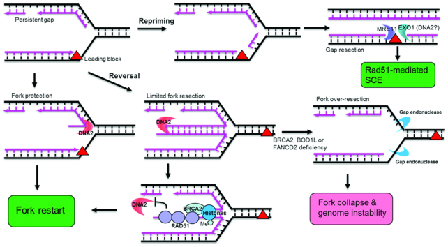
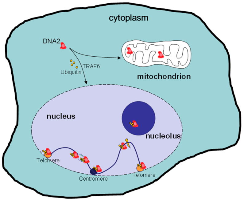
DNA2 nuclease/helicase is a structure-specific nuclease, 5'-to-3' helicase, and DNA-dependent ATPase. It is involved in multiple DNA metabolic pathways, including Okazaki fragment maturation, replication of 'difficult-to-replicate' DNA regions, end resection, stalled replication fork processing, and mitochondrial genome maintenance. The participation of DNA2 in these different pathways is regulated by its interactions with distinct groups of DNA replication and repair proteins and by post-translational modifications. These regulatory mechanisms induce its recruitment to specific DNA replication or repair complexes, such as DNA replication and end resection machinery, and stimulate its efficient cleavage of various structures, for example, to remove RNA primers or to produce 3' overhangs at telomeres or double-strand breaks. Through these versatile activities at replication forks and DNA damage sites, DNA2 functions as both a tumor suppressor and promoter. In normal cells, it suppresses tumorigenesis by maintaining the genomic integrity. Thus, DNA2 mutations or functional deficiency may lead to cancer initiation. However, DNA2 may also function as a tumor promoter, supporting cancer cell survival by counteracting replication stress. Therefore, it may serve as an ideal target to sensitize advanced DNA2-overexpressing cancers to current chemo- and radiotherapy regimens.


Figures

Figure 1.
Figure 1.
Schematic of the DNA2–DNA complex, elucidating its overall structure and the proposed mechanism for substrate binding and cleavage. Mouse DNA2 has an overall cylindrical shape. A narrow central tunnel is formed within the nuclease domain at the base and is extended by a β-barrel motif and a helicase 1A domain in the middle. A helicase 2A domain sits atop the tunnel. Several residues important for DNA substrate binding are indicated along the tunnel. The central tunnel is too narrow to allow dsDNA to access the active center in the nuclease domain. However, ssDNA can enter the tunnel through the bottom or top and thread to the other side. This schematic is based on previously published crystal structure information (35), with the two Ca2+ ions in the crystal structure replaced by two Mg2+ ions to reflect the role of Mg2+ as a co-factor for DNA2 catalysis.
Figure 2.
Figure 2.
Sequential actions of DNA2 and FEN1 to remove a long RNA–DNA flap during Okazaki fragment maturation. RPA binds to long 5′ RNA-DNA flaps generated by Pol δ/PCNA and/or PIF1. The flap-bound RPA inhibits the action of FEN1 on the flaps and simultaneously recruits and stimulates DNA2 to cleave at the middle of the ssDNA strand, generating a shorter (∼8 nt) 5′ flap. FEN1 then dislodges DNA2 and cleaves at the junction between the ssDNA and dsDNA strands. DNA2 can also function alone to process long flaps. The RPA-mediated sequential actions of DNA2 and FEN1 or of DNA2 alone produce ligatable DNA nicks that can be joined to form intact lagging strand DNA.
Figure 3.
Figure 3.
Roles of DNA2 in resolving DNA secondary structures, as typified by G4s, to facilitate DNA replication and repair. DNA2 endonuclease activity can directly excise G4s obstructing DNA replication fork movement. The resulting ssDNA gap is repaired by high-fidelity SSB repair. DNA2 can also remove G4s from DSBs to enable efficient end resection. The resolution of G4s by DNA2 is particularly crucial in the presence of G4 stabilizers that inhibit G4 unwinding by other helicases.
Figure 4.
Figure 4.
Multiple functions of DNA2 in stalled replication fork processing. DNA2 can participate in stalled replication fork protection, limited resection, or over-resection, depending on the nature of the fork and the availability of other fork-protecting factors. To protect a replication fork, the 5′ nuclease activity of DNA2 may cleave the 5′ ssDNA flap, preventing the reversal of nascent DNA, which can lead to the formation of potentially deleterious regressed fork structures. If regressed forks do arise, however, the 5′ nuclease activity of DNA2 may conduct limited 5′ resection, generating a 3′ ssDNA overhang to facilitate the helicase-driven regression of the regressed fork structure to restart the fork. Meanwhile, BRCA2 may be recruited to the regressed fork structure to limit the action of DNA2 to avoid over-resection. In addition, SET1A, in complex with BOD1L, catalyzes H3K4 methylation, which facilitates FANCD2-mediated histone assembly on the regressed fork and stabilizes the RAD51 filament on the nascent DNA to protect the fork before restarting. In the absence of BRCA2 or BOD1L, extensive degradation of the regressed fork by DNA2 and other nucleases may occur, leading to fork collapse and genome instability. In addition, the lesion that blocks leading strand DNA synthesis may be bypassed by the repriming process. The resulting gap will be processed and repaired post replication. The role of DNA2 in processing of the gap is undefined.
Figure 5.
Figure 5.
Mitochondrial localization and TRAF6-mediated nuclear translocation of human DNA2. Like its yeast counterpart, human DNA2 migrates into both mitochondria and nuclei. However, unlike scDNA2, human DNA2 has no NLS and translocates into nuclei via an NLS-independent mechanism that depends on TRAF6-mediated polyubiquitination. Once in the nucleus, DNA2 can localize to telomeres, centromeres or the nucleolus to facilitate the DNA replication of these difficult-to-replicate regions. DNA2 can also be recruited stalled replication forks or DSBs to facilitate the repair of these intermediate structures via the HDR pathway.

References

    1. Ogawa T., Okazaki T.. Discontinuous DNA replication. Annu. Rev. Biochem. 1980; 49:421–457. - PubMed
    1. Zheng L., Shen B.. Okazaki fragment maturation: nucleases take centre stage. J. Mol. Cell Biol. 2011; 3:23–30. - PMC - PubMed
    1. Konstantinopoulos P.A., Ceccaldi R., Shapiro G.I., D’Andrea A.D.. Homologous recombination deficiency: exploiting the fundamental vulnerability of ovarian cancer. Cancer Discov. 2015; 5:1137–1154. - PMC - PubMed
    1. Sogo J.M., Lopes M., Foiani M.. Fork reversal and ssDNA accumulation at stalled replication forks owing to checkpoint defects. Science. 2002; 297:599–602. - PubMed
    1. Mirkin E.V., Mirkin S.M.. Replication fork stalling at natural impediments. Microbiol. Mol. Biol. Rev. 2007; 71:13–35. - PMC - PubMed

Publication types

MeSH terms