Review
. 2020 Jan;77(1):19-33.
doi: 10.1007/s00018-019-03366-0. Epub 2019 Nov 21.

Poly(ADP-ribose) polymerase enzymes and the maintenance of genome integrity

Affiliations
Review

Poly(ADP-ribose) polymerase enzymes and the maintenance of genome integrity

Travis Eisemann et al. Cell Mol Life Sci. 2020 Jan.

Abstract

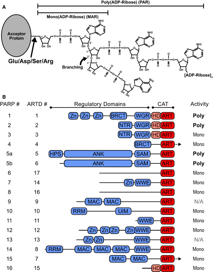
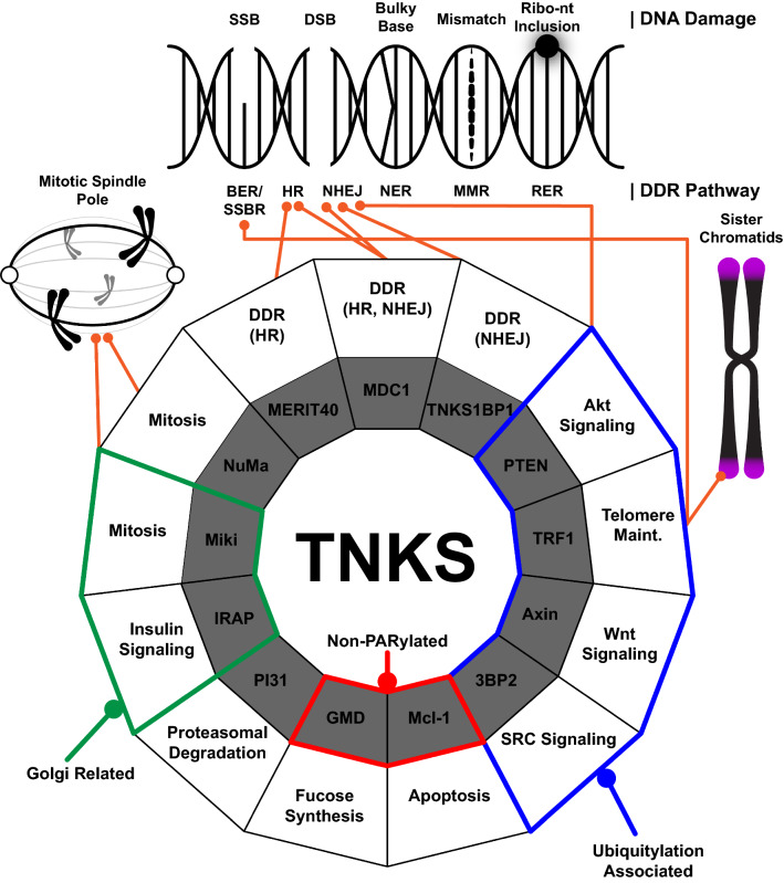
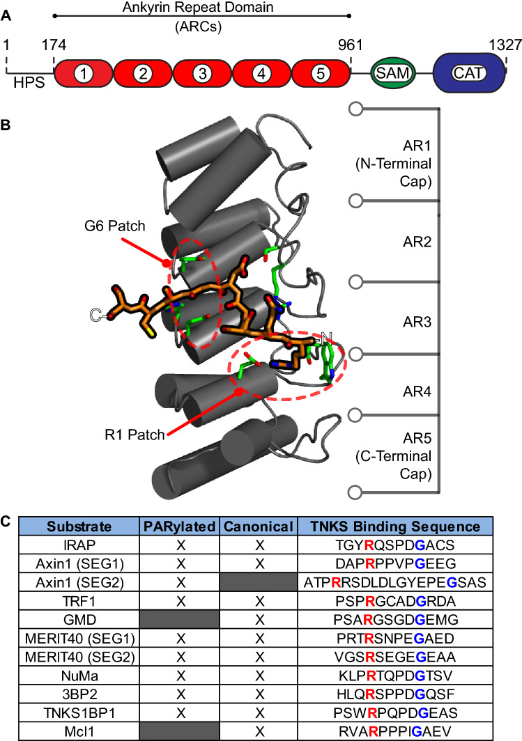
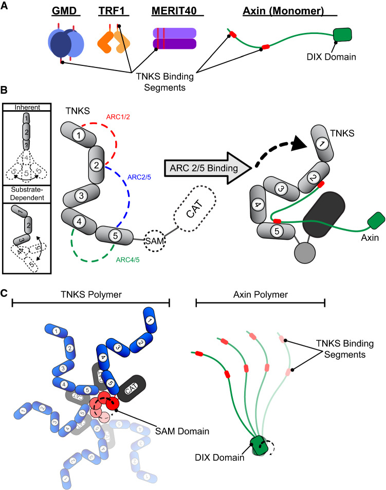
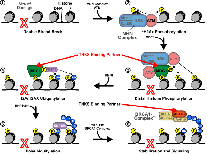
DNA damage response (DDR) relies on swift and accurate signaling to rapidly identify DNA lesions and initiate repair. A critical DDR signaling and regulatory molecule is the posttranslational modification poly(ADP-ribose) (PAR). PAR is synthesized by a family of structurally and functionally diverse proteins called poly(ADP-ribose) polymerases (PARPs). Although PARPs share a conserved catalytic domain, unique regulatory domains of individual family members endow PARPs with unique properties and cellular functions. Family members PARP-1, PARP-2, and PARP-3 (DDR-PARPs) are catalytically activated in the presence of damaged DNA and act as damage sensors. Family members tankyrase-1 and closely related tankyrase-2 possess SAM and ankyrin repeat domains that regulate their diverse cellular functions. Recent studies have shown that the tankyrases share some overlapping functions with the DDR-PARPs, and even perform novel functions that help preserve genomic integrity. In this review, we briefly touch on DDR-PARP functions, and focus on the emerging roles of tankyrases in genome maintenance. Preservation of genomic integrity thus appears to be a common function of several PARP family members, depicting PAR as a multifaceted guardian of the genome.

Keywords: DDR, DNA damage response; PAR, poly(ADP-ribose); PARP, poly(ADP-ribose) polymerase; Tankyrase.


Figures

Fig. 1
Fig. 1
ADP-ribose and PARP superfamily overview. a Representation of mono- and poly(ADP-ribose) (MAR and PAR, respectively) as posttranslational modifications (PTMs). b Domain schematic of PARP superfamily members. PARP catalytic (CAT) regions are shown in pink/red, regulatory domains are shown in blue. Family members are identified by their current “PARP” numbering as well as their “ARTD” classification (ADP-ribosyltransferases diphtheria toxin-like) numbering. Arrows indicate regions that extend C-terminal to the CAT domains
Fig. 2
Fig. 2
TNKS-binding partners and their respective functions. Types of DNA damage and their respective repair pathways (top), and wheel depicting the many known functions of TNKS (bottom) are shown. The types of DNA damage depicted include single and double strand breaks (SSB and DSB, respectively), bulky base, mismatch, and ribonucleotide (Ribo-nt) inclusion. Repair pathways include base excision repair (BER), single strand break repair (SSBR), homologous recombination (HR), non-homologous end joining (NHEJ), nucleotide excision repair (NER), mismatch repair (MMR), and ribonucleotide excision repair (RER). Binding partners are represented by the inner ring of the wheel (in gray), and their associated functions are represented in the outer ring of the wheel (in white). Non-PARylated binding partners are outlined in red. Related TNKS functions are grouped into Golgi-related (green outline), ubiquitylation associated (blue outline), and preservation of genomic integrity (orange lines)
Fig. 3
Fig. 3
TNKS interaction with TNKS-binding motifs. a Schematic of TNKS domains with human tankyrase-1 amino acid numbering shown. b Structure of a representative ankyrin repeat cluster (ARC) (in gray) bound to a peptide representing the binding partner IRAP (orange, PDB #5JHQ [70]). The ARC consists of five ankyrin repeats (AR). Residues that interact with the IRAP peptide are shown in green. The patches that coordinate binding to the R1 or G6 residues of the TNKS-binding motif are circled in red. c Table showing the TNKS-binding motifs of several TNKS-binding partners. Peptides with the canonical binding motif are noted with an “X” in the “Canonical” column. Binding partners that are modified with PAR are noted with an “X” in the “PARylated” column. The amino acid sequences of the binding motifs are shown, with the residues that bind at position R1 or G6 in red or blue text, respectively
Fig. 4
Fig. 4
TNKS interaction with structurally dissimilar binding partners. a Structural heterogeneity of TNKS-binding partners. The schematics indicate the varying multimeric quaternary structures of TNKS-binding partners. Each unique TNKS-binding motif is indicated, and all TNKS-binding motifs are drawn in red. b (left) The expected flexibility of the ankyrin repeat domain and the relative orientations of ARCs is shown schematically. The five ARCs are numbered 1–5. (middle) ARC pairs that function together in binding to Axin1 are shown in the context of the complete TNKS domain structure. (right) A proposed model for the roles of TNKS structural characteristics in binding partner interaction and potential delivery to the catalytic domain (CAT). c Schematic of TNKS and Axin polymers, illustrating the potential for multivalent contacts between the polymeric proteins
Fig. 5
Fig. 5
A model for recruitment of TNKS-binding partners in the DSBR pathway. Following a double strand break (1), the MRN complex and ATM colocalize to the site of damage (2). MDC1 binds, recruiting additional ATM molecules and resulting in the phosphorylation of distal histones (3). MDC1 also recruits RNF8, which ubiquitylates histones H2A and H2AX (4). RNF168 then binds the ubiquitylated histones, yielding polyubiquitylated histones (5). The BRCA1 complex is then recruited through an interaction between RAP80 and polyubiquitin (6). MERIT40 functions as a scaffold that stabilizes the BRCA1 complex, facilitating signaling for downstream repair factors. TNKS-binding partners MDC1 and MERIT40 are designated by red arrows Figure adapted from [149, 150]

References

    1. Hoeijmakers JHJ. DNA damage, aging, and cancer. N Engl J Med. 2009;361:1475–1485. - PubMed
    1. Ciccia A, Elledge SJ. The DNA damage response: making it safe to play with knives. Mol Cell. 2010;40:179–204. doi: 10.1016/j.molcel.2010.09.019. - DOI - PMC - PubMed
    1. Wei H, Yu X. Functions of PARylation in DNA damage repair pathways. Genom Proteom Bioinform. 2016;14:131–139. doi: 10.1016/j.gpb.2016.05.001. - DOI - PMC - PubMed
    1. Aredia F, Scovassi AI. Poly(ADP-ribose): a signaling molecule in different paradigms of cell death. Biochem Pharmacol. 2014;92:157–163. doi: 10.1016/j.bcp.2014.06.021. - DOI - PubMed
    1. Schuhwerk H, Atteya R, Siniuk K, Wang ZQ. PARPing for balance in the homeostasis of poly(ADP-ribosyl)ation. Semin Cell Dev Biol. 2017;63:81–91. doi: 10.1016/j.semcdb.2016.09.011. - DOI - PubMed

LinkOut - more resources