. 2020 May;23(2):179-192.
doi: 10.1007/s10456-019-09696-8. Epub 2019 Nov 21.

The potassium channel Kcne3 is a VEGFA-inducible gene selectively expressed by vascular endothelial tip cells

Affiliations

The potassium channel Kcne3 is a VEGFA-inducible gene selectively expressed by vascular endothelial tip cells

Ron A Deckelbaum et al. Angiogenesis. 2020 May.

Abstract

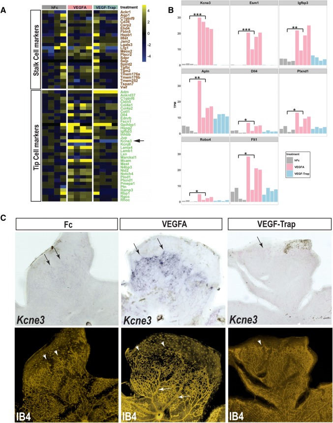
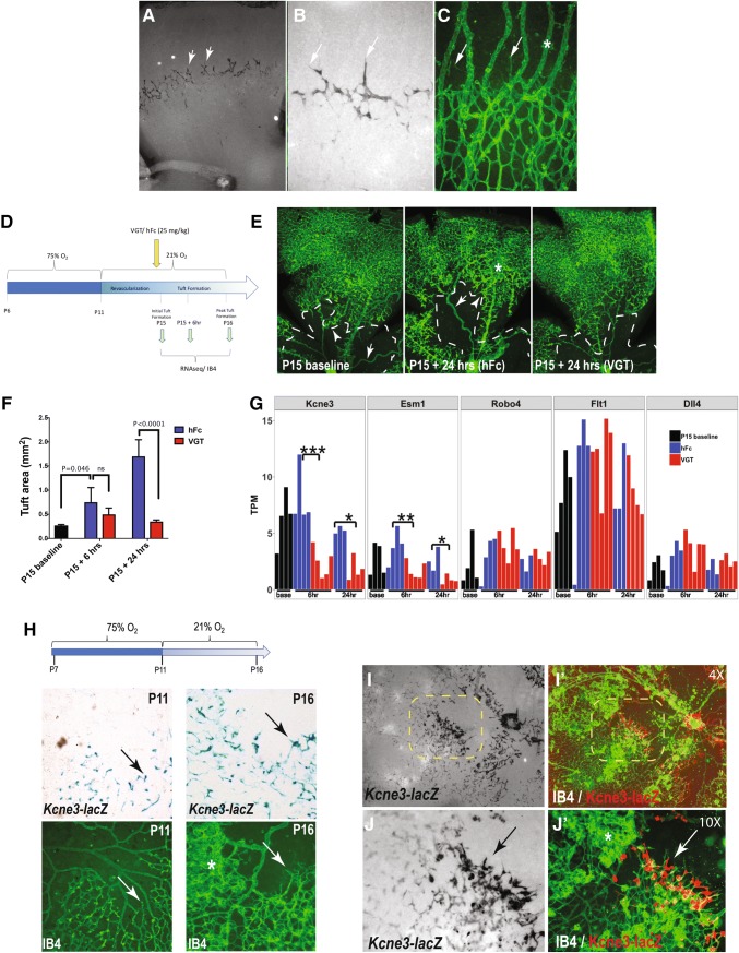
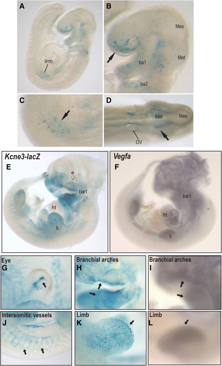
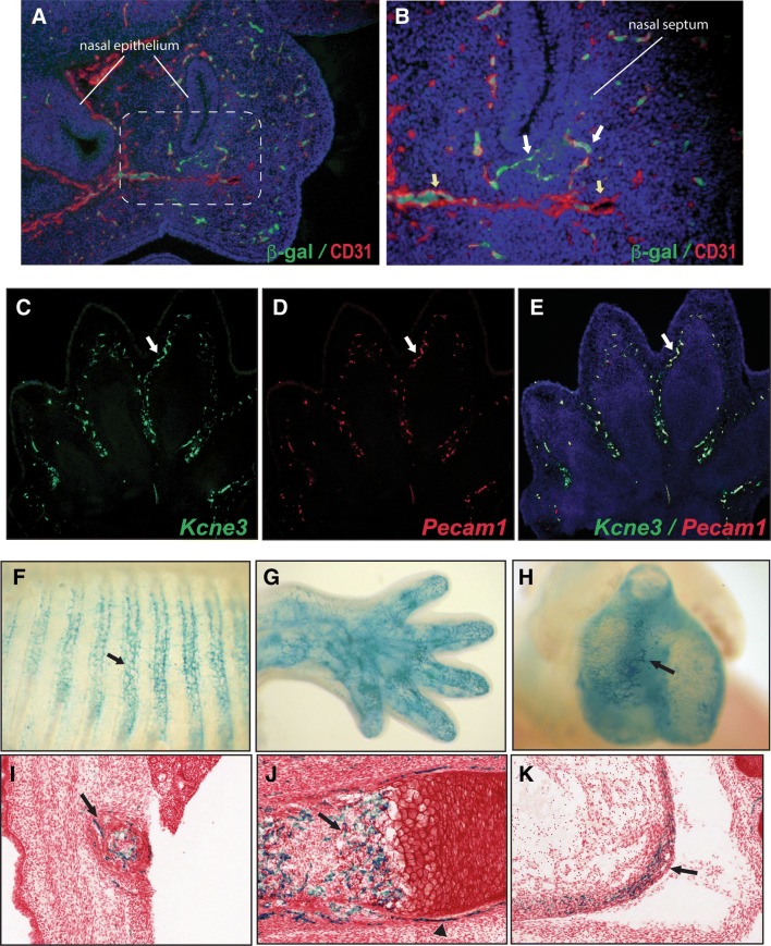
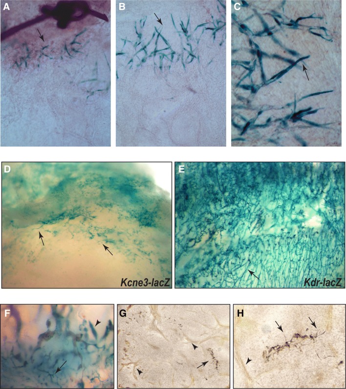
Angiogenesis is largely driven by motile endothelial tip-cells capable of invading avascular tissue domains and enabling new vessel formation. Highly responsive to Vascular Endothelial Growth-Factor-A (VEGFA), endothelial tip-cells also suppress angiogenic sprouting in adjacent stalk cells, and thus have been a primary therapeutic focus in addressing neovascular pathologies. Surprisingly, however, there remains a paucity of specific endothelial tip-cell markers. Here, we employ transcriptional profiling and a lacZ reporter allele to identify Kcne3 as an early and selective endothelial tip-cell marker in multiple angiogenic contexts. In development, Kcne3 expression initiates during early phases of angiogenesis (E9) and remains specific to endothelial tip-cells, often adjacent to regions expressing VEGFA. Consistently, Kcne3 activation is highly responsive to exogenous VEGFA but maintains tip-cell specificity throughout normal retinal angiogenesis. We also demonstrate endothelial tip-cell selectivity of Kcne3 in several injury and tumor models. Together, our data show that Kcne3 is a unique marker of sprouting angiogenic tip-cells and offers new opportunities for investigating and targeting this cell type.

Keywords: Endothelial tip-cell; Kcne3; Retinal angiogenesis; VEGFA.


Figures

Fig. 1
Fig. 1
Kcne3 is transcriptionally regulated by VEGFA in retinal endothelial cells. a Heat map displaying the expression profiles of previously defined endothelial stalk-cell or tip-cell genes, in postnatal day 8 (P8) mouse retinas 24-h following intravitreal injection of hFc, VEGFA, or VEGF-Trap. Scale shown is log2 transformed fold-change, relative to the median of hFc-treated controls. The analysis of N = 4 mice per treatment group is shown. b Comparative expression of Kcne3 to cognate endothelial and ETC genes—Esm1, Igfbp3, Apln, Dll4, Plxnd1, Robo4, and Flt1, in retinas exposed to hFc, VEGFA, or VEGF-Trap. ***PBH-corrected = 8 × 10−40 − 5 × 10−38, **PBH-corrected = 4.6 × 10−10 − 1.7 × 10−7, *PBH-corrected = 6.5 × 10−4 − 2.8 × 10−3. cTop panel: whole mount ISH analysis of Kcne3 mRNA expression in retinal preparations from P7 mice, 24-h following intravitreal injection of hFc, VEGFA, or VEGF-Trap (representative samples from N = 4 mice per treatment group). Weak Kcne3 signal is observed in ETCs and ESCs within the leading angiogenic front of hFc-injected eyes (left panel, arrows), but is dramatically increased in VEGFA-injected eyes (middle panel, arrows). By comparison, no Kcne3 signal is observed in VEGF-Trap-injected eyes (right panel). Lower panel: sequential fluorescent isolectin (IB4-cy3) staining reveals significant vessel dilation in VEGFA-injected eyes (middle panel; arrows), and also shows that the majority of Kcne3+ cells localize to the peripheral aspects of the vascular plexus (middle panels, arrow and arrowheads)
Fig. 2
Fig. 2
Kcne3 is activated in ETCs during normal and pathogenic retinal angiogenesis. ac β-galactosidase staining of P7 retina from heterozygous Kcne3–lacZ reporter mice at low and high-power showing specific expression in ETCs and weaker expression in ESCs along the angiogenic front (arrows a, b). c Comparative fluorescent isolectin (IB4) showing the overall vascular pattern compared to that in b; staining shows that Kcne3lacZ is absent from hyaloid vessels (asterisk), but labels endothelial cells at the vascular front. d Schematic illustration of regimen for short-term OIR experiments. At the end of the hyperoxic phase (P11), single dose intravitreal injections of hFc or VEGF-Trap (VGT) was performed. Retinal samples were analyzed at baseline, 6-h, and 24-h from the start of neovascular tuft formation at P15 in normoxia. For each independent time point, analysis was performed on N = 3-5 mice per treatment group. e IB4-labeled retinal vessels at baseline and 24-h after return to normoxia, showing a reduction in the vaso-obliterative area (dotted line) and in neovascular tuft formation (asterisk) following VEGF-Trap delivery. f Quantitative image analysis of neovascular tuft area at baseline, 6-, and 24-h following return to normoxia. g Relative mRNA expression of selected endothelial markers, expressed as transcripts per million (TPM), derived from an RNAseq analysis of retinas subjected to OIR. Downregulation in expression levels of Kcne3 and Esm1 are observed under VEGF-Trap treatment conditions (compared to hFc), but other EC-specific genes are not significantly affected. h Analysis of Kcne3–lacZ reporter expression under standard OIR conditions (schematic). Comparative lacZ- and IB4-stained retina highlights the detection of Kcne3 reporter activity in putative ETCs bordering the avascular zone that forms following a 4-day exposure to 75% O2, and which intensifies during 5-days upon return to normoxia (arrows). Note the absence of lacZ + cells in neovascular tufts (asterisk). i, j’ Low and high-power images of Kcne3lacZ P16 retina, 5-days following the return to normoxia, detecting β-galactosidase and IB4+ vessels. Kcne3lacZ+ ETCs inundate the vascular-avascular interface (arrows), but are largely absent from neovascular tufts (asterisk). ***Pvalue = 1.2 × 10−5, **Pvalue = 6.4 × 10−5, *Pvalue = 6.5 × 10−4
Fig. 3
Fig. 3
Kcne3 is an early ETC marker during embryonic angiogenesis. ad Analysis of β-galactosidase activity in homozygous Kcne3lacZ embryos at E9.0 (21 somites), showing expression in nascent endothelial sprouts of the emerging forelimb bud (a, c), branchial arches and nasal prominence (B, arrow), and close proximity to mid- and hind-brain structures (d arrow). el Comparative of Kcne3lacZ reporter activity and Vegfa mRNA (WMISH) in embryos at E10.5. Kcne3lacZ reporter is specific to presumptive ETCs and endothelial sprouts forming the hyaloid vessels (g), microvasculature in branchial arches and facial prominence (h), intersomitic vasculature (j), and limb (k). Vegfa is detected in mesenchyme of all cephalic structures (f, i), heart (f), and limb (l). e eye, li limb, ht heart, ba1 first branchial arch
Fig. 4
Fig. 4
Kcne3 localizes to ECs associated with the developing skeleton. a, b Immunodetection of β-galactosidase (green) and CD31 (red) within the nasal prominence of a Kcne3lacZ embryo at E10.5 (“b” is a high-power view of dotted area in “a”). Kcne3lacZ+ ECs are detected in microvascular sprouts within the condensing mesenchyme of the nasal septum (white arrows b), but are absent from lumenized vessels (yellow arrows b; Note: autofluorescent red blood cells are present in vessel lumen). ce Double fluorescent In Situ Hybridization (ISH) employing RNAscope technology to simultaneously detect Kcne3 (green) and Pecam1 (red) mRNA in the distal limb of a wild-type mouse embryo at E13.5. Kcne3 specifically localizes to all Pecam1+ ECs surrounding the cartilaginous digital elements. fk β-galactosidase expression in Kcne3lacZ mice at E15.5 showing robust staining in microvascular ECs surrounding skeletal anlagen including ribs (f, i), distal limb (g), and femur (j). At this stage, Kcne3LacZ+ ECs are also in the coronary microvasculature of the heart (h, k)
Fig. 5
Fig. 5
Kcne3 is expressed by endothelial tip cells during pathogenic angiogenesis. acKcne3lacZ reporter expression in the corneal suture injury model. Seven days after suture insertion into the central cornea of Kcne3lacZ reporter mice, β-galactosidase is detected in ETCs immediately adjacent to the suture implantation site (arrows), but not in any other vascular structures emanating from the limbal arcade. d LLC cells allografted subcutaneously into Kcne3lacZ homozygous mice result in prominent host-derived lacZ expression within edges of the tumor microvasculature (d, arrows), but not in larger venules or arterioles. e LLC tumor allograft in Kdr-lacZ mouse showing broad expression throughout the capillary network (arrow). f Subcutaneous teratomas derived from Kcne3lacZ ES cells showing specific β-galactosidase staining in microvascular (arrow) and epithelial structures (arrowhead). g, h Low- and high-power images of sectioned Kcne3lacZ ES tumors showing the immuno-detection of CD31 in endothelial cells of lumenized blood vessels and capillaries (brown, arrowheads), while β-galactosidase is expressed solely by CD31+ ETCs (black, arrows)

Similar articles

Cited by

References

    1. Carmeliet P, Jain RK. Angiogenesis in cancer and other diseases. Nature. 2000;407(6801):249–257. - PubMed
    1. Carmeliet P, Jain RK. Molecular mechanisms and clinical applications of angiogenesis. Nature. 2011;473(7347):298–307. - PMC - PubMed
    1. Duh EJ, Sun JK, Stitt AW. Diabetic retinopathy: current understanding, mechanisms, and treatment strategies. JCI Insight. 2017;2(14):e93751. - PMC - PubMed
    1. Potente M, Gerhardt H, Carmeliet P. Basic and therapeutic aspects of angiogenesis. Cell. 2011;146(6):873–887. - PubMed
    1. Roca C, Adams RH. Regulation of vascular morphogenesis by Notch signaling. Genes Dev. 2007;21(20):2511–2524. - PubMed

MeSH terms

Substances