Review
. 2019 Dec;67(12):1965-1973.
doi: 10.4103/ijo.IJO_2036_19.

Circumscribed choroidal hemangioma: An overview of clinical manifestation, diagnosis and management

Affiliations
Review

Circumscribed choroidal hemangioma: An overview of clinical manifestation, diagnosis and management

Mrittika Sen et al. Indian J Ophthalmol. 2019 Dec.

Abstract

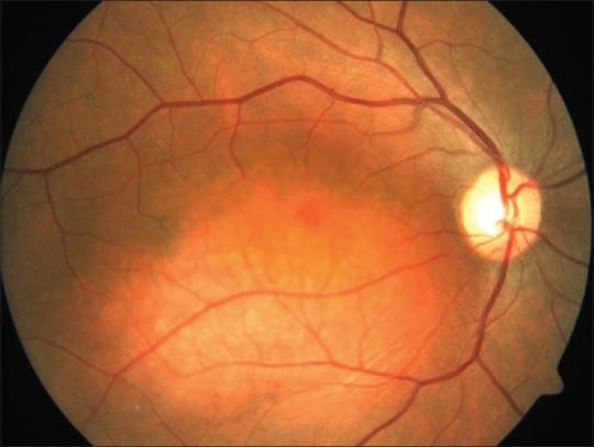
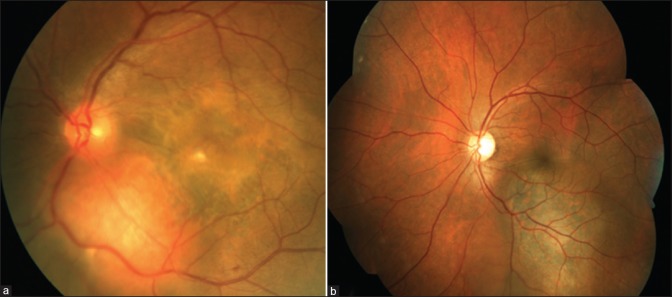
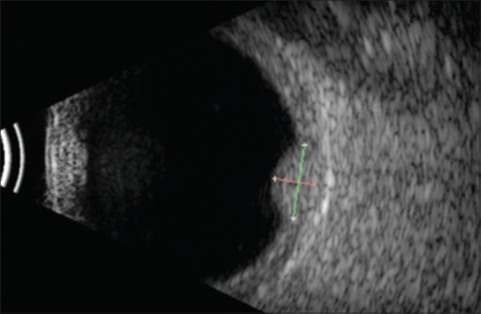
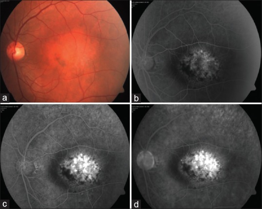
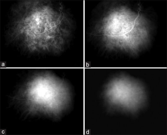
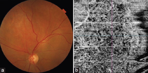
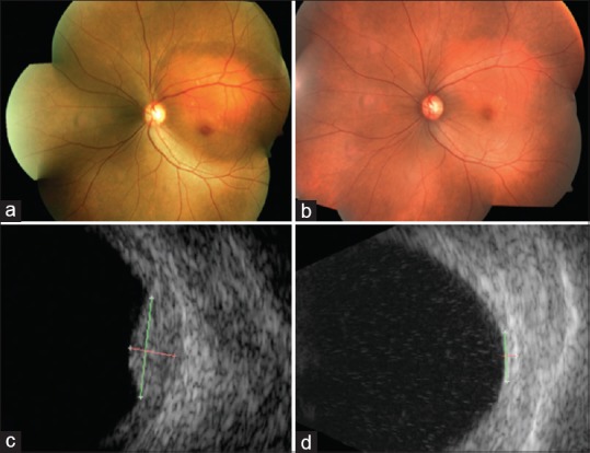
Circumscribed choroidal hemangioma is a benign vascular tumor which presents in middle-aged adults with progressive diminution of vision, metamorphopsia, floaters, and visual field defects. Diagnosis is based on the characteristic clinical features. It is an orange-red, usually solitary, tumor situated in the posterior pole. The visual symptoms are because of the associated subretinal fluid, cystoid macular edema, and, in long-standing cases, retinal pigment epithelium changes, subretinal fibrosis and retinoschisis. It must be distinguished from the more ominous amelanotic melanoma and choroidal metastasis. Diagnostic tools such as ultrasound, fundus fluorescein angiography, indocyanine green angiography, and optical coherence tomography are helpful in cases with diagnostic dilemma. Treatment is indicated in symptomatic cases. The management of choroidal hemangioma has evolved over the years beginning with laser photocoagulation to transpupillary thermotherapy, photodynamic therapy, plaque brachytherapy and external beam radiotherapy. No one therapeutic option holds superiority over the other. In this article, we review the epidemiology, clinical manifestations and treatment of the circumscribed variant of choroidal hemangioma.

Keywords: Brachytherapy; choroidal hemangioma; photodynamic therapy; subretinal fluid; transpupillary thermotherapy.


Conflict of interest statement

None

Figures

Figure 1
Figure 1
Fundus photograph of a 26-year-old gentleman with circumscribed choroidal hemangioma showing a well-defined orange-red mass situated in the posterior pole involving the fovea
Figure 2
Figure 2
Fundus photographs of (a) circumscribed choroidal hemangioma with subfoveal fluid, fibrous metaplasia over the fovea and (b) choroidal melanoma with overlying orange pigments
Figure 3
Figure 3
Ultrasound B-scan of a circumscribed choroidal hemangioma showing an acoustically solid dome-shaped mass with echogenicity like that of the surrounding normal choroid
Figure 4
Figure 4
(a) Fundus photograph of a macular circumscribed choroidal hemangioma and the fundus fluorescein angiography showing (b) early lacy hyperfluorescence (30 s), (c) intense hyperfluorescence (1.42 min) that persists in (d) the late phase (15 min)
Figure 5
Figure 5
ICG angiography in a case of circumscribed choroidal hemangioma. The arterial phase shows filling of intratumoral vessels on a hypofluorescent tumoral background (a). During the venous phase, the tumor reaches maximal intensity of fluorescence (b), with superimposed hyper- and hypofluorescent spots. The late phases (c and d) show a hypofluorescent lesion with sparse hyperfluorescent caverns. Early hyperfluorescence and late hyporfluorescence on ICG is termed “washout phenomenon,” typical of choroidal hemangioma
Figure 6
Figure 6
(a) Fundus photograph of the left eye showing circumscribed choroidal hemangioma superior to the disc with subretinal fluid at the macula and (b) optical coherence tomography angiography showing a dense vascular network in the choriocapillary layer. (figure reprinted with permission from Dr Mahesh Shanmugam, Konana VK, Shanmugam PM, Ramanjulu R, Mishra KD, Sagar P. Optical coherence tomography angiography features of choroidal hemangioma. Indian journal of ophthalmology. 2018 Apr;66(4):581.)[28]
Figure 7
Figure 7
(a) Fundus photograph and (c) ultrasound of a 52-year-old lady with juxtapapillary circumscribed choroidal hemangioma of the left eye with surrounding subretinal fluid involving the fovea causing diminution of vision (20/400). She was treated with Ruthenium-106 plaque brachytherapy with apex dose of 40 Gy. Six months after the treatment, (b) the lesion had regressed as a placoid scar with (d) no measurable mass on ultrasound and with complete resolution of subretinal fluid and visual acuity of 20/40

References

    1. Mashayekhi A, Shields CL. Circumscribed choroidal hemangioma. Curr Opin Ophthalmol. 2003;14:142–9. - PubMed
    1. Shields CL, Honavar SG, Shields JA, Cater J, Demirci H. Circumscribed choroidal hemangioma: Clinical manifestations and factors predictive of visual outcome in 200 consecutive cases. Ophthalmology. 2001;108:2237–48. - PubMed
    1. Krohn J, Rishi P, Frøystein T, Singh AD. Circumscribed choroidal haemangioma: Clinical and topographical features. Br J Ophthalmo. 2019;103:1448–1452. - PMC - PubMed
    1. Schalenbourg A, Piguet B, Zografos L. Indocyanine green angiographic findings in choroidal hemangiomas: A study of 75 cases. Ophthalmologica. 2000;214:246–52. - PubMed
    1. Sanborn GE, Augsburger JJ, Shields JA. Treatment of circumscribed choroidal hemangiomas. Ophthalmology. 1982;89:1374–80. - PubMed