

Case Reports
doi: 10.4103/ijo.IJO_1928_18.
Optical coherence tomography angiography-based analysis of intrinsic vasculature in juxtapapillary melanoma after ruthenium-106 plaque brachytherapy
Affiliations
- PMID: 31755470
- PMCID: PMC6896575
- DOI: 10.4103/ijo.IJO_1928_18
Item in Clipboard
Case Reports
Optical coherence tomography angiography-based analysis of intrinsic vasculature in juxtapapillary melanoma after ruthenium-106 plaque brachytherapy
Indian J Ophthalmol.
2019 Dec.
Abstract
In this case report, we demonstrate the use of optical coherence tomography angiography (OCTA) as a tool to evaluate intrinsic vasculature in a case of juxtapapillary melanoma which underwent ruthinium.106 plaque brachytherapy. In this case, OCTA could demonstrate a decrease in caliber and density of the intrinsic vasculature of the tumor post brachytherapy.
Keywords: Angiography; OCTA; brachytherapy; juxtapapillary; melanoma; response; tumor; vasculature.
Conflict of interest statement
None
Figures
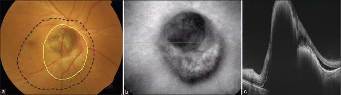
(a) Color photograph of right eye showing a brownish, elevated lesion over disc with U-shaped extension below the disc(yellow line). Blood vessels over the lesion can be seen dipping into the tumor. Subretinal fluid was noted around the lesion which involved the fovea (black dotted line). (b) Infrared reflectance image of right eye showing the extent of the lesion. (c) Optical coherence tomography of right eye disc showing a bilobed elevated lesion with high surface reflectivity with back shadowing. Subretinal fluid can be seen adjacent to the lesion
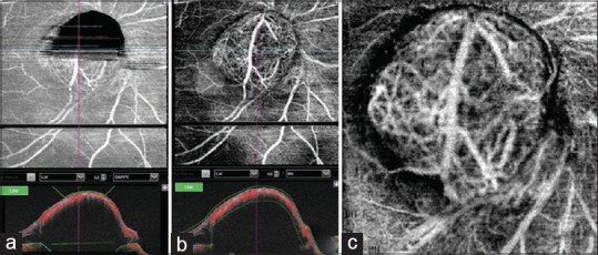
(a) Optical coherence tomography angiography (OCTA) after automatic segmentation. Upper half of the lesion over the disc appears black due to segmentation artifact. (b) OCTA of the same lesion after manual segmentation. (c) OCTA 6 × 6 mm protocol showing large retinal vessel over the tumor with its branches entering the tumor with interlacing of tumor vasculature
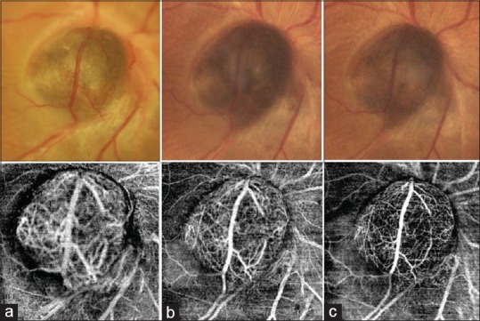
Serial color photograph and corresponding 6 × 6 mm OCTA scans at (a) presentation, (b) 3 months after plaque brachytherapy, and (c) 6 months after plaque brachytherapy. Serial photographs show significant decrease in size, color, and caliber of the overlying vessel. Prominent vascular branches seen at presentation could not be noted after brachytherapy indicating decrease in the vascularity of the tumor. Serial OCTA of the lesion demonstrated significant decrease in the caliber and density of the intrinsic vasculature of the tumor
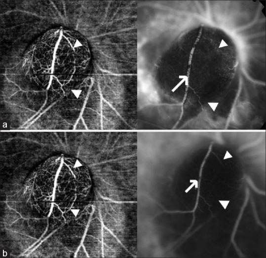
(a) OCTA at 6 months of follow-up with corresponding fluorescein angiography (FA). Note that the vessel seen on OCTA is not seen on FA (white arrow head) and large vessel traversing the tumor is partially obscured by pigment clumps on FA (white arrow). Leakage can be noted around the lesion. (b) OCTA and corresponding indocyanine green angiography (ICG) at 6 months of follow-up. The vascular branches and the intrinsic vasculature were better visualized with OCTA when compared with ICG (white arrow head). Vessel traversing the tumor was segmentally obscured by overlying pigment clumps on ICG (white arrow)
Similar articles
-
Choroidal melanoma suspect. Conservative treatment and evolution. Case report.Rom J Ophthalmol. 2016 Oct-Dec;60(4):264-269. Rom J Ophthalmol. 2016. PMID: 29450360 Free PMC article.
-
Intravitreal aflibercept for the treatment of radiation-induced macular edema after ruthenium 106 plaque radiotherapy for choroidal melanoma.Graefes Arch Clin Exp Ophthalmol. 2019 Jul;257(7):1547-1554. doi: 10.1007/s00417-019-04347-6. Epub 2019 May 12. Graefes Arch Clin Exp Ophthalmol. 2019. PMID: 31081526
-
Blue-Light Fundus Autofluorescence Imaging following Ruthenium-106 Brachytherapy for Choroidal Melanoma.Ophthalmologica. 2020;243(4):303-315. doi: 10.1159/000504715. Epub 2020 Jan 15. Ophthalmologica. 2020. PMID: 31940652
-
Predicting local control of choroidal melanomas following ¹⁰⁶Ru plaque brachytherapy.Br J Ophthalmol. 2011 Feb;95(2):166-70. doi: 10.1136/bjo.2009.176198. Epub 2010 Oct 1. Br J Ophthalmol. 2011. PMID: 20889528 Review.
-
PSEUDO UVEAL MELANOMA CAUSED BY OPTIC DISK DRUSEN WITH JUXTAPAPILLARY CHOROIDAL NEOVASCULAR MEMBRANE.Retin Cases Brief Rep. 2016 Spring;10(2):168-70. doi: 10.1097/ICB.0000000000000218. Retin Cases Brief Rep. 2016. PMID: 26444522 Free PMC article. Review.
Cited by
-
Importance of Optical Coherence Tomography and Optical Coherence Tomography Angiography in the Imaging and Differentiation of Choroidal Melanoma: A Review.Cancers (Basel). 2022 Jul 10;14(14):3354. doi: 10.3390/cancers14143354. Cancers (Basel). 2022. PMID: 35884415 Free PMC article. Review.
References
-
- Shields CL, Shields JA. Recent developments in the management of choroidal melanoma. Curr Opin Ophthalmol. 2004;15:244–51. - PubMed
-
- Emara K, Weisbrod DJ, Sahgal A, McGowan H, Jaywant S, Michaels H, et al. Stereotactic radiotherapy in the treatment of juxtapapillary choroidal melanoma. Int J Radiat Oncol Biol Phys. 2004;59:94–100. - PubMed
-
- Weinhaus RS, Seddon JM, Albert DM, Gragoudas ES, Robinson N. Prognostic factor study of survival after enucleation for juxtapapillary melanomas. Arch Ophthalmol. 1985;103:1673–7. - PubMed
-
- Schachat AP. Ryan's retina. In: Boldt CH, Houston SK, Markoe AM, Murray TG, editors. Brachytherapy for Choroidal Melanoma. 6th ed. Elsevier; 2018. pp. 2566–81.
Publication types
MeSH terms
Substances
LinkOut - more resources
Full Text Sources
Medical
