Review
. 2020 Feb 10:82:21-43.
doi: 10.1146/annurev-physiol-021119-034453. Epub 2019 Nov 22.

Cardiac Pacemaker Activity and Aging

Affiliations
Review

Cardiac Pacemaker Activity and Aging

Colin H Peters et al. Annu Rev Physiol. .

Abstract

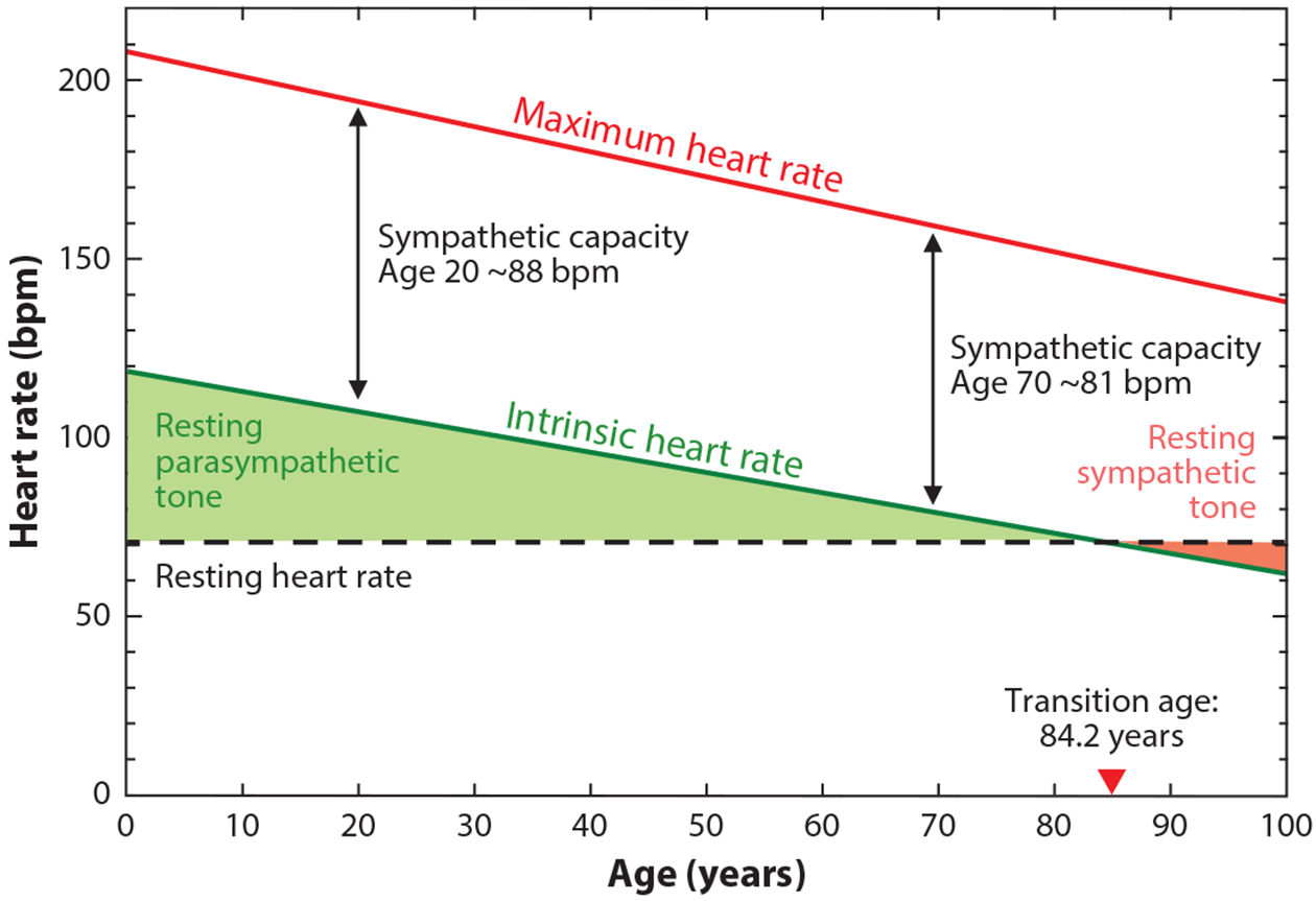
A progressive decline in maximum heart rate (mHR) is a fundamental aspect of aging in humans and other mammals. This decrease in mHR is independent of gender, fitness, and lifestyle, affecting in equal measure women and men, athletes and couch potatoes, spinach eaters and fast food enthusiasts. Importantly, the decline in mHR is the major determinant of the age-dependent decline in aerobic capacity that ultimately limits functional independence for many older individuals. The gradual reduction in mHR with age reflects a slowing of the intrinsic pacemaker activity of the sinoatrial node of the heart, which results from electrical remodeling of individual pacemaker cells along with structural remodeling and a blunted β-adrenergic response. In this review, we summarize current evidence about the tissue, cellular, and molecular mechanisms that underlie the reduction in pacemaker activity with age and highlight key areas for future work.

Keywords: aging; cardiac pacemaking; intrinsic heart rate; maximum heart rate; sinoatrial node.


Figures

Figure 1
Figure 1
Similar declines in maximum and intrinsic heart rates with no change in resting heart rate necessitate an increase in sympathetic tone with age. Maximum heart rate (mHR; red line) is plotted as 208 – (0.7 × age); data from Reference . Intrinsic heart rate (green line) is plotted as 118 – (0.57 × age); data from Reference . Resting heart rate (dashed black line) is plotted as a constant 70 beats per minute (bpm) throughout life. The red arrowhead indicates an average transition age (84.2 years) beyond which sympathetic tone at rest (pink shading) predominates over parasympathetic tone at rest (green shading) in order to maintain resting heart rate. Sympathetic capacity (black double arrows), the difference between intrinsic and maximum heart rate, decreases slightly with age; however, the majority of the age-dependent decline in mHR is caused by the decline in intrinsic heart rate.
Figure 2
Figure 2
The cardiac conduction system and sinoatrial node myocytes. (a) The cardiac conduction system visualized in a heart from a contactin2-eGFP reporter mouse. The pacemaker depolarization initiated by the sinus node propagates through the atria to the atrioventricular (AV) node and then on to the ventricles via the His-Purkinje network. Adapted from Reference under the terms of the Creative Commons Attribution (CC BY) License, http://creativecommons.org/licenses/by/4.0. (b) Sinoatrial node myocyte (SAM) morphology. SAMs are long, thin cells with few striations. Shown is a mouse SAM in the recording chamber of an electrophysiology rig. Note the patch pipette entering from the right that contacts the cell near its center. Adapted from Reference under the terms of the Creative Commons Attribution-ShareAlike (CC BY-SA) License, https://creativecommons.org/licenses/by-sa/4.0. (c) Rich sympathetic innervation of SAMs. Co-immunolabeling of SAMs and sympathetic nerve fibers with antibodies against HCN4 (red) and tyrosine hydroxylase (green), respectively. Image on right shows enlargement of boxed area from image on left. E. Larson and C. Proenza, unpublished data.
Figure 3
Figure 3
Characteristic waveform of sinoatrial node action potentials. (a) Schematic illustration of a sinoatrial action potential (top) and its first derivative (bottom) indicating key features of the action potential. (b) Summary of action potential waveform parameter descriptions. Adapted from Reference . Copyright 2017, Elsevier.
Figure 4
Figure 4
Mechanisms of the age-dependent decline in maximum heart rate (mHR). Shown are known (black boxes and arrows, color-coded to corresponding subpanels) and hypothesized (gray boxes and arrows) mechanisms associated with reduced pacemaker function in aging as described in this review. mHR is decreased primarily due to a reduction in intrinsic heart rate (iHR), as well as a reduction in the response to β-adrenergic receptor (βAR) stimulation. (a) iHR is decreased in part as a result of reduced action potential (AP) firing rate in sinoatrial myocytes (SAMs) from aged mice (red) compared to young mice (black). (b) The decreased SAM AP firing rate is caused in turn by a reduced diastolic depolarization rate (DDR), a hyperpolarized maximum diastolic potential (MDP), and an increased AP duration. The decrease in iHR is also accompanied by a reduction in the number of SAMs and fibrosis and structural remodeling of the sinoatrial node (SAN). Panels a and b adapted from Reference . (c) Increased diffuse fibrosis in a SAN section from an aged human heart (right), as indicated by increased blue-green staining compared to a section from a young heart (left). Adapted from Reference under the terms of the Creative Commons Attribution (CC BY) License, http://creativecommons.org/licenses/by/4.0. ICa,L, ICa,T, and If currents have been demonstrated to be reduced in aged mouse SAMs. (d) Hyperpolarized voltage dependence of activation of If in excised inside-out membrane patches from SAMs from young (black) or old (red) mice. Adapted from Reference under the terms of the Creative Commons Attribution-ShareAlike (CC BY-SA) License, https://creativecommons.org/licenses/by-sa/4.0. (e) Reduced ICa,L current density in SAMs from aged mice. Adapted from Reference . We speculate that INa, INCX, and IK may also be decreased with age (gray boxes). Reduced expression of NCX1, RYR2, SERCA2, and Cx43 transcripts and protein has been observed in a variety of species. (f) Reduced relative mRNA abundance of RYR2 and SERCA2 in SAN of old (red) compared to young (black) rats, normalized to expression in atrial muscle. Adapted with permission from Reference . Copyright 2011, John Wiley & Sons. We believe that aging may also reduce expression of many other channels and signaling molecules (gray boxes). Asterisks in panels e and f indicate significant age-dependent differences (p < 0.05).

References

    1. Homer. 1990. Iliad. London: Penguin
    1. Robinson S. 1938. Experimental studies of physical fitness in relation to age. Eur. J. Appl. Physiol 10(3):251–323
    2. Seminal study showing age-dependent declines in mHR.

    1. Robinson S, Dill DB, Tzankoff SP, Wagner JA, Robinson RD. 1975. Longitudinal studies of aging in 37 men. J. Appl. Physiol 38(2):263–67 - PubMed
    1. Heath GW, Hagberg JM, Ehsani AA, Holloszy JO. 1981. A physiological comparison of young and older endurance athletes. J. Appl. Physiol 51(3):634–40 - PubMed
    1. Tanaka H, Monahan KD, Seals DR. 2001. Age-predicted maximal heart rate revisited. J. Am. Coll. Cardiol 37(1):153–56 - PubMed

Publication types

LinkOut - more resources