. 2020 Feb:203:110859.
doi: 10.1016/j.jinorgbio.2019.110859. Epub 2019 Sep 13.

Evaluation of the regulatory model for Ni2+ sensing by Nur from Streptomyces coelicolor

Affiliations

Evaluation of the regulatory model for Ni2+ sensing by Nur from Streptomyces coelicolor

Olivia M Manley et al. J Inorg Biochem. 2020 Feb.

Abstract

Streptomyces coelicolor is a soil-dwelling bacterium that is medically important due to its ability to produce several antibiotics, and nickel accumulation within this organism has been shown to prevent the production of the antibiotic undecylprodigiosin. The transcriptional repressor important in regulation of nickel uptake is the homodimeric Nur, a member of the Fur family. Nur contains two metal-binding sites per monomer: the M-site and the Ni-site. The work described here seeks to determine the roles of each of the metal-binding sites to establish a model of Nur activity through mutational studies, metal titrations, and fluorescence anisotropy. Through these studies, a model of Nur activity is proposed in which femtomolar metal binding to one M-site of Nur prompts DNA-binding, and metal binding to the second M-site fully activates the protein. Evidence is provided that shows cooperative metal binding to the Ni-site, but this process dampens affinity for promoter DNA.


Figures

Figure 1:
Figure 1:
The ribbon structure of dimeric Nur. One Nur monomer is shown in black and the other in white. Metal-coordinating residues of the M- and Ni-sites are shown. The flexible loop proposed to be stabilized by metal binding at the M-site is highlighted in yellow.
Figure 2.
Figure 2.
Competition metal binding titration between MagFura2 and Nur. 10 μM each Nur dimer and MF2 were titrated with 1 mM Ni2+ in 25 mM Tris 100 mM NaCl pH 8.0. Panel A shows the raw spectra, with the inital spectrum shown in red and the spectrum at the conclusion of the titration shown in blue. Panel B shows the absorbance plotted vs. [Ni2+] overlaid with the best fit to the data according to a competition model that included two independent sites per Nur dimer and MF2.
Figure 3:
Figure 3:
DNA binding of Nur. The saturation of DNA by WT-Nur (black), ΔM-Nur (red), ΔNi-Nur (blue), and WT-Nur with 10 mM supplementary Ni2+ (green) with fluorescein-labeled sodF promoter.
Figure 4
Figure 4
Relative Zn2+ concentration overlaid on a gel filtration (red) trace of Nur with excess EDTA with (bottom) or without (top) sodF promoter DNA. The blue and black lines in the bottom panel represent the elution profile of the sodF promoter DNA and the Nur protein, respectively, for comparison.
Figure 5.
Figure 5.
A: 10 nM sodF and 5 μM Nur incubated in the presence (black squares) or absence (red circles) of 1 mM EDTA. B: Ni2+ titrated into Nur-sodF complex that has been incubated with 150 μM EDTA. The red line is a best fit according to the model described in the text where KNi-Nur = 4.7 × 10¹⁴ (Kd = 2.13 fM). The fractional saturation is calculated from fluorescence anisotropy values determined for each time point.
Figure 6:
Figure 6:. Nur docked to a model of the sodF promoter.
A: The recognition helix of Nur, which interacts with the major groove of DNA and nonspecifically with the backbone, is highlighted in yellow. The inset shows a magnified view of His70, His72, His 126, and Glu63. Potential hydrogen bonds are shown in green [7,27]. B: Binding isotherms of wild type (WT) and E63A Nur proteins titrated into the sodF promoter in the presence of absence of 10 μM Ni2+.
Figure 7:
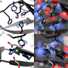
Figure 7:. Magnified views of the metal-binding sites in the crystal structure of Nur.
The backbone of the protein is shown in black, and solvent ligands that coordinate metal at the Ni-site are shown in light grey. Heteroatoms are also colored, with red being oxygen and blue being nitrogen. A: The M-site with metal-coordinating residues shown. B: The M-site with the surface of the protein shown with 30% transparency, showing that the M-site metal is shielded from solvent. C: The Ni-site with metal-coordinating residues shown. Solvent molecules from the crystallization matrix, ethylene glycol and malonate coordinate nickel at the Ni-site. D: The Ni-site with the surface of the protein shown with 30% transparency, showing that the Ni-site is exposed to solvent. Solvent molecules ethylene glycol and malonate are shown in grey.

References

    1. Mulrooney SB, Hausinger RP, Nickel uptake and utilization by microorganisms., FEMS Microbiol. Rev 27 (2003) 239–261. - PubMed
    1. Stohs SJ, Bagchi D, Oxidative mechanisms in the toxicity of metal ions., Free Radic. Biol. Med 18 (1995) 321–336. - PubMed
    1. Macomber L, Hausinger RP, Mechanisms of nickel toxicity in microorganisms., Metallomics. 3 (2011) 1153–1162. doi:10.1039/c1mt00063b. - DOI - PMC - PubMed
    1. Abbas AS, Edwards C, Effects of Metals on Streptomyces coelicolor Growth and Actinorhodin Production, Appl. Environ. Microbiol. 56 (1990) 675–680. http://www.ncbi.nlm.nih.gov/pmc/articles/PMC183404/. - PMC - PubMed
    1. Kim HM, Ahn B-E, Lee J-H, Roe J-H, Regulation of a nickel-cobalt efflux system and nickel homeostasis in a soil actinobacterium Streptomyces coelicolor, Metallomics. 7 (2015) 702–709. doi:10.1039/C4MT00318G. - DOI - PubMed

Publication types

LinkOut - more resources