Review
. 2020 Jan;59(1):39-44.
doi: 10.1016/j.clinimag.2019.08.006. Epub 2019 Oct 25.

Follicular pancreatitis: A rare pancreatic inflammatory pseudotumor

Affiliations
Review

Follicular pancreatitis: A rare pancreatic inflammatory pseudotumor

W James Tom et al. Clin Imaging. 2020 Jan.

Abstract

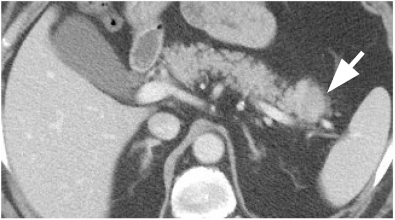
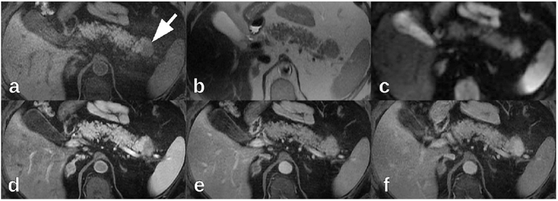
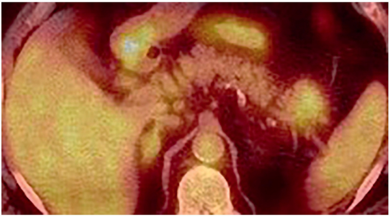
Inflammatory pseudotumors imitate neoplasms on imaging but actually represent focal inflammation. We report a case of follicular pancreatitis, which is a recently recognized distinct form of mass-forming focal chronic pancreatitis pathologically characterized by lymphoid infiltration with abundant reactive germinal centers. In our patient, follicular pancreatitis manifested as a pancreatic tail mass that was resected due to imaging findings, which were suggestive of pancreatic malignancy. We performed a literature review of this rare condition and present a summary of reported imaging findings. The most distinguishing feature from pancreatic adenocarcinoma is the enhancement pattern, as follicular pancreatitis enhances more than the surrounding pancreatic parenchyma on delayed post-contrast images which is unusual for pancreatic adenocarcinoma. If this benign diagnosis is suggested on imaging, unnecessary surgery and its potential complications may be avoided.

Keywords: Chronic pancreatitis; Follicular pancreatitis; Mass-forming pancreatitis.


Conflict of interest statement

Declarations of interest

None. The funding source was not involved in study design, data collection, data analysis, data interpretation, writing, or the decision to submit for publication.

Figures

Figure 1:
Figure 1:
Contrast-enhanced abdominal CT in the portal venous phase demonstrated a 2.8 cm hypodense pancreatic tail mass (arrow).
Figure 2:
Figure 2:
On 1.5 T MRI, the pancreatic tail mass (arrow) was (a) hypointense on axial non-contrast fat-suppressed T1-weighted images, (b) isointense on axial T2-weighted images, and (c) hyperintense on axial diffusion-weighted imaging (b=600 mm2/s). On axial post-contrast fat-suppressed T1-weighted imaging (gadobutrol), the pancreatic tail mass was (d) hypointense in the late hepatic arterial phase, (e) isointense in the portal venous phase, and (f) hyperintense in delayed phases.
Figure 3:
Figure 3:
Endoscopic ultrasound demonstrated a well-defined, hypoechoic, solid-appearing pancreatic tail mass.
Figure 4:
Figure 4:
On 18F-FDG PET/CT, the pancreatic tail mass had elevated FDG uptake (SUVmax 4.8).
Figure 5:
Figure 5:
Hematoxylin and eosin stained slides of the pancreatic tail mass demonstrated scattered lymphoid follicles with reactive germinal centers and atrophic changes of the pancreatic acini and ducts at (a) 40x magnification and (b) 100x magnification.

Similar articles

References

    1. Klöppel G, Chronic pancreatitis, pseudotumors and other tumor-like lesions, Modern Pathology 20 (2007) S113–S131. doi:10.1038/modpathol.3800690. - DOI - PubMed
    1. Klöppel G, Adsay NV, Chronic pancreatitis and the differential diagnosis versus pancreatic cancer, Archives of Pathology & Laboratory Medicine 133 (3) (2009) 382–387. doi:10.1043/1543-2165-133.3.382. URL https://www.archivesofpathology.org/doi/abs/10.1043/1543-2165-133.3.382 - DOI - DOI - PubMed
    1. Coakley FV, Hanley-Knutson K, Mongan J, Barajas R, Bucknor M, Qayyum A, Pancreatic imaging mimics: Part 1, imaging mimics of pancreatic adenocarcinoma, American Journal of Roentgenology 199 (2) (2012) 301–308. doi:10.2214/AJR.11.7907. - DOI - PubMed
    1. Raman SP, Salaria SN, Hruban RH, Fishman EK, Groove pancreatitis: Spectrum of imaging findings and radiology-pathology correlation, American Journal of Roentgenology 201 (1) (2013) W29–W39. doi:10.2214/AJR.12.9956. - DOI - PMC - PubMed
    1. Kwon JH, Kim JH, Kim SY, Byun JH, Kim HJ, Hong S-M, Lee M-G, Lee SS, Imaging and clinical features of xanthogranulomatous pancreatitis: an analysis of 10 cases at a single institution, Abdominal Radiology 43 (12) (2018) 3349–3356. doi:10.1007/s00261-018-1630-0. - DOI - PubMed