. 2019 Nov 20;20(23):5839.
doi: 10.3390/ijms20235839.

Nucleoplasmic Reticulum Formation in Human Endometrial Cells is Steroid Hormone Responsive and Recruits Nascent Components

Affiliations

Nucleoplasmic Reticulum Formation in Human Endometrial Cells is Steroid Hormone Responsive and Recruits Nascent Components

Lior Pytowski et al. Int J Mol Sci. .

Abstract

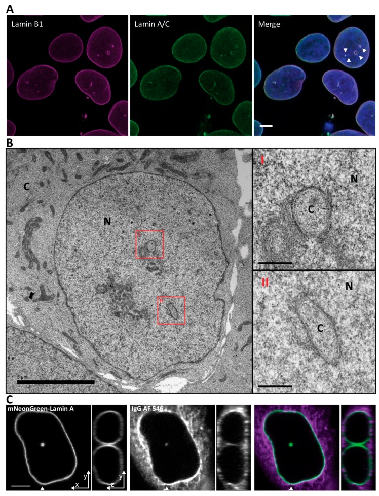
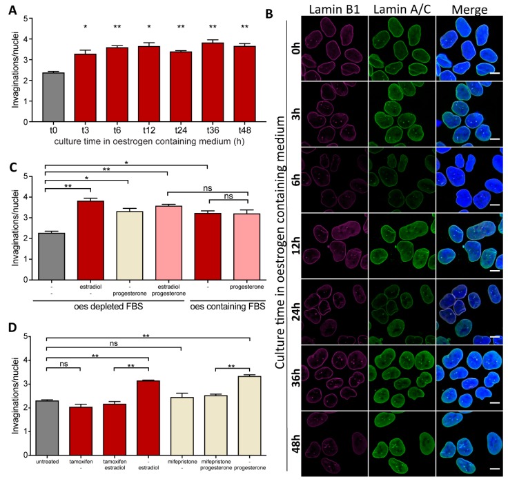
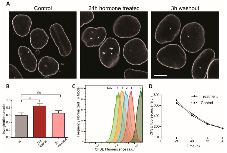
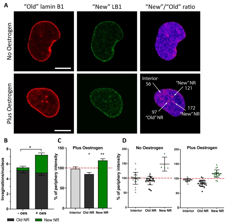
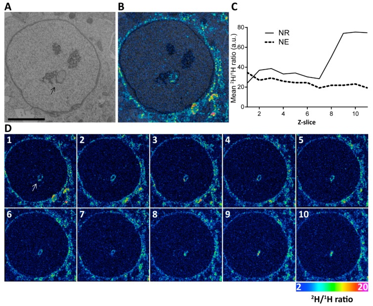
The nuclei of cells may exhibit invaginations of the nuclear envelope under a variety of conditions. These invaginations form a branched network termed the nucleoplasmic reticulum (NR), which may be found in cells in pathological and physiological conditions. While an extensive NR is a hallmark of cellular senescence and shows associations with some cancers, very little is known about the formation of NR in physiological conditions, despite the presence of extensive nuclear invaginations in some cell types such as endometrial cells. Here we show that in these cells the NR is formed in response to reproductive hormones. We demonstrate that oestrogen and progesterone are sufficient to induce NR formation and that this process is reversible without cell division upon removal of the hormonal stimulus. Nascent lamins and phospholipids are incorporated into the invaginations suggesting that there is a dedicated machinery for its formation. The induction of NR in endometrial cells offers a new model to study NR formation and function in physiological conditions.

Keywords: nuclear architecture; nucleoplasmic reticulum; reproductive cycle.


Conflict of interest statement

Authors declare no conflicts of interest with the contents of this article.

Figures

Figure 1
Figure 1
Nuclei of Ishikawa cells contain invaginations of the nuclear envelope forming a nucleoplasmic reticulum. (A) Ishikawa cells immunostained with anti-Lamin B1 and anti Lamin A/C. Scale bar 5 µm. (B) Electron microscopy micrograph of a high-pressure frozen Ishikawa cell. Scale bar is 5 µm for low magnification nucleus and 0.5 µm for high magnification insets. N, nucleus; C, cytoplasm. (C) Cytoplasmic loading by IgG AF 546 to visualize the cytoplasmic core of the invagination. Arrowhead indicates position of orthogonal projection in the panels to the right. Scale bar 5 µm.
Figure 2
Figure 2
Formation of NR tubules in endometrial cells is hormone responsive. (A) NR formation time-course in Ishikawa cells in response to oestrogen. An F-test rejects the null hypothesis (slope = 0) between t0, t3 and t6 (p = 0.005) while this null hypothesis cannot be rejected for later time-point (t 12 to t 48) (p > 0.5). (B) Immunostained Ishikawa cells imaged at different intervals after addition of oestrogen to the culture medium. Scale bar 10 µm. (C) NR abundance in Ishikawa cells in response to estradiol and/or progesterone treatment in medium containing either oestrogen-stripped FBS (oestrogen-depleted FBS) or regular FBS (oestrogen-containing FBS). (D) NR abundance in Ishikawa cells treated for 72 h with estradiol or progesterone and their respective antagonists. Data from 2 independent experiments, 100 nuclei each; mean ± SEM; * for p-value < 0.05, ** for p-value < 0.01, ns for non-significant.
Figure 3
Figure 3
NR formation in endometrial cells is reversible and cell cycle independent. (A) NR induction in Ishikawa cells treated with oestrogen and progesterone for 24h and then subjected to 3 h washout in hormone-free media. Nuclei immunostained with anti-Lamin B1-Cy5. Scale bar 10 µm. (B) NR abundance quantification. Three replicates total, n = 242, 262, 203, respectively. ** for p < 0.01; ns for non-significant. Error bars represent SEM. (C) Hormone addition or removal does not affect cell division rate. Normalised frequency of CFSE fluorescence of Ishikawa cells measured by flow cytometry. Hormones were added for 48 h then removed for another 48 h. Average cell number per profile = 28,000. Individual flow cytograms in Supplementary Figure S1. (D) Average CFSE loss over time of treated and control samples reveal non-significant difference in cell proliferation rates.
Figure 4
Figure 4
Nascent lamin B1 is incorporated in newly formed invaginations. (A) Confocal microscopy of Ishikawa cells expressing lamin B1- Maple3. Indicated are the “old” (red channel) and “new” (green channel) lamin protein pools. Ratiometric image of “New”/”Old” is provided with indication of ratio values for selected ROIs around the features arrowed. (B) Evaluation of invagination abundance per nucleus in Ishikawa cells with (+ oes) or without oestrogen (-oes) treatment. (C) Pixel intensities of the ROIs defined in based on the ratiometric images and normalised to the signal at the nuclear rim showing increased incorporation of nascent lamin B1 at the newly forming NR channels; results from three independent experiments, 35 cells in total; mean ± SD; ** p-value < 0.001; * p-value < 0.05. (D) An example data plot from a single experiment showing distribution of “New”/”Old” lamin B1 ratio at different nuclear structures and normalised to the nuclear rim ratio with or without oestrogen.
Figure 5
Figure 5
Nascent phospholipids are incorporated in the forming NR during estradiol stimulation. (A) Representative backscattered electron micrograph of an Ishikawa nucleus pulse labelled with deuterated choline. Arrow points to an NR channel. Scale bar 5 µm. (B) Overlay of the electron micrograph and NanoSIMS image showing deuterium enrichment at the invagination. (C) Quantification of the mean 2H/1H ratio in the NR versus the NE rim for the ion-beam eroded z-series through this cell. The average 2H/1H ratio for two regions of interest (total NE and NR) are shown for each z plane. This shows increasing nascent deuterated choline signal in the tip of the NR invagination where no lumen is visible. See also Supplementary Movie S1. (D) Panel of individual sections of 3D reconstruction. Color scale of NanoSIMS images 2–20 equals 0.02–0.2% of 2H/1H ratio.

Similar articles

Cited by

References

    1. Prunuske A.J., Ullman K.S. The nuclear envelope: Form and reformation. Curr. Opin. Cell Biol. 2006;18:108–116. doi: 10.1016/j.ceb.2005.12.004. - DOI - PMC - PubMed
    1. Stewart C.L., Roux K.J., Burke B. Blurring the boundary: The nuclear envelope extends its reach. Science. 2007;318:1408–1412. doi: 10.1126/science.1142034. - DOI - PubMed
    1. Grossman E., Medalia O., Zwerger M. Functional architecture of the nuclear pore complex. Annu. Rev. Biophys. 2012;41:557–584. doi: 10.1146/annurev-biophys-050511-102328. - DOI - PubMed
    1. Schirmer E.C., Foisner R. Proteins that associate with lamins: Many faces, many functions. Exp. Cell Res. 2007;313:2167–2179. doi: 10.1016/j.yexcr.2007.03.012. - DOI - PubMed
    1. Fricker M., Hollinshead M., White N., Vaux D. Interphase nuclei of many mammalian cell types contain deep, dynamic, tubular membrane-bound invaginations of the nuclear envelope. J. Cell Biol. 1997;136:531–544. doi: 10.1083/jcb.136.3.531. - DOI - PMC - PubMed

LinkOut - more resources