

Longitudinal Detection of Radiation-Induced Peripapillary and Macular Retinal Capillary Ischemia Using OCT Angiography
- PMID: 31757690
- PMCID: PMC7065970
- DOI: 10.1016/j.oret.2019.10.001
Longitudinal Detection of Radiation-Induced Peripapillary and Macular Retinal Capillary Ischemia Using OCT Angiography
Abstract
Purpose: To study longitudinal changes in retinal capillary circulation in eyes treated with iodine 125 (I125) plaque brachytherapy for uveal melanoma using OCT angiography (OCTA).
Design: Longitudinal prospective study of 21 patients undergoing treatment for uveal melanoma with I125 plaque brachytherapy. Eyes with melanoma were imaged with OCTA before treatment and at 12-month intervals until 2 years after brachytherapy.
Participants: After institutional review board approval, participants were enrolled prospectively from an academic ocular oncology clinic.
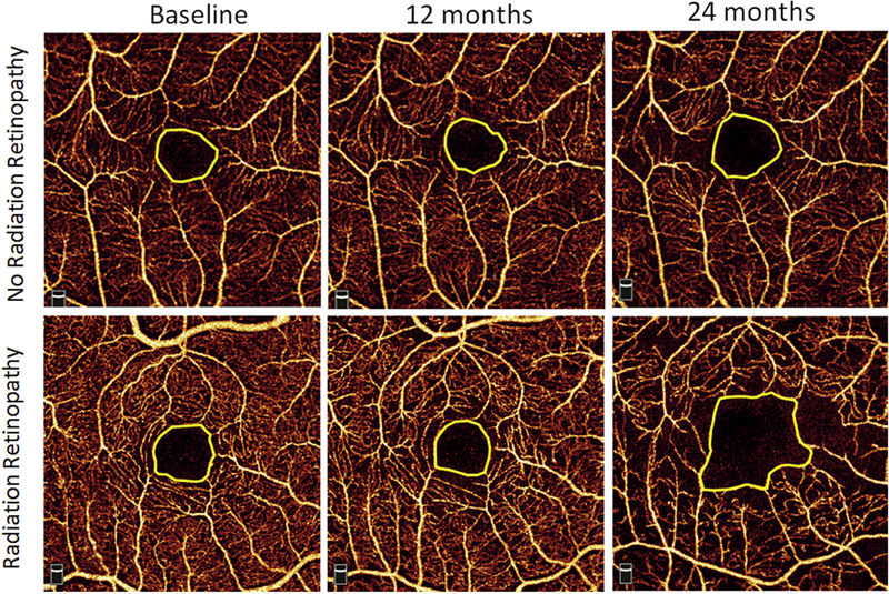
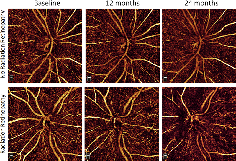
Methods: Peripapillary (4.5 × 4.5-mm) and macular (3 × 3-mm) OCTA scans were acquired with AngioVue (Optovue, Inc, Fremont, CA).
Main outcome measures: The peripapillary nerve fiber layer plexus capillary density (NFLP_CD), macular superficial vascular complex vessel density (mSVC_VD), and foveal avascular zone (FAZ) area were calculated.
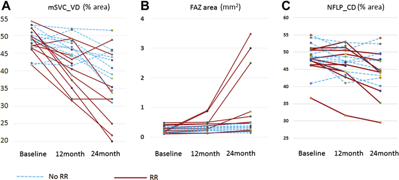
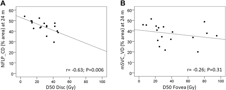
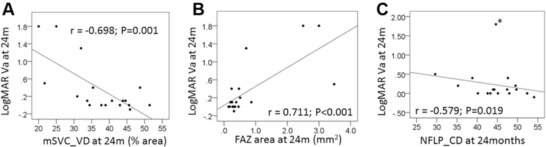
Results: Before treatment, no significant difference was found in the NFLP_CD, mSVC_VD, or FAZ area between eyes with melanoma and normal fellow eyes. By 24 months, 11 eyes had developed clinical signs of radiation retinopathy, radiation optic neuropathy, or both. In treated eyes, the NFLP_CD (48.4±4.1%) was reduced at 12 months (46.7±5.0%; P = 0.04, Wilcoxon signed-rank test) and 24 months (44.5±6.1%; P < 0.001). Similarly, the mSVC_VD (48.4 2±3.6%) was reduced in treated eyes at 12 months (43.5±5.9%; P = 0.01) and 24 months (37.4±9.1%; P < 0.001). The FAZ area (0.26±0.11 mm2) increased in treated eyes at 12 months (0.35±0.22 mm2; P = 0.009) and 24 months (0.81±1.03 mm2; P = 0.001). When only eyes with clinically evident radiation changes were evaluated, the changes in NFLP_CD, mSVC_VD, and FAZ area were more pronounced. OCT angiography measurements correlated with both radiation dose and visual acuity. The mSVC_VD measured at 12 months was found to predict the development of clinically apparent radiation retinopathy within 1 year.
Conclusions: OCT angiography demonstrated early emergence of peripapillary and macular capillary vasculature changes after I125 plaque brachytherapy. OCT angiography provided a quantitative measurement of retinal capillary changes associated with ischemia that correlated with visual acuity and radiation dose and may predict future development of radiation-induced retinal toxicity.
Copyright © 2019 American Academy of Ophthalmology. Published by Elsevier Inc. All rights reserved.
Figures
References
-
- Singh AD, Turell ME, Topham AK. Uveal melanoma: trends in incidence, treatment, and survival. Ophthalmology. 2011;118:1881e1885. - PubMed
-
- Melia BM, Abramson DH, Albert DM, et al. Collaborative Ocular Melanoma Study (COMS) randomized trial of I-125 brachytherapy for medium choroidal melanoma. I. Visual acuity after 3 years. COMS report no. 16. Ophthalmology. 2001;108:348e366. - PubMed
-
- Gunduz K, Shields CL, Shields JA, et al. Radiation retinopathy following plaque radiotherapy for posterior uveal melanoma. Arch Ophthalmol. 1999;117:609e614. - PubMed
Publication types
MeSH terms
Grants and funding
LinkOut - more resources
Full Text Sources
Medical
