Randomized Controlled Trial
. 2020 Feb;4(2):154-163.
doi: 10.1016/j.oret.2019.09.001. Epub 2019 Sep 10.

Longitudinal Panretinal Leakage and Ischemic Indices in Retinal Vascular Disease after Aflibercept Therapy: The PERMEATE Study

Affiliations
Randomized Controlled Trial

Longitudinal Panretinal Leakage and Ischemic Indices in Retinal Vascular Disease after Aflibercept Therapy: The PERMEATE Study

Natalia Figueiredo et al. Ophthalmol Retina. 2020 Feb.

Abstract

Purpose: To characterize the longitudinal panretinal retinal vascular dynamics in diabetic macular edema (DME) and retinal vein occlusion (RVO) over a 12-month period while being treated with intravitreal aflibercept injections (IAIs).

Design: Prospective open-label study (clinicaltrials.gov identifier, NCT02503540).

Participants: Thirty-one treatment-naive eyes with foveal-involving retinal edema secondary to DME and RVO.

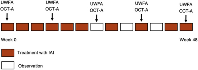
Methods: Participants received 2 mg IAI every 4 weeks for the first 6 months, followed by 2 mg every 8 weeks. Ultra-widefield fluorescein angiography (UWFA; California Optos [Optos, Dunfermline, United Kingdom]) and spectral-domain OCT (Cirrus; Zeiss, Oberkochen, Germany) scans were obtained and analyzed using a novel quantitative assessment platform. Visual acuity, central subfield thickness, and adverse events also were collected.

Main outcome measures: The primary end point was the mean change in panretinal leakage index at month 12 from baseline as measured by UWFA.

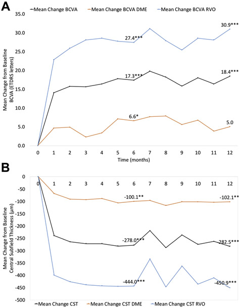
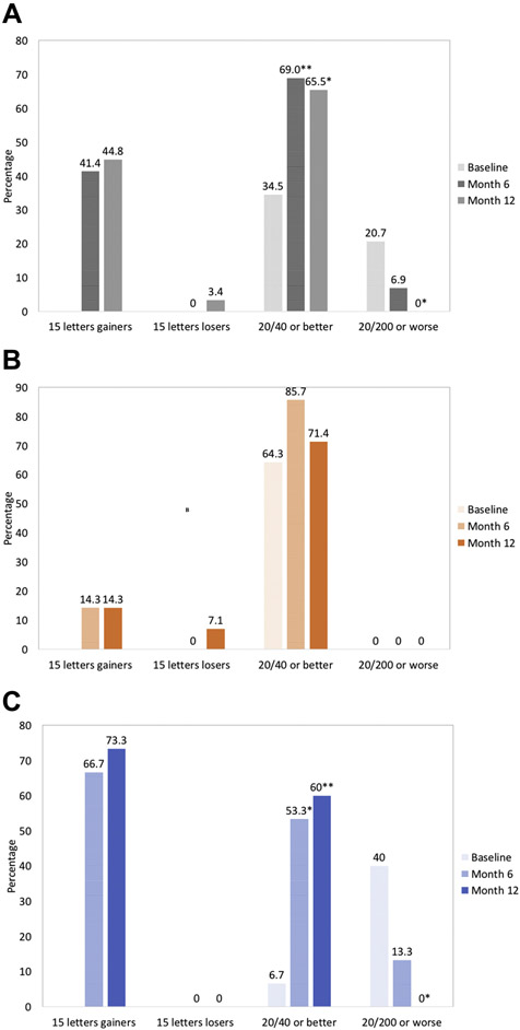
Results: Mean age was 67.1 years. At month 12, visual acuity significantly improved by a mean of 18.4±21.4 letters (P < 0.0001), and central subfield thickness also improved significantly, with a mean reduction of 301.3±250.3 μm (P < 0.0001). Mean panretinal leakage index improved significantly, decreasing from 3.4% at baseline to 0.5% at month 6 (P <0.0001) and 0.4% at month 12 (P < 0.0001). Panretinal ischemic index did not demonstrate any significant change but showed a nonsignificant increase from 5.5% at baseline to 6.1% at month 6 (P = 0.315) and 8.7% at month 12 (P = 0.193). Eyes with DME showed a decrease in leakage index from 3.5±2.7% at baseline to 1.6±0.8% at month 12 (P = 0.018) and overall stability in ischemic index from 5.0±4.1% at baseline to 4.7±3.5% at month 12 (P = 0.689). Participants with RVO showed a decrease in leakage index from 3.3±1.1% at baseline to 0.02±0.03% at 12 months (P < 0.0001) and a nonsignificant increase in ischemic index from 5.9±4.5% at baseline to 12.6±9.8% at month 12 (P = 0.172).

Conclusions: Intravitreal aflibercept injections resulted in a dramatic reduction in panretinal leakage index. Panretinal ischemic index did not improve and trended toward worsening.


Conflict of interest statement

Conflict of Interest Statement: JPE and RPS are consultants for Alcon, Allegro and Genentech/Roche. JPE is also a consultant for Thrombogenics, Novartis and Aerpio. JPE and RPS receive research support from Regeneron and Genentech. SKS receives research support from Allergan. No other specific conflicts of interest exist related to this study for any of the other authors.

Figures

Figure 1:
Figure 1:
Study Design. Additional treatment with intravitreal IAI injection at observation visits if criteria met. OCT-A is optional at month 0, 3, 6, 9 and 12.
Figure 2:
Figure 2:
Ultra-widefield fluorescein angiograms from two representative PERMEATE subjects. A-C, Late-phase UWFA image of a patient diagnosed with CRVO. UWFA with overlay of the leakage in red (B), and binary mask containing the segmented leakage (C). D-F, Early phase UWFA of a patient diagnosed with PDR. UWFA with ischemia overlay in green (E), and binary mask containing the segmented ischemia (F).
Figure 3:
Figure 3:
UWFA images from two PERMEATE subjects. The left column is the baseline visit, middle column shows month-6, and the right column is month-12. A-C, Late-phase UWFA images of a subject diagnosed with CRVO. D-F, Leakage overlay in red. G-I, Early-phase angiograms of a subject with PDR and a close-up view of the inferonasal quadrant presenting an area of ischemia which remains the same over the three time points. J-L, Ischemia overlay in green.
Figure 4:
Figure 4:
Percentage of mean leakage index values (Top) and mean ischemic index values (Bottom) from baseline to month 12. Black lines show overall results; orange lines show results for subjects diagnosed with DME; blue lines show results for patients diagnosed with RVO. *P < 0.05, **P < 0.01, and ***P < 0.0001 versus baseline.
Figure 5:
Figure 5:
Graphs showing the functional and anatomical outcomes of PERMEATE subjects. (Top) Mean change in best-corrected visual acuity (BCVA) from baseline to month 12. (Bottom) Mean change in central (OCT) subfield thickness (CST) at each study visit. Black lines represent overall results; orange lines are values for patients with DME; blue lines represent patients with RVO. *P < 0.05, **P < 0.01, and ***P < 0.0001 versus baseline.
Figure 6:
Figure 6:
Additional key secondary endpoints. Bar graph showing functional outcomes after 6 months and 12 months of treatment for the full analysis set (A), patients diagnosed with DME (B), and patients diagnosed with RVO (C). Proportion of eyes that gained and lost ≥ 15 letters from baseline; proportion of eyes that had a best-corrected visual acuity (BCVA) ≥ 20/40 and ≤20/200. *P < 0.05, **P < 0.01, and ***P < 0.0001 versus baseline.

References

    1. Whiting DR, Guariguata L, Weil C, Shaw J. IDF Diabetes Atlas: Global estimates of the prevalence of diabetes for 2011 and 2030. Diabetes Res Clin Pract. 2011. ;94(3):311–321. doi: 10.1016/j.diabres.2011.10.029 - DOI - PubMed
    1. Cheung N, Mitchell P, Wong TY. Diabetic retinopathy. Lancet. 2010;376(9735):124–136. doi:10.1016/S0140-6736(09)62124-3 - DOI - PubMed
    1. Tan GS, Cheung N, Simó R, Cheung GCM, Wong TY. Diabetic macular oedema. Lancet Diabetes Endocrinol. 2017;5(2):143–155. doi:10.1016/S2213-8587(16)30052-3 - DOI - PubMed
    1. Campochiaro PA. Anti-vascular endothelial growth factor treatment for retinal vein occlusions. Ophthalmologica. 2012;227(SUPPL. 1):30–35. doi:10.1159/000337157 - DOI - PubMed
    1. Lee J, Moon BG, Cho AR, Yoon YH. Optical Coherence Tomography Angiography of DME and Its Association with Anti-VEGF Treatment Response. Ophthalmology. 2016;123(11):2368–2375. doi:10.1016/j.ophtha.2016.07.010 - DOI - PubMed

Publication types

MeSH terms

Substances

Associated data