. 2020 Mar;199(3):303-313.
doi: 10.1111/cei.13401. Epub 2019 Dec 9.

IL-1-dependent electrophysiological changes and cardiac neural remodeling in a mouse model of Kawasaki disease vasculitis

Affiliations

IL-1-dependent electrophysiological changes and cardiac neural remodeling in a mouse model of Kawasaki disease vasculitis

M Abe et al. Clin Exp Immunol. 2020 Mar.

Abstract

Kawasaki disease (KD) is the leading cause of acquired heart disease in children. In addition to coronary artery abnormalities, aneurysms and myocarditis, acute KD is also associated with echocardiogram (ECG) abnormalities in 40-80% of patients. Here, we show that these ECG changes are recapitulated in the Lactobacillus casei cell wall extract (LCWE)-induced KD vasculitis mouse model. LCWE-injected mice developed elevated heart rate and decreased R wave amplitude, with significant differences in prolonged ventricular repolarization. LCWE-injected mice developed cardiac ganglion inflammation, that may affect the impulse-conducting system in the myocardium. Furthermore, serum nerve growth factor (NGF) was significantly elevated in LCWE-injected mice, similar to children with KD vasculitis, associated with increased neural remodeling of the myocardium. ECG abnormalities were prevented by blocking interleukin (IL)-1 signaling with anakinra, and the increase in serum NGF and cardiac neural remodeling were similarly blocked in Il1r1-/- mice and in wild-type mice treated with anakinra. Thus, similar to clinical KD, the LCWE-induced KD vasculitis mouse model also exhibits electrophysiological abnormalities and cardiac neuronal remodeling, and these changes can be prevented by blocking IL-1 signaling. These data support the acceleration of anti-IL-1 therapy trials to benefit KD patients.


Conflict of interest statement

None to declare.

Figures

Figure 1
Figure 1
Lactobacillus cell wall extract (LCWE) induces electrophysiological abnormalities in an interleukin (IL)‐1‐dependent manner. Echocardiogram (ECG) parameters were measured in wild‐type (WT) mice following injection of phosphate‐buffered saline (PBS), LCWE or LCWE with anakinra. (a) Heart rate per min (HR) 1 week following injection. (b) R amplitude 1 week following injection. (c) QTc interval (QTc = QT/(RR)1/2) 1 week following injection. (d) QTc interval measured 2 weeks post‐injection. **P < 0·01 by one‐way analysis of variance (anova) with Bonferroni post‐test analysis; n = 5, 4 and 5 for PBS, LCWE and LCWE + IL‐1Ra groups, respectively.
Figure 2
Figure 2
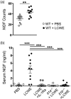
Nerve growth factor (NGF) is induced by Lactobacillus cell wall extract (LCWE) and is (IL)‐1‐dependent. (a) RNA seq analysis of heart tissues. mRNA expression counts of NGF in heart tissues are shown from phosphate‐buffered saline (PBS) and LCWE‐injected mice 1 week post‐injection (n = 10/group). (b) Mice were injected with PBS or LCWE and serum was obtained 1 week later. NGF was measured by enzyme‐linked immunosorbent assay (ELISA). PBS (n = 6), LCWE (n = 10), LCWE + IL‐1Ra (n = 10), Il‐1α −/− + LCWE (n = 10) and Il‐1β −/− + LCWE (n = 10). *≤ 0·05; **≤ 0·01 by Student’s t‐test (a) or one‐way analysis of variance (anova) with Tukey’s post‐hoc test used for multiple comparison (b).
Figure 3
Figure 3
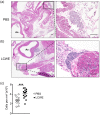
Kawasaki disease (KD) vasculitis is associated with ganglionitis. Mice were injected with phosphate‐buffered saline (PBS) or Lactobacillus cell wall extract (LCWE) and killed 1 week after injection. (a,b) Representative haematoxylin and eosin (H&E)‐stained heart sections showing ganglia from PBS (a) or LCWE‐injected (b) wild‐type (WT) mice 1 week post‐LCWE injection. Images are representative of n = 5 per group and were taken at ×2 and ×20 magnification. Scale bar = 100 μm. (c) Quantification of infiltrating cells in the heart ganglia of PBS and LCWE‐injected mice 1 week post‐LCWE injection (n = 11–15). ***≤ 0·01 by Student’s t‐test. Ao = aorta.
Figure 4
Figure 4
Immune cells infiltrate the cardiac ganglia during Lactobacillus cell wall extract (LCWE)‐induced Kawasaki disease (KD) vasculitis. Mice were injected with phosphate‐buffered saline (PBS) or LCWE and killed 1 week later. Immunofluorescence was performed on frozen sections to determine immune infiltrate constituents. (a) Isotype control image of ganglia. (b) Isotype control image of coronary lesion. (c,d) Ganglion (c) and coronary lesion staining (d) for CD45.1 (leukocyte common antigen, red arrows) and CD19 (B lymphocytes marker, green arrows). (e,f) Ganglion (e) and coronary lesion staining (f) for CD3 (T cell marker, red arrows) and F4/80 (macrophage/monocyte marker, green arrows). (g,h) Ganglion (g) and coronary lesion staining (h) CD11b‐ (myeloid cells, green arrows) and for CD11c (dendritic cell and macrophages, red arrows). Scale bars 10 µm for ganglia and 25 µm for coronary or magnification as indicated.
Figure 5
Figure 5
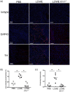
Neural remodeling in myocardium after Lactobacillus cell wall extract (LCWE)‐induced myocarditis is (IL)‐1‐dependent. wild‐type (WT) and Il1r1−/− mice were injected with LCWE and killed 2 weeks later. Immunofluorescence was performed on frozen section. (a) Immunofluorescence staining of the neuronal sprouting marker growth‐associated protein 43 (GAP43) and the sympathetic neuronal tyrosine hydrogenase (TH) in the myocardium. Scale bars, 100 µm. (b,c) Quantification of staining density of GAP43 (b) and TH (c) in the myocardium. n = 4, 5 and 7 for WT + phosphate‐buffered saline (PBS), WT + LCWE and Il1r−/− + LCWE groups, respectively. P < 0·05 by one‐way analysis of variance (anova) with Bonferroni post‐test analysis.

References

    1. Kawasaki T, Kosaki F, Okawa S, Shigematsu I, Yanagawa H. A new infantile acute febrile mucocutaneous lymph node syndrome (MLNS) prevailing in Japan. Pediatrics 1974; 54:271–6. - PubMed
    1. McCrindle BW, Rowley AH, Newburger JW et al, on behalf of the American Heart Association Rheumatic Fever, Endocarditis, Kawasaki Disease, Committee of the Council on Cardiovascular Disease in the Young; Council on Cardiovascular and Stroke Nursing; Council on Cardiovascular Surgery and Anesthesia ; Council on Epidemiology and Prevention . Diagnosis, treatment, and long‐term management of Kawasaki disease: a scientific statement for health professionals from the American Heart Association. Circulation 2017; 135:e927–99. - PubMed
    1. Newburger JW, Takahashi M, Burns JC. Kawasaki disease. J Am Coll Cardiol 2016; 67:1738–49. - PubMed
    1. Rowley AH. Is Kawasaki disease an infectious disorder? Int J Rheum Dis 2018; 21:20–5. - PMC - PubMed
    1. Rowley AH. Finding the cause of Kawasaki disease: a pediatric infectious diseases research priority. J Infectious Dis 2006; 194:1635–7. - PubMed

Publication types

MeSH terms