. 2020 Mar 15;106(4):857-866.
doi: 10.1016/j.ijrobp.2019.11.010. Epub 2019 Nov 20.

High-Dose Radiation Increases Notch1 in Tumor Vasculature

Affiliations

High-Dose Radiation Increases Notch1 in Tumor Vasculature

Debarshi Banerjee et al. Int J Radiat Oncol Biol Phys. .

Abstract

Purpose: The aim of this study is to characterize the effects of high-dose radiation therapy (HDRT) on Notch signaling components of the tumor vasculature.

Methods and materials: Human umbilical vein endothelial cells monolayers were exposed to different single fraction doses of irradiation; ribonucleic acid RNA was isolated and polymerase chain reaction was performed for Notch signaling components. The vascular response to radiation therapy was examined in a xenograft model of neuroblastoma. Tumors were treated with 0 Gy, 2 Gy, and 12 Gy single fraction doses and analyzed by double immunofluorescence staining for Notch1, Notch ligands Jagged1 and Dll4, and the endothelial cell (EC) marker endomucin. To assess the role of Notch in vivo, NGP xenograft tumors expressing Fc or Notch1-1-24-decoy (a novel Notch inhibitor) were treated with 0 Gy and 12 Gy. Immunofluorescence staining for endomucin and endomucin/αSMA was performed to analyze the effect of combination treatment on tumor EC and endothelial-to-mesenchymal-transition (EndMT), respectively.

Results: In human umbilical vein endothelial cells monolayers doses ≥8 Gy increased expression of NOTCH1, JAG1, and Notch target genes HEY1 and HEY2 as early as 6 hours after irradiation. In vivo, 12 Gy significantly increased Notch1 and Jagged1 in tumor ECs compared with 0 Gy or 2 Gy after 72 hours. Combining HDRT with Notch inhibition using the Notch1-1-24-decoy resulted in a greater loss of EC coverage of tumor vessels than HDRT alone at 6 hours and 72 hours post treatment. Notch inhibition reduced EndMT induced by HDRT, as indicated by diminished αSMA staining in ECs.

Conclusions: HDRT induced Notch1 expression and increased Notch1 signaling in the endothelial component of tumor vasculature, which was not observed with lower doses. This increase in Notch1 activation might protect tumor vessels from HDRT induced damage and regulate EndMT process.


Figures

Fig. 1.
Fig. 1.
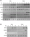
High-dose radiation therapy induces NOTCH1 and JAGGED1 signaling in human umbilical vein endothelial cells (HUVEC). (A) Reverse transcriptase-polymerase chain reaction analysis of messenger ribonucleic acid (mRNA) of Notch components after irradiation. HUVEC monolayers were treated with different single fraction irradiation doses. RNA was isolated at 6 hours and 72 hours, reverse transcribed and polymerase chain reaction was performed for Notch receptors and ligands. Increased NOTCH1 message was observed at 6 hours and 72 hours after irradiation with doses ≥8 Gy. JAGGED1 mRNA level was slightly increased, at 6 hours after ≥8 Gy, and markedly increased at 72 hours after ≥10 Gy doses. Concomitant with NOTCH1 mRNA increase, target genes HEY1 and HEY2 mRNA levels were also elevated. ACTB (β-Actin) was used as internal control. (B) Immunoblot analyses for Notch components after irradiation. HUVEC were exposed to different irradiation doses for 6 hours and 72 hours and cell lysates were immunobloted with NOTCH1, JAGGED1, HEY2, and β-Actin antibodies. Irradiation doses ≥8 Gy increased NOTCH1 protein expression at 6 hours and 72 hours. JAGGED1 level is elevated only at 72 hours after 12 Gy. HEY2 was also markedly increased at 6 hours and 72 hours after irradiation with 12 Gy.
Fig. 2.
Fig. 2.
High-dose radiation increases Notch1 expression in tumor endothelial cells. Representative images double immunofluorescence staining for Notch1 (red) and endomucin (green) of NGP tumors at 6 hours (A) and 72 hours (D) after 0 Gy, 2 Gy, and 12 Gy single fraction dose treatment. Increased Notch1 immunostaining colocalizing (arrows) with endomucin staining was observed at 6 hours and 72 hours after 12 Gy. Quantification of Notch1 (+) vessels, normalized by total number of endomucin (+) vessels, per field (magnification, 20x) in NGP tumors at 6 hours (B) and 72 hours (E) after 12 Gy. Quantification of amount of endomucin, as % area, per field (magnification, 20X) at 6 hours (C) and 72 hours (F). *P < .05, **P < .01, ***P < .001. Mean ± SD. Bar, 100 μm.
Fig. 3.
Fig. 3.
High-dose radiation induces endothelial Jagged1 expression in tumor vessel in vivo. (A) Double immunofluorescence staining of Jagged1 (red) colocalizing (arrows) with endomucin (green) in tumors at 72 hours after 0 Gy, 2 Gy, and 12 Gy. (B) Quantification of Jagged1 (+) vessels, normalized by endomucin (+) vessels, in tumors per field (magnification, 20X). Mean ± SD. *P < .05. Bar, 50 μm.
Fig. 4.
Fig. 4.
High-dose radiation increases nuclear Hey2 expression in tumor endothelial cell at 72 hours (A) Triple immunofluorescence of DAPI (blue), endomucin (green), and Hey2 (red). Very little Hey2 staining was detected in the DAPI (+) nuclei (dotted arrow) of endomucin (+) endothelial cells (ECs) at 72 hours after 0 Gy and 2 Gy. 12 Gy single fraction dose increased Hey2 staining (solid arrow) in the nuclei of endomucin (+) EC in vessel. Hey2 was localized in the nuclei and endomucin staining was observed around the nuclei. Insets represent the same portion (dotted box) of the tumor. Bar, 50μm. (B) Quantification of Hey2(+) nuclei of endomucin (+) EC in the vessels, normalized by total number of DAPI(+) nuclei in the endomucin (+) EC in vessels, in tumors per field (magnification, 40X). Mean ± SD. ***P <.001.
Fig. 5.
Fig. 5.
Combining Notch inhibition with high-dose radiation therapy decreases endothelial cell coverage of tumor. (A) Representative images of endomucin immunofluorescence (green) of Fc + 0 Gy control N11–24-decoy + 0 Gy, Fc + 12 Gy, and N11–24-decoy + 12 Gy tumors (NGP xenograft) at 6 hours. Bar = 100 μm. (B) Quantification of endomucin immunofluorescence, at 6 hours, as area percentage. N11–24-decoy + 0 Gy and Fc + 12 Gy tumors had decreased endomucin than Fc + 0 Gy control. N11–24-decoy + 12 Gy tumors had an additive decrease in endomucin area fraction. (C) Immunofluorescence detection of endomucin in tumors at 72 hours. (D) Quantification of endomucin, as area percentage, at 72 hours also shows greater loss of endothelial cells post N11–24-decoy + 12 Gy treatments. Mean ± SD. **P <.05, ***P <.001. Bar, 100μm.
Fig. 6.
Fig. 6.
Increased Notch1 activity is associated with endothelial-to-mesenchymal transition (A) human umbilical vein endothelial cells monolayers were exposed to different doses of irradiation and cells lysate were analyzed at 72 hours by immunoblot for VE-Cadherin and cleaved NOTCH1. 8 Gy and 12 Gy doses, but not 2 Gy, increased cleaved NOTCH1 and decreased VE-Cadherin. (B) Notch inhibition reduced radiation-induced EndMT in tumor vasculature. Double immunofluorescence of endomucin and αSMA in tumors. 12 Gy increased endomucin and αSMA colocalization (arrows) in NGP tumors at 72 hours post 12 Gy. Combining high-dose radiation therapy with N11–24-decoy treatment decreased colocalization. Bar, 50 μm. (C) Quantification of αSMA (+) vessels, normalized by endomucin (+) vessels, in tumors per field (magnification, 20X). Mean ± SD. ***P <.001.

References

    1. Lo SS, Fakiris AJ, Chang EL, et al. Stereotactic body radiation therapy: A novel treatment modality. Nat Rev Clin Oncol 2010;7:44–54. - PubMed
    1. Brown LC, Lester RA, Grams MP, et al. Stereotactic body radiotherapy for metastatic and recurrent ewing sarcoma and osteosarcoma. Sarcoma 2014;2014:418270. - PMC - PubMed
    1. Garcia-Barros M, Paris F, Cordon-Cardo C, et al. Tumor response to radiotherapy regulated by endothelial cell apoptosis. Science 2003; 300:1155–1159. - PubMed
    1. Song CW, Kim MS, Cho LC, et al. Radiobiological basis of SBRT and SRS. Int J Clin Oncol 2014;19:570–578. - PubMed
    1. Kirkpatrick JP, Meyer JJ, Marks LB. The linear-quadratic model is inappropriate to model high dose per fraction effects in radiosurgery. Semin Radiat Oncol 2008;18:240–243. - PubMed

Publication types

MeSH terms