. 2020 Feb:58:101260.
doi: 10.1016/j.trim.2019.101260. Epub 2019 Nov 22.

TLR2 and NODs1 and 2 cooperate in inflammatory responses associated with renal ischemia reperfusion injury

Affiliations

TLR2 and NODs1 and 2 cooperate in inflammatory responses associated with renal ischemia reperfusion injury

Sashi G Kasimsetty et al. Transpl Immunol. 2020 Feb.

Abstract

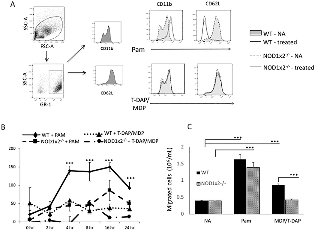
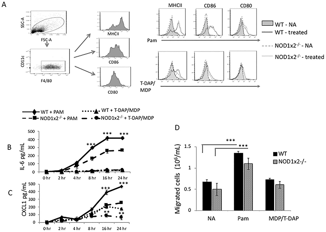
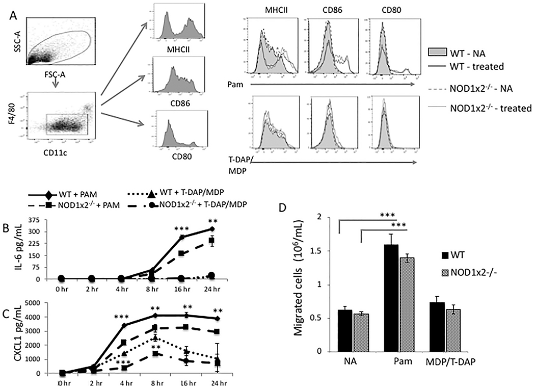
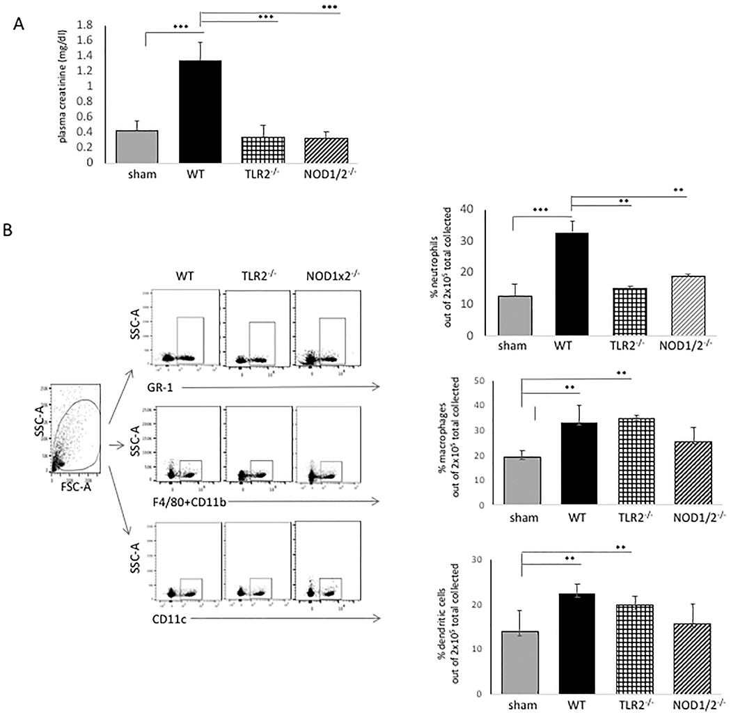
Pattern recognition receptors (PRRs) are potent triggers of tissue injury following renal ischemia/reperfusion injury (IRI). Specific PRRs, such as the toll-like receptor 2 (TLR2) and the nucleotide-binding oligomerization domain-like receptors (NLRs) NOD1 and NOD2 are promising targets to abrogate inflammatory injury associated with renal IRI. Several recent reports have shown there is crosstalk between TLRs and NODs, which might boost inflammatory responses to tissue injury. This study examined the relative roles of TLR2 and NODs 1 and 2 in activation of myeloid cells that contribute to inflammation after renal IRI. We found that TLR2 and NOD1 and 2 signaling induces neutrophil, macrophage and dendritic cell migration in vitro, however their blockade only decreases neutrophil infiltration into ischemic kidneys. The results of this study suggest that future therapies targeted to innate immune blockade should consider that either TLR2 or NOD1/2 blockade could decrease neutrophil inflammation following an ischemic insult to the kidney, however blockade of these PRRs would not likely impact infiltration of dendritic cells or macrophages. Developing rational approaches that target innate immunity in IRI-induced acute kidney injury requires an understanding of the relative role of PRRs in directing inflammation in the kidney.

Keywords: Acute kidney injury; Ischemia/reperfusion injury; NOD1; NOD2; TLR2.


Conflict of interest statement

Declaration of Competing Interest

The authors declare that the research was conducted in the absence of any commercial or financial relationships that could be construed as a potential conflict of interest.

Figures

Fig. 1.
Fig. 1.
Effect of TLR2 and NOD1/2 agonists (PAM and T-DAP/MDP) on neutrophil activation. Panel A. Expression of CD11b and CD62L on cell surface of neutrophils from WT or NOD1x2−/− mice stimulated with either the TLR2 agonist PAM (1 μg/mL) or combined NOD1 and NOD2 agonists T-DAP/MDP (10 μg/mL). Panel B. Secretion of CXCL1 in supernatants over time from cultures of WT or NOD1x2−/− neutrophils stimulated with PAM (1 μg/mL) or T-DAP/MDP (10 μg/mL). Error bars represent SDs of 3 samples/gp. Panel C. Neutrophil migration detected in a transwell assay comparing WT vs NOD1x2−/− neutrophils. Neutrophils were first cultured for 24 h with either the TLR2 agonist PAM (1 μg/mL) or the combined NOD1/2 agonists T-DAP/MDP (10 μg/mL). Migration to CXCL8 (250 ng/mL) in the lower chamber was assessed at 4 h. Error bars represent SD of three samples. Each of the graphs represent one of three identical experiments. *** p < .001.
Fig. 2.
Fig. 2.
Effect of TLR2 and NOD1/2 agonists on macrophage activation. Panel A. Expression of MHC class II, CD86 and CD80 on cell surface of macrophages derived from bone marrows of WT or NOD1x2−/− mice stimulated overnight with either the TLR2 agonist PAM (1 μg/mL) or the combined NOD1/2 agonists T-DAP + MDP (10 μg/mL) and analyzed by FACS. Panels B and C. IL-6 and CXCL1 (respectively) detected in supernatants over time of cultures of WT vs. NOD1x2−/− macrophages stimulated with PAM (1 μg/mL). Error bars represent SD of triplicates. Panel D. Macrophage migration detected in a transwell assay comparing WT vs NOD1x2−/− macrophages. Macrophages were cultured for 24 h with either the TLR2 agonist PAM (1 μg/mL) or the combined NOD1 and NOD2 agonists T-DAP/MDP (10 μg/mL). Migration to CXCL8 (250 ng/mL) in the lower chamber was assessed at 4 h. Error bars represent SD of three samples. In each of the experiments error bars represent SD of triplicates. Each graph represents one of three identical experiments. In Panel B **p < .01, ***p < .001 (between WT and NOD1x2−/−, PAM treated). In Panel C ** p < .01 and ***p < .001 (between WT and NOD1x2−/−, PAM treated – top two lines of graph) and ** p < .01 and ***p < .001 (between WT and NOD1x2−/−.T-DAP/MDP treated – bottom two lines of graph).
Fig. 3.
Fig. 3.
Effect of TLR2 and NOD1 x NOD2 agonists on dendritic cell activation. Panel A. Expression of MHC Class II, CD86, and CD80 on cell surface of dendritic cells derived from spleens of WT or NOD1x2−/− mice stimulated overnight with either the TLR2 agonist PAM (1 μg/mL) or combined NOD1/2 agonists T-DAP + MDP (10 μg/mL) and analyzed by FACS. Panel B. Detection of IL-6 or CXCL1 (Panel C) over time in culture supernatants of WT vs. NOD1x2−/− dendritic cells stimulated overnight with PAM (1 μg/mL). Panel D. Dendritic cell migration detected in a transwell assay comparing WT vs NOD1x2−/− dendritic cells. Dendritic cells were cultured for 24 h with either the TLR2 agonist PAM (1 μg/mL) or the combined NOD1/2 agonists T-DAP + MDP (10 μg/mL). Migration to CXCL8 (250 ng/mL) in the lower chamber was assessed at 4 h. Error bars represent SD of three samples. Each graph represents one of three identical experiments. **p < .01, ***p < .001.
Fig. 4.
Fig. 4.
Effect of TLR2 and NOD1x2 deficiency in renal IRI. WT vs. TLR2−/− or NOD1x2−/− mice were treated with 25 min bilateral renal artery clamping followed by 24 h reperfusion. At end of reperfusion, plasma creatinine was detected and the kidneys were harvested and single cell suspension of each tissue prepared. The suspension was stained for inflammatory infiltrates and analyzed by FACS using florescence antibodies to GR-1 (neutrophils); F4/80 and CD11b (macrophages); CD11c (dendritic cells). Error bars represent standard deviations of triplicate samples. **p < .01, *** p < .001.

References

    1. Zuk A, Bonventre JV, Acute kidney injury, Annu. Rev. Med 67 (2016) 293–307. - PMC - PubMed
    1. Boros P, Bromberg JS, New cellular and molecular immune pathways in ischemia/reperfusion injury, Am. J. Transplant 6 (2006) 652–658. - PubMed
    1. Devarajan P, Cellular and molecular derangements in acute tubular necrosis, Curr. Opin. Pediatr 17 (2005) 193–199. - PubMed
    1. Dagher PC, Herget-Rosenthal S, Ruehm SG, et al. , Newly developed techniques to study and diagnose acute renal failure, J. Am. Soc. Nephrol 14 (2003) 2188–2198. - PubMed
    1. Li C, Jackson RM, Reactive species mechanisms of cellular hypoxia-reoxygenation injury, Am J Physiol Cell Physiol 282 (2002) C227–C241. - PubMed

Publication types

MeSH terms