. 2020 Apr;159(4):1640-1653.e18.
doi: 10.1016/j.jtcvs.2019.09.121. Epub 2019 Oct 17.

Multiday maintenance of extracorporeal lungs using cross-circulation with conscious swine

Affiliations

Multiday maintenance of extracorporeal lungs using cross-circulation with conscious swine

Ahmed E Hozain et al. J Thorac Cardiovasc Surg. 2020 Apr.

Abstract

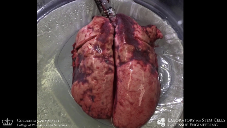
Objectives: Lung remains the least-utilized solid organ for transplantation. Efforts to recover donor lungs with reversible injuries using ex vivo perfusion systems are limited to <24 hours of support. Here, we demonstrate the feasibility of extending normothermic extracorporeal lung support to 4 days using cross-circulation with conscious swine.

Methods: A swine behavioral training program and custom enclosure were developed to enable multiday cross-circulation between extracorporeal lungs and recipient swine. Lungs were ventilated and perfused in a normothermic chamber for 4 days. Longitudinal analyses of extracorporeal lungs (ie, functional assessments, multiscale imaging, cytokine quantification, and cellular assays) and recipient swine (eg, vital signs and blood and tissue analyses) were performed.

Results: Throughout 4 days of normothermic support, extracorporeal lung function was maintained (arterial oxygen tension/inspired oxygen fraction >400 mm Hg; compliance >20 mL/cm H2O), and recipient swine were hemodynamically stable (lactate <3 mmol/L; pH, 7.42 ± 0.05). Radiography revealed well-aerated lower lobes and consolidation in upper lobes of extracorporeal lungs, and bronchoscopy showed healthy airways without edema or secretions. In bronchoalveolar lavage fluid, granulocyte-macrophage colony-stimulating factor, interleukin (IL) 4, IL-6, and IL-10 levels increased less than 6-fold, whereas interferon gamma, IL-1α, IL-1β, IL-1ra, IL-2, IL-8, IL-12, IL-18, and tumor necrosis factor alpha levels decreased from baseline to day 4. Histologic evaluations confirmed an intact blood-gas barrier and outstanding preservation of airway and alveolar architecture. Cellular viability and metabolism in extracorporeal lungs were confirmed after 4 days.

Conclusions: We demonstrate feasibility of normothermic maintenance of extracorporeal lungs for 4 days by cross-circulation with conscious swine. Cross-circulation approaches could support the recovery of damaged lungs and enable organ bioengineering to improve transplant outcomes.

Keywords: acute lung injury; airway lavage; alveolar recruitment; bronchoalveolar lavage fluid; chimerism; cross-circulation; ex vivo lung perfusion; extracorporeal membrane oxygenation; infrared thermography; lung bioengineering; lung transplantation; medical thermography; normothermic organ perfusion; organ shortage; regenerative medicine; swine model; tissue engineering; transplantation; whole organ bioreactor.


Figures

None
Graphical abstract
None
Multiday normothermic support system for extracorporeal lungs using cross-circulation.
Figure 1
Figure 1
Experimental overview of multiday extracorporeal lung support system. A, Lungs were explanted from donor swine and the pulmonary artery (PA) and pulmonary vein (PV) were cannulated. Extracorporeal lungs were maintained in a humidified, normothermic preservation chamber and connected to a mechanical ventilator (V) via endotracheal intubation (T). Recipient swine were cannulated with a dual-lumen cannula via the right internal jugular (RIJ) vein. After recovery from anesthesia, recipient swine were placed in a custom enclosure for the duration of the procedure. The extracorporeal circuit contained a centrifugal pump (P) that cross-circulated whole blood between the recipient swine and the extracorporeal lungs. In-line sensors (S) monitored the hemodynamic stability of the recipient swine and extracorporeal lungs during multiday normothermic support. B, Experimental timeline. Extracorporeal lungs were maintained by normothermic cross-circulation (XC) and periodically assessed for 4 days. BAL, Bronchoalveolar lavage.
Figure 2
Figure 2
Experimental setup of multiday extracorporeal lung support system using a swine recipient. A, A dual-lumen cannula was placed in the right internal jugular (RIJ) vein under fluoroscopic guidance. White arrows indicate cannula. B, The cannula was tunneled along the lateral aspect of the right neck and secured to the dorsum of recipient swine. C and D, Following initiation of cross-circulation, cannulated recipient swine were transferred to a Panepinto sling where they remained elevated from the enclosure floor for safe recovery from anesthesia and extubation. E, Once conscious, recipient swine were lowered into a custom enclosure where they remained for the duration of the procedure. SVC, Superior vena cava; R, right; IVC, inferior vena cava.
Video 1
Video 1
Normothermic maintenance of extracorporeal swine lungs with multiday lung support system. Video available at: https://www.jtcvs.org/article/S0022-5223(19)32146-4/fulltext.
Video 2
Video 2
Transfer of swine recipient in Panepinto sling to custom enclosure. Video available at: https://www.jtcvs.org/article/S0022-5223(19)32146-4/fulltext.
Video 3
Video 3
Active enrichment of recipient swine in custom enclosure throughout multiday lung support. Video available at: https://www.jtcvs.org/article/S0022-5223(19)32146-4/fulltext.
Figure 3
Figure 3
Stability of circuit parameters during multiday extracorporeal lung support. A, Pressure. B, Flow. C, Transpulmonary pressure gradient (TPG), which is the difference between pulmonary artery and pulmonary vein pressures. D, Temperature. E, pH. F, Lactate. Dotted lines define target range. Values are presented as mean ± standard deviation.
Figure 4
Figure 4
Maintenance of extracorporeal lung function for 4 days using multiday lung support system. A, Arterial oxygen tension/inspired oxygen fraction. B, Dynamic compliance. C, Peak inspiratory pressure (PIP). D, Lung weight. Values are presented as mean ± standard deviation.
Figure 5
Figure 5
Multiscale analyses of extracorporeal lung maintenance and integrity. A, Photographic appearance. B, Thermographic appearance. C, Radiographic appearance. D, Bronchoscopic evaluation of extracorporeal lungs throughout 4 days of normothermic support, including aerated regions (dotted lines), and areas of local consolidation (stars). Microscopic analyses of bilateral lower lobes by hematoxylin and eosin staining. E, Low magnification. F, High magnification. G, Transmission electron microscopy revealed type II pneumocytes (black arrows) with visible lamellar bodies containing surfactant, and intact alveolar epithelial barrier (white arrows) of type I pneumocytes.
Figure 6
Figure 6
Quantification of airway cytokines and evaluation of lung injury during multiday extracorporeal lung support. Bronchoalveolar lavage fluid concentrations. A, Interferon gamma (IFNγ). B, Tumor necrosis factor alpha (TNFα). C, Interleukin (IL) 1β. D, IL-6. E, IL-8. F, IL-10. G, Lung injury scoring by blinded histopathologic review. Values are presented as mean ± standard deviation. PMN, Polymorphonuclear cells.
Figure 7
Figure 7
Cellular integrity and function in extracorporeal lungs after 4 days of normothermic support. Pentachrome staining. A, Large airways at low magnification with preserved airway mucosa (AM), smooth muscle (SM), and cartilage plate (CP). B, Large airways at high magnification with outstanding preservation of airway cilia (AC), pseudostratified epithelium (PE), and basement membrane (BM). C, Alveoli with intact blood–gas barrier and perfused venule and alveolar capillaries (arrows). D, Immunohistochemical staining was used to confirm retention of alpha smooth muscle actin (αSMA) around small airways (stars). E, Immunohistochemical staining was used to confirm retention of vascular endothelial (VE)-cadherin by endothelial cells in large and small vessels throughout the pulmonary vascular tree. F, Cell viability throughout the lung parenchyma was confirmed by pervasive uptake of carboxyfluorescein succinimidyl ester (CFSE). Star indicates alveolar space. G, Metabolic activity of lung parenchyma. Dotted lines indicate normal range of metabolic activity of healthy swine lungs in vivo. H, Changes in peak inspiratory pressure after administration of nebulized methacholine (arrow). Values are presented as mean ± standard deviation. PIP, Peak inspiratory pressure.
Figure 8
Figure 8
Multiday maintenance of extracorporeal lungs using cross-circulation with conscious swine: Experimental overview and results. A, Extracorporeal lungs were maintained using cross-circulation with conscious swine for a duration of 4 days. Lungs were placed in a normothermic organ preservation chamber and ventilated. Functional, biochemical, and multimodal imaging analyses were used to enable continuous monitoring of extracorporeal lungs and swine recipients. B, At day 4, lungs demonstrated maintenance of respiratory function, intact blood–gas barrier, cellular viability, and outstanding preservation of airway and alveolar architecture. PIP, Peak inspiratory pressure.
Figure E1
Figure E1
Maintenance of extracorporeal circuit parameters by height adjustments of extracorporeal lungs in response to changes in recipient swine position. A, Target ranges of extracorporeal circuit parameters. B, Representative photographs of recipient positions: upright and prone. C, Heights of circuit elements corresponding to recipient position. D, Extracorporeal circuit diagrams demonstrating changes in extracorporeal lung height corresponding to changes in recipient position in order to maintain target circuit parameters. PA, Pulmonary artery; PV, pulmonary vein.
Figure E2
Figure E2
Custom enclosure for recipient swine. Stainless steel enclosure featured width-adjustable sidewall to ensure swine recipients remained comfortable and secure throughout multi-day procedures. The open top of the enclosure enabled easy access to recipient swine, cannula site, and circuit components. Immediately following recipient feeding, urination, and defecation, the excreta pan was removed, thoroughly cleaned, and replaced to minimize the presence of waste in the custom enclosure.
Figure E3
Figure E3
Histologic evaluation of the upper lobes of extracorporeal lungs throughout 4 days of normothermic support. A, Subpleural regions. B, Parenchyma. C, Pulmonary airways. D, vessels. Dotted lines outline surface of visceral pleura.
Figure E4
Figure E4
Randomized lung sampling and scoring rubric of lung injury score. A, Lung map used for randomized tissue sampling showing lungs were divided into 5 lobes. B, Tissue sample locations at each time point. Sample bias was avoided by predetermining tissue sampling location before the start of all experiments. C, Scoring rubric of lung injury scores. RUL, Right upper lobe; RML, right middle lobe; RLL, right lower lobe; LUL, left upper lobe; LLL, left lower lobe; PMN, polymorphonuclear cells; hpf, high-power field.
Figure E5
Figure E5
Preprocedure swine behavior training program. A, Target training with clicker (red) to encourage swine to enter custom enclosure. B, Throughout the behavior training program, recipient swine comfort and familiarity were maintained while the width of the side wall (stars) of the custom enclosure was incrementally decreased, limiting the ability of recipient swine to rotate during the procedure. C, Active enrichment was provided throughout behavior training. D, Positive reinforcement was provided throughout behavior training.
Figure E6
Figure E6
Histologic evaluation by hematoxylin and eosin stain of recipient swine lung after 4 days of normothermic support. A, Low-power microscopy of parenchyma. B, High-power microscopy of alveoli. C, Airway. D, Vessels. E, Comparison of lung injury scores at day 4 of multiday support to lung injury scores of recipient lung at the end of the procedure. PMN, Polymorphonuclear cells.
None

Comment in

References

    1. Ware L.B., Wang Y., Fang X., Warnock M., Sakuma T., Hall T.S. Assessment of lungs rejected for transplantation and implications for donor selection. Lancet. 2002;360:619–620. - PubMed
    1. Singer J., Chen J., Blanc P.D., Leard L.E., Kukreja J., Chen H. A thematic analysis of quality of life in lung transplant: the existing evidence and implications for future directions. Am J Transplant. 2013;13:839–850. - PMC - PubMed
    1. Kugler C., Gottlieb J., Warnecke G., Schwarz A., Weissenborn K., Barg-Hock H. Health-related quality of life after solid organ transplantation: a prospective, multiorgan cohort study. Transplant J. 2013;96:316–323. - PubMed
    1. Pinezich M., Vunjak-Novakovic G. Bioengineering approaches to organ preservation ex vivo. Exp Biol Med. 2019;244:630–645. - PMC - PubMed
    1. Guibert E.E., Petrenko A.Y., Balaban C.L., Somov A.Y., Rodriguez J.V., Fuller B.J. Organ preservation: current concepts and new strategies for the next decade. Transfus Med Hemotherapy. 2011;38:125–142. - PMC - PubMed

E-References

    1. O'Neill J.D., Guenthart B.A., Kim J., Chicotka S., Queen D., Fung K. Cross-circulation for extracorporeal support and recovery of the lung. Nat Biomed Eng. 2017;1:0037.
    1. Guenthart B.A., O'Neill J.D., Kim J., Queen D., Chicotka S., Fung K. Regeneration of severely damaged lungs using an interventional cross-circulation platform. Nat Commun. 2019;10:1985. - PMC - PubMed
    1. Rampersad S.N. Multiple applications of alamar blue as an indicator of metabolic function and cellular health in cell viability bioassays. Sensors. 2012;12:12347. - PMC - PubMed
    1. Roman M., Gjorgjimajkoska O., Neil D., Nair S., Colah S., Parmar J. Comparison between cellular and acellular perfusates for ex vivo lung perfusion in a porcine model. J Heart Lung Transplant. 2015;34:978–987. - PubMed
    1. Jung K., Renukaradhya G.J., Alekseev K.P., Fang Y., Tang Y., Saif L.J. Porcine reproductive and respiratory syndrome virus modifies innate immunity and alters disease outcome in pigs subsequently infected with porcine respiratory coronavirus: implications for respiratory viral co-infections. J Gen Virol. 2009;90(Pt 11):2713–2723. - PMC - PubMed

Publication types