. 2019 Nov 27;179(6):1357-1369.e16.
doi: 10.1016/j.cell.2019.10.035. Epub 2019 Nov 21.

Transient Protein-RNA Interactions Guide Nascent Ribosomal RNA Folding

Affiliations

Transient Protein-RNA Interactions Guide Nascent Ribosomal RNA Folding

Olivier Duss et al. Cell. .

Abstract

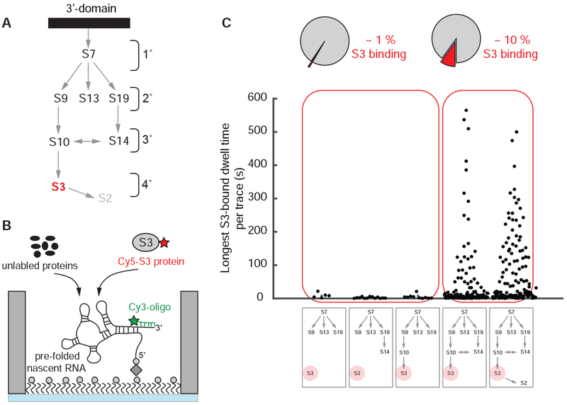
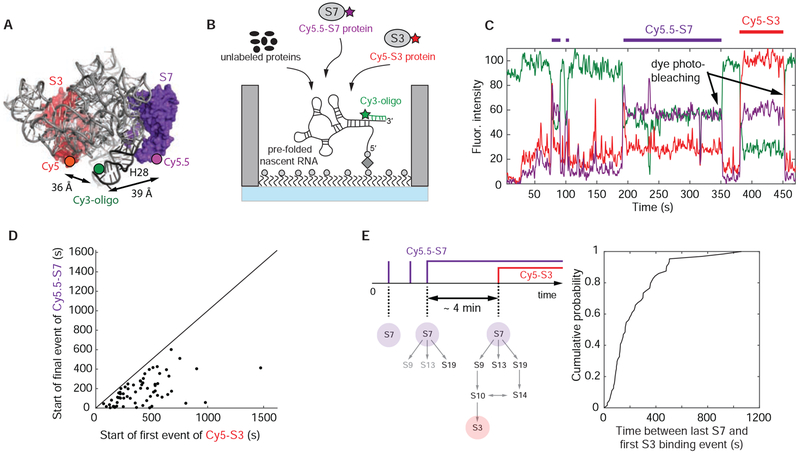
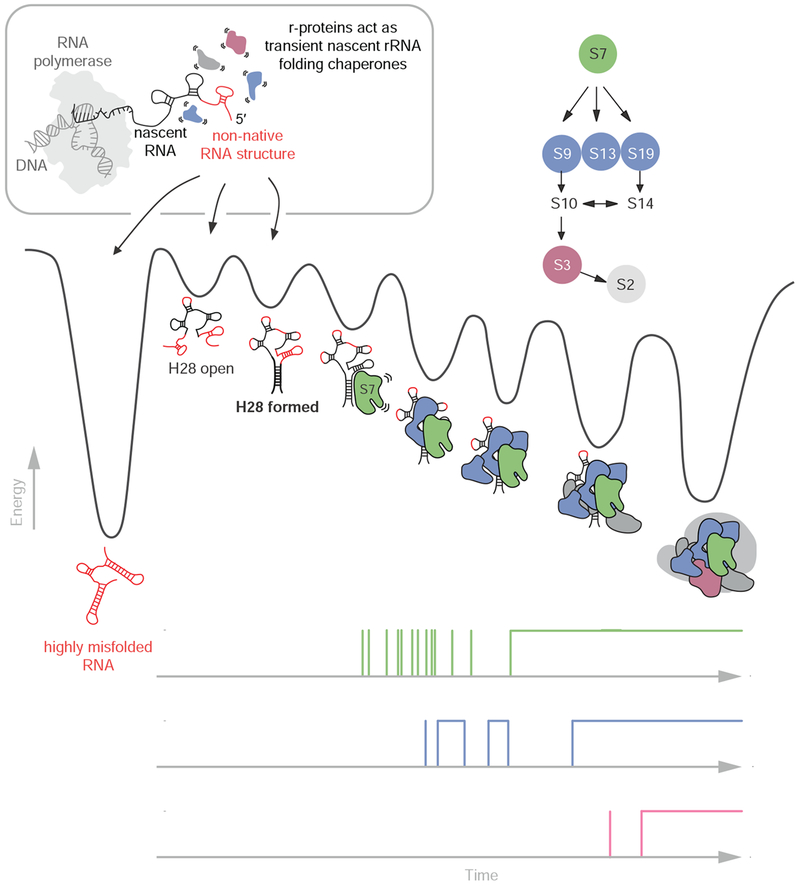
Ribosome assembly is an efficient but complex and heterogeneous process during which ribosomal proteins assemble on the nascent rRNA during transcription. Understanding how the interplay between nascent RNA folding and protein binding determines the fate of transcripts remains a major challenge. Here, using single-molecule fluorescence microscopy, we follow assembly of the entire 3' domain of the bacterial small ribosomal subunit in real time. We find that co-transcriptional rRNA folding is complicated by the formation of long-range RNA interactions and that r-proteins self-chaperone the rRNA folding process prior to stable incorporation into a ribonucleoprotein (RNP) complex. Assembly is initiated by transient rather than stable protein binding, and the protein-RNA binding dynamics gradually decrease during assembly. This work questions the paradigm of strictly sequential and cooperative ribosome assembly and suggests that transient binding of RNA binding proteins to cellular RNAs could provide a general mechanism to shape nascent RNA folding during RNP assembly.

Keywords: RNA biology; RNA chaperones; RNA misfolding; co-transcriptional RNA folding; cooperativity; protein-RNA dynamics; protein-RNA interactions; ribonucleoprotein assembly; ribosome assembly; single-molecule fluorescence microscopy.


Conflict of interest statement

DECLARATION OF INTERESTS

The authors declare no competing interests.

Figures

Figure 1.
Figure 1.. Real-time S7 encounters with nascent RNA.
(A) Secondary structure of E. coli S7 binding site. (B) 3-dimensional structure of E. coli 30S ribosomal subunit with labeling sites (PDB accession code: 4V9P); the Cy3 dye of the labeled DNA oligonucleotide binding to the 3’-end of the nascent RNA is modeled into the structure. (C and D) Single-molecule fluorescence approach (Duss et al., 2018). (E) Representative single-molecule trace with fluorescence intensity (top) and FRET efficiency (bottom); transcription (TC; orange), Cy5-S7 (red), Cy3-oligo (green). See also Figure S1.
Figure 2.
Figure 2.. Inefficient and slow nascent RNA folding.
(A and B) Nascent 3’domain rRNA folding is inefficient (A) and slow (B) in absence of other r-proteins. In (B), the time from full transcription till appearance of the first r-protein binding event with a duration of >2 s at 20 °C is represented. (C) Smaller nascent RNAs fold more efficiently. Δ147 stands for the truncation of 147 nt compared to the full-length 3’domain construct and other constructs are named accordingly. Concentrations: 20 nM Cy5-S7 or 25 nM Cy5-S15. Data with S15 in (A and B) is from reference (Duss et al., 2018). Number of molecules analyzed (n) = 172, 138, 64, 144, 92 (A), (n) = 46, 40 (B) and (n) = 138, 86, 119, 89 (C). See also Figure S2 and Data S2.
Figure 3.
Figure 3.. Correlation of nascent H28 formation with S7 binding.
(A) Experimental approach to detect nascent H28 formation and correlate it with S7 binding. (B and C) Representative single-molecule traces for nascent RNA molecules with H28 not formed (B) and H28 formed with subsequent Cy5.5-S7 binding (C). TC stands for transcription. (D and E) H28 formation efficiency (D) and S7 binding competency of H28-containing RNA molecules (E) for different constructs and conditions. (F) Model for nascent H28 formation and correlation to S7 protein binding. All the experiments were initiated by delivering 500 μM NTPs, 100 nM Cy3-oligo, 100 nM Cy3.5-oligo, 5 nM Cy5.5-S7 and different combinations of unlabeled 3’domain r-proteins (400 nM each) or non-cognate binder S15 (2 μM) to the stalled transcription complex. Number of molecules analyzed in (D and E) (n) = 100, 60, 72, 115, 147, 139, 115. See also Figure S3 and Data S2.
Figure 4.
Figure 4.. 3’-proteins chaperone and stabilize rRNA.
(A) Temperature dependence of the long S7-bound lifetime phase shown for Δ270 truncation construct and the full 3’domain RNA in absence of any other r-proteins. The error bars represent the 95 % confidence intervals of the fit. (B) Nomura ribosome assembly map. (C) Experimental setup. (D) The probability of stable S7 incorporation increases with the presence of secondary binding r-proteins; temperature = 35 °C; [Cy5-S7] = 20 nM; [unlabeled r-proteins] = 400 nM, [S15] = 2 μM. (E and F) Simplified single-molecule traces for S7 binding at 35 °C in absence (E) or presence (F) of S9, S13 and S19. (G) Tertiary interactions between H41 and H42 are required for stable S7 binding. The area of the dots is proportional to the populations of the two lifetime phases. The error bars represent the 95 % confidence intervals of the fit. (H) Model on how RNA tertiary interactions and r-proteins progressively stabilize S7. Number of molecules analyzed in (D) (n) = 101, 120, 144, 115, 111, 141, 98, 149, 121, 57, 57. See also Figures S4, S5 and Data S2.
Figure 5.
Figure 5.. Assembly of entire 3’domain.
(A) Nomura ribosome assembly map. (B) Experimental setup: 400 nM unlabeled S7, 100 nM Cy5-S3 and 400 nM all other unlabeled 3’domain r-proteins were injected; 35 °C. (C) All r-proteins binding upstream of S3 are required for stable S3 binding. Number of molecules analyzed in (C) (n) = 505, 1017, 924, 2699, 1453. See also Figures S6, S7 and Data S2.
Figure 6.
Figure 6.. Correlating S7 with S3 binding.
(A) 3-dimensional structure of E. coli 30S ribosomal subunit with labeling sites for Cy5-S3 and Cy5.5-S7 (PDB accession code: 4V9P); the Cy3 dye of the labeled DNA oligonucleotide binding to the 3’-end of the nascent RNA is modeled into the structure. (B) Experimental setup: 100 nM Cy5-S3, 5 nM Cy5.5-S7 and 400 nM of all the other unlabeled 3’domain r-proteins were injected; 35 °C. (C) Representative single-molecule trace with Cy5-S3 (red), Cy5.5-S7 (purple) and Cy3-oligo (green). The trace was not corrected for fluorescence spectral bleed-through from the Cy5 into the Cy5.5 channel and the reverse. (D) The first Cy5-S3 binding event always follows the final Cy5.5-S7 binding event demonstrating sequential ribosome assembly. (E) The assembly process from stable S7 incorporation until 3’domain completion is slow, indicating that RNA conformational changes are also required later in assembly. Number of molecules analyzed in (D and E) (n) = 64. See also Figure S7 and Data S2.
Figure 7.
Figure 7.. Model for nascent 3’domain assembly in absence of assembly factors.
Nascent RNA molecules can either misfold (shown in red) into stable non-native structures (deep valley on the left) or fold into RNA molecules that eventually transition into S7 binding competent conformations. The r-proteins chaperone the rRNA folding process early in assembly prior to their stable incorporation into the growing RNP particle. Nascent RNA folding and assembly are slow due to the rugged energy landscape. Progressive binding of r-proteins facilitates folding and stabilizes the nascent RNA. The single-molecule traces on the bottom demonstrate the decreasing protein binding dynamics during assembly.

Comment in

References

    1. Abeysirigunawardena SC, Kim H, Lai J, Ragunathan K, Rappe MC, Luthey-Schulten Z, Ha T, and Woodson SA (2017). Evolution of protein-coupled RNA dynamics during hierarchical assembly of ribosomal complexes. Nature communications 8, 492. - PMC - PubMed
    1. Adilakshmi T, Bellur DL, and Woodson SA (2008). Concurrent nucleation of 16S folding and induced fit in 30S ribosome assembly. Nature 455, 1268–1272. - PMC - PubMed
    1. Aitken CE, Marshall RA, and Puglisi JD (2008). An oxygen scavenging system for improvement of dye stability in single-molecule fluorescence experiments. Biophys J 94, 1826–1835. - PMC - PubMed
    1. Boyle J, Robillard GT, and Kim SH (1980). Sequential folding of transfer RNA. A nuclear magnetic resonance study of successively longer tRNA fragments with a common 5′ end. J Mol Biol 139, 601–625. - PubMed
    1. Buenrostro JD, Araya CL, Chircus LM, Layton CJ, Chang HY, Snyder MP, and Greenleaf WJ (2014). Quantitative analysis of RNA-protein interactions on a massively parallel array reveals biophysical and evolutionary landscapes. Nature biotechnology 32, 562–568. - PMC - PubMed

Publication types