. 2019 Nov 27;179(6):1306-1318.e18.
doi: 10.1016/j.cell.2019.10.018. Epub 2019 Nov 21.

The Hyaluronidase, TMEM2, Promotes ER Homeostasis and Longevity Independent of the UPRER

Affiliations

The Hyaluronidase, TMEM2, Promotes ER Homeostasis and Longevity Independent of the UPRER

Robert Thomas Schinzel et al. Cell. .

Abstract

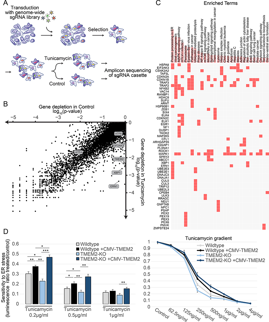
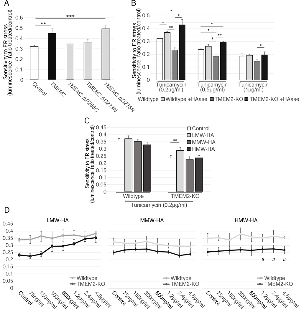
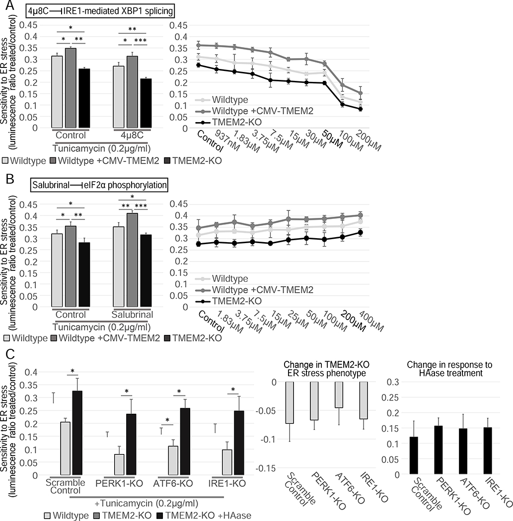
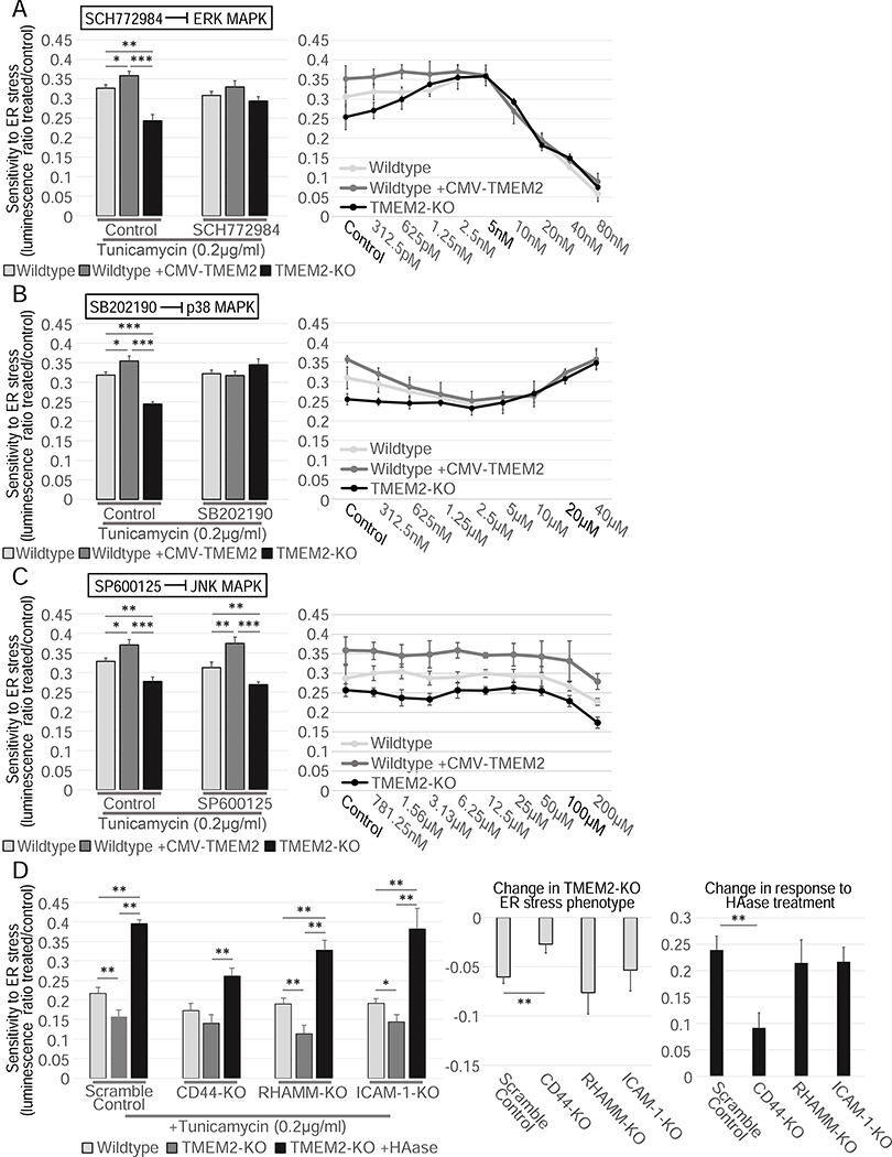
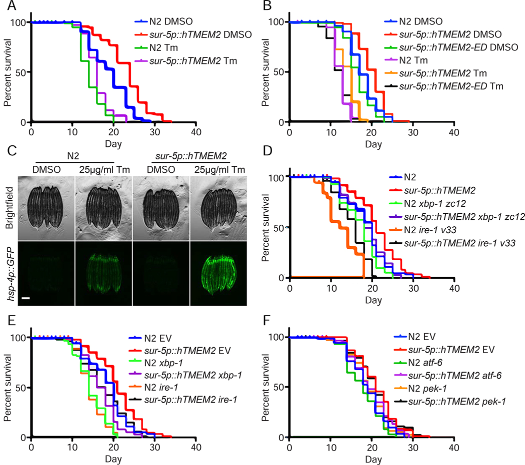
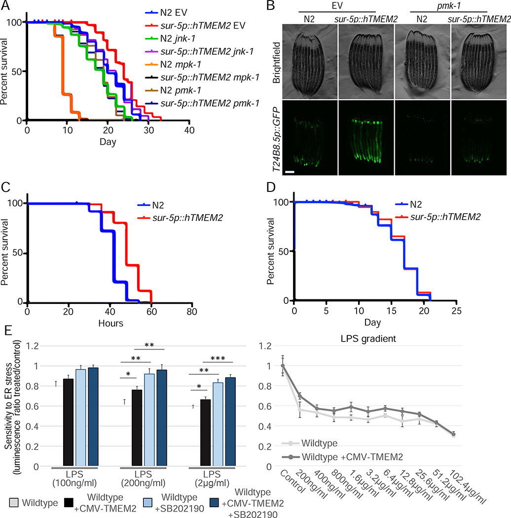
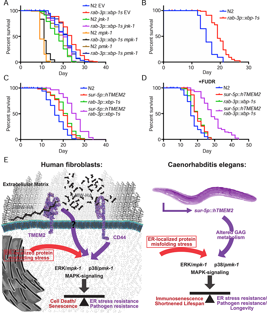
Cells have evolved complex mechanisms to maintain protein homeostasis, such as the UPRER, which are strongly associated with several diseases and the aging process. We performed a whole-genome CRISPR-based knockout (KO) screen to identify genes important for cells to survive ER-based protein misfolding stress. We identified the cell-surface hyaluronidase (HAase), Transmembrane Protein 2 (TMEM2), as a potent modulator of ER stress resistance. The breakdown of the glycosaminoglycan, hyaluronan (HA), by TMEM2 within the extracellular matrix (ECM) altered ER stress resistance independent of canonical UPRER pathways but dependent upon the cell-surface receptor, CD44, a putative HA receptor, and the MAPK cell-signaling components, ERK and p38. Last, and most surprisingly, ectopic expression of human TMEM2 in C. elegans protected animals from ER stress and increased both longevity and pathogen resistance independent of canonical UPRER activation but dependent on the ERK ortholog mpk-1 and the p38 ortholog pmk-1.

Keywords: CRISPR-Cas9; MAPK signaling; aging; endoplasmic reticulum; extracellular matrix; glucosaminoglycan; immune response; stress response.


Conflict of interest statement

Competing Financial Interests/Declaration of Interests

The authors declare no competing interests.

Figures

Figure 1:
Figure 1:
Whole genome CRISPR-KO library screen identifies TMEM2 as a potent modulator of ER stress sensitivity. A) Screen outline: Human immortalized fibroblasts were transduced with a genome-wide sgRNA lentiviral library and cultured for two weeks to maximize genome editing and target protein depletion. Cells were then split into control and Tunicamycin treatment and harvested after 3 weeks for sequencing (as described in detail in STAR METHODS) B) Comparison of gene depletion p-values between control and Tunicamycin-treated cells (individual depletion/enrichment are available in Supplemental Table 1) C) Enrichment analysis of the top differentially depleted genes (using 10% FDR as a cutoff) using the EnrichR online tool (https://amp.pharm.mssm.edu/Enrichr/). D) Viability and proliferation of Wildtype and clonal TMEM2-KO human immortalized fibroblast in the presence of Tunicamycin-induced ER stress with or without CMV-TMEM2 overexpression. Results are relative to untreated control to adjust for variability in initial cell number between cell lines. Cell density at the endpoint of a 5 day treatment period was determined via CellTiter-Glo analysis (CTG) (as described in detail in STAR METHODS); (n=3). Statistical Analysis: One-way ANOVA analysis with post-hoc Bonferroni-Holm analysis
Figure 2:
Figure 2:
TMEM2’s enzymatic breakdown of HMW-HA to LMW-HA is responsible for the ER stress phenotype. A) The CMV-TMEM2 plasmid was altered through site-directed mutagenesis in order to disrupt HAase enzymatic activity of the gene. ER stress resistance was then measured through CTG analysis. The ER stress resistance was then compared between the constructs with no or a neutral mutation (ΔD275N) of the gene, and two lines in which the HAase function of TMEM2 was diminished (ΔP265C; ΔD273N); (n=3). B) Resistance to Tunicamycin-induced ER stress was measured in Wildtype and TMEM2-KO human fibroblasts in the presence and absence of supplemented hyaluronidase (HAase) (Concentration in bar graph: HAase 5U/ml). All HAase concentrations tested (0.6U/ml – 160U/ml) were equally able to evoke this phenotype. C-D) Wildtype and TMEM2-KO cells were exposed to low molecular weight hyaluronan (LMW-HA; <20kDa in molecular weight) medium molecular weight hyaluronan (MMW-HA; 200–1000kDa) or high molecular weight hyaluronan (HMW-HA; >1000kDa). The concentration of LMW-HA, MMW-HA and HMW-HA in the bar graphs was limited to 600ng/ml, since HMW-HA did not go fully into solution at higher concentrations (marked with #).
Figure 3:
Figure 3:
The TMEM2 ER stress phenotype is independent the three canonical UPRER pathways. A-B) The resistance to Tunicamycin-induced ER stress of Wildtype, TMEM2-KO, and CMV-TMEM2-overexpressing cells was determined in the presence of A) an inhibitor of IRE1-mediated XBP1 splicing, 4μ8C (Concentration in bar graphs: 4μ8C 50μM; HAase 5U/ml) or B) an inhibitor of eIF2α phosphorylation, Salubrinal (Concentration in bar graphs: Salubrinal 200μM; HAase 5U/ml) through CTG analysis; (n=3). C) In Wildtype and TMEM2-KO cells, the UPRER pathway components IRE1, PERK1 and ATF6 were each targeted via CRISPR/CAS9-mediated gene disruption (see STAR METHODS for details). Each cell line was then cultured in the presence and absence of Tunicamycin (200ng/ml) and HAase (5U/ml) for 5 days, and the cell density at the endpoint of the experiment was measured by CTG analysis; (n=3). The additional graphs highlight the on the TMEM2-KO ER stress phenotype and the response to HAase due to the targeting of the UPRER pathways. Statistical Analysis: One-way ANOVA analysis with post-hoc Bonferroni-Holm analysis
Figure 4:
Figure 4:
TMEM2 mediates ER stress resistance through the MAPK pathway components, ERK and p38, and the cell surface receptor CD44. ER stress resistance was measured in the presence of A) the ERK inhibitor SCH772984 (Concentration in bar graphs: SCH770984 5nM, HAase 5U/ml), B) the p38 MAPK pathway inhibitor SB202190 (Concentration in bar graphs: SB202190 10μM, HAase 5U/ml) or the C) JNK MAPK pathway inhibitor SP600125 (Concentration in bar graphs: SP600125 5μM, HAase 5U/ml); (n=3) D) In Wildtype and TMEM2-KO cells, the cell-surface receptors CD44, RHAMM, and ICAM-1 were targeted via CRISPR/CAS9-mediated gene disruption (see STAR METHODS for details). Each cell line was then cultured in the presence and absence of Tunicamycin (200ng/ml) and HAase. ER stress resistance was measured through CTG analysis (n=3). The additional graphs highlight the impact on the TMEM2-KO ER stress phenotype and the response to HAase due to the targeting of the receptors. Statistical Analysis: One-way ANOVA analysis with post-hoc Bonferroni-Holm analysis.
Figure 5:
Figure 5:
hTMEM2 overexpression extends lifespan in C. elegans independent of canonical UPRER pathways. A) Lifespans were measured in Wildtype (N2) and sur-5p::hTMEM2 worms grown on EV RNAi on 1% DMSO and 25μg/ml Tunicamycin (Tm) from Day 1 (D1) as described in STAR METHODS. Data is representative of four independent trials. B) Lifespans were measured in Wildtype (N2), sur-5p::hTMEM2, and an enzymatic dead version of hTMEM2 (sur-5p::hTMEM2-ED, carrying R265C, D273N, D286N mutations; this overexpression line is an extrachromosomal array) using similar methods as A. Data is representative of three independent trials. C) Fluorescent micrographs of Wildtype (N2) and sur-5p::hTMEM2 animals expressing the UPRER reporter, hsp-4p::GFP. Animals were treated with DMSO or 25μg/ml Tunicamycin (Tm) at L4, and imaged at D1 as described in STAR METHODs. Data is representative of four independent trials. D) Lifespans were measured in Wildtype and sur-5p::hTMEM2 animals carrying either Wildtype alleles of xbp-1 and ire-1 or mutant alleles, xbp-1(zc12) or ire-1(v33), on EV RNAi. Data is representative of three independent trials. E) Lifespans were measured in Wildtype and sur-5p::hTMEM2 animals grown on EV, xbp-1, or ire-1 RNAi from hatch. Data is representative of three independent trials. F) Lifespans were measured in Wildtype and sur-5p::hTMEM2 animals grown on EV, atf-6, or pek-1 RNAi from hatch. Data is representative of two independent trials. All statistics for lifespans were performed using Log-Rank (Mantel-Cox) test using PRISM, and are available in Supplemental Table 2.
Figure 6:
Figure 6:
hTMEM2 overexpression promotes immunity and extends lifespan through mpk-1/pmk-1. A) Lifespans were measured in Wildtype and sur-5p::hsf-1 animals on EV, jnk-1, mpk-1, or pmk-1 RNAi from hatch. Data is representative of three independent trials. B) Fluorescent micrographs of Wildtype (N2) and sur-5p::hTMEM2 animals expressing the immune response reporter, T24B8.5p::GFP. Animals were grown on EV or pmk-1 RNAi as described in STAR METHODS. Data is representative of three independent trials. C) Survival was scored in Wildtype (N2) and sur-5p::hTMEM2 animals exposed to PA14 infection at L4. Survival was scored every 6 hours as described in STAR METHODS. Data is representative of two independent trials. D) Lifespans were measured in Wildtype (N2) and sur-5p::hTMEM2 animals grown on dead EV RNAi from hatch. Bacteria were killed by UV irradiation, as described in STAR METHODS. All statistics for C-D were performed using Log-Rank (Mantel-Cox) test using PRISM, and are available in Supplemental Table 2. E) Wildtype and CMV-TMEM2 overexpressing human fibroblasts were exposed to lipopolysaccharides (LPS) derived from the E. coli bacteria strain (O111:B4). Resistance to the presence of LPS was measured through CTG to determine the cell density after 5 days of exposure; (n=3). Statistical analysis: One-way ANOVA test with a post-hoc Bonferroni-Holm analysis
Figure 7:
Figure 7:
Lifespan extension through canonical xbp-1s signaling is not dependent on mpk-1/pmk-1 and is distinct from hTMEM2. A) Lifespans were measured in Wildtype (N2) and rab-3p::xbp-1s animals on EV, jnk-1, mpk-1, or pmk-1 RNAi from hatch. Data is representative of three independent trials. B) Lifespans were measured in Wildtype (N2) and rab-3p::xbp-1s animals grown on dead EV RNAi from hatch as per 5D. Data is representative of two independent trials. C-D) Lifespans were measured in Wildtype (N2), sur-5p::hTMEM2, rab-3p::xbp-1s, and sur-5p::hTMEM2/rab-3p::xbp-1s animals in the absence (C) and presence (D) of FUDR. Animals were grown on EV RNAi from hatch, and the assay was either performed on standard EV plates (C) or moved to FUDR containing plates at L4 for (D) - see STAR Methods for details. E) Graphical representation of the key insight generated by this work. In human fibroblasts, TMEM2 breaks down HMW-HA into LMW-HA within the ECM. Through interaction with CD44, LMW-HA influences ERK/mpk1- and p38/pmk-1-mediated MAPK-signaling. This in turn alters ER stress resistance and pathogen resistance in human fibroblasts. In C. elegans, TMEM2-mediated changes to glycosaminoglycan (GAG) metabolism cause a shift in MAPK signaling. This in turn alters the response to ER stress and with it, changes pathogen resistance and the lifespan of the animals.

Comment in

References

    1. Adamson B, Norman TM, Jost M, Cho MY, Nuñez JK, Chen Y, Villalta JE, Gilbert LA, Horlbeck MA, Hein MY, Pak RA, Gray AN, Gross CA, Dixit A, Parnas O, Regev A, Weissman JS, 2016. A Multiplexed Single-Cell CRISPR Screening Platform Enables Systematic Dissection of the Unfolded Protein Response. Cell 167, 1867–1882.e21. 10.1016/j.cell.2016.11.048 - DOI - PMC - PubMed
    1. Bain J, McLauchlan H, Elliott M, Cohen P, 2003. The specificities of protein kinase inhibitors: an update. Biochem. J 371, 199–204. 10.1042/BJ20021535 - DOI - PMC - PubMed
    1. Bain J, Plater L, Elliott M, Shpiro N, Hastie CJ, McLauchlan H, Klevernic I, Arthur JSC, Alessi DR, Cohen P, 2007. The selectivity of protein kinase inhibitors: a further update. Biochem. J 408, 297–315. 10.1042/BJ20070797 - DOI - PMC - PubMed
    1. Brown MK, Naidoo N, 2012. The endoplasmic reticulum stress response in aging and age-related diseases. Front. Physiol 3, 263 10.3389/fphys.2012.00263 - DOI - PMC - PubMed
    1. Chanmee T, Ontong P, Itano N, 2016. Hyaluronan: A modulator of the tumor microenvironment. Cancer Lett. 375, 20–30. 10.1016/j.canlet.2016.02.031 - DOI - PubMed

Publication types