. 2019 Dec:50:122-134.
doi: 10.1016/j.ebiom.2019.10.060. Epub 2019 Nov 21.

Epigenetically upregulated GEFT-derived invasion and metastasis of rhabdomyosarcoma via epithelial mesenchymal transition promoted by the Rac1/Cdc42-PAK signalling pathway

Affiliations

Epigenetically upregulated GEFT-derived invasion and metastasis of rhabdomyosarcoma via epithelial mesenchymal transition promoted by the Rac1/Cdc42-PAK signalling pathway

Chunxia Liu et al. EBioMedicine. 2019 Dec.

Erratum in

Abstract

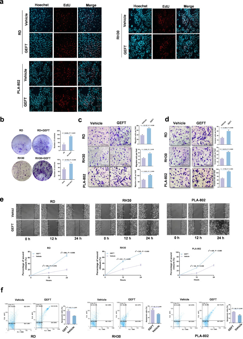
Background: Metastasis of rhabdomyosarcoma (RMS) is the primary cause of tumour-related deaths. Previous studies have shown that overexpression of the guanine nucleotide exchange factor T (GEFT) is correlated with a poorer RMS prognosis, but the mechanism remains largely unexplored.

Methods: We focused on determining the influence of the GEFT-Rho-GTPase signalling pathway and the epithelial-mesenchymal transition (EMT) or mesenchymal-epithelial transition (MET) on RMS progression and metastasis by using RMS cell lines, BALB/c nude mice and cells and molecular biology techniques.

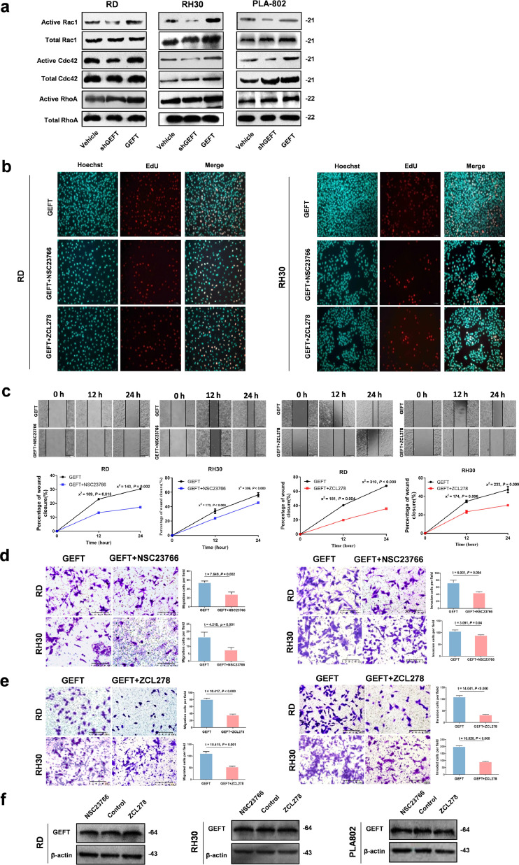
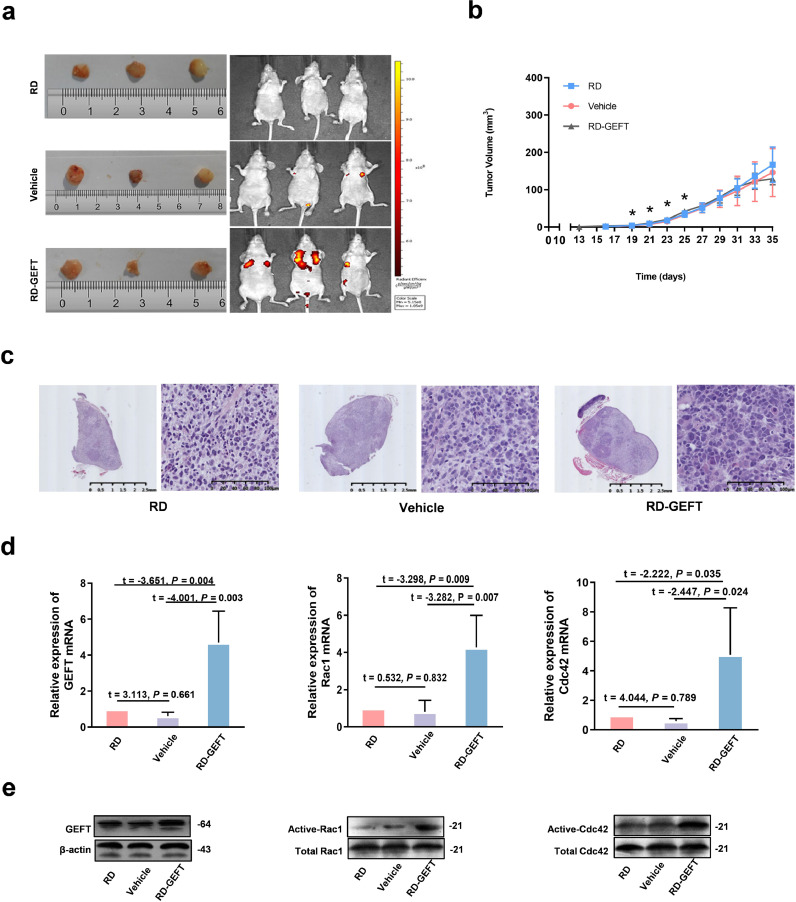
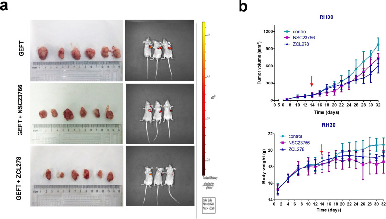
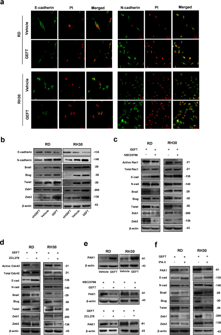
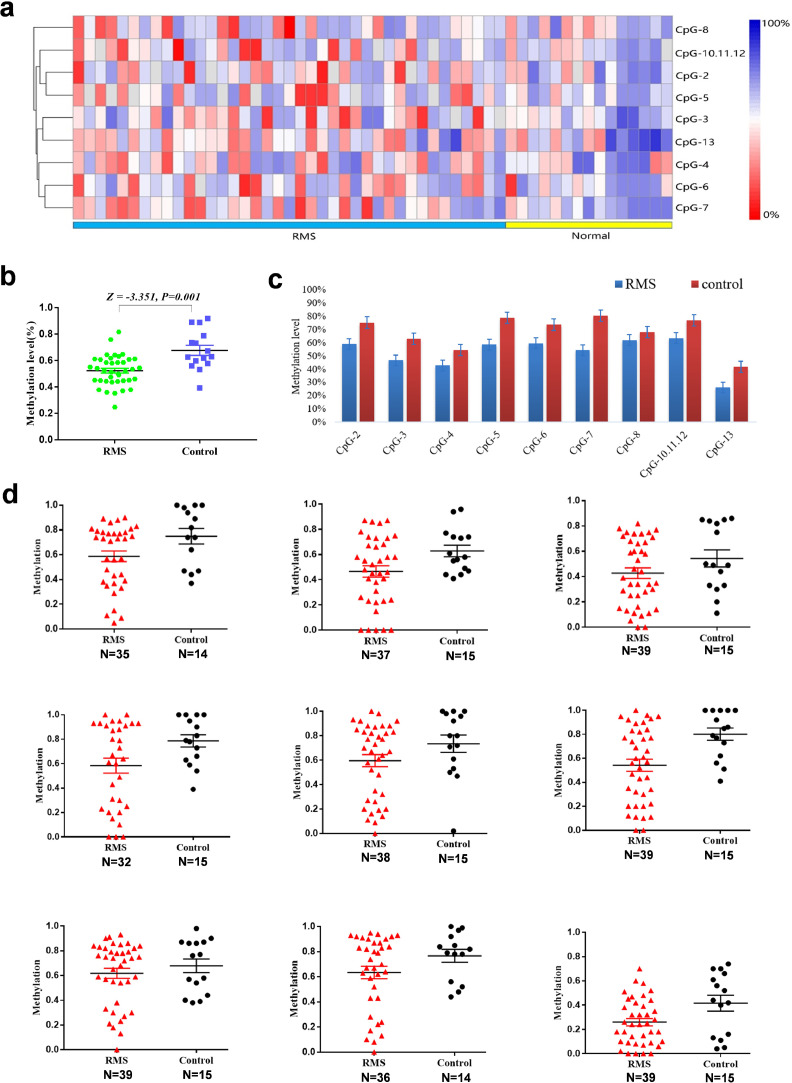
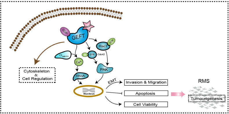
Findings: GEFT promotes RMS cell viability, migration, and invasion; GEFT also inhibits the apoptosis of RMS cells and accelerates the growth and lung metastasis of RMS by activating the Rac1/Cdc42 pathways. Interestingly, GEFT upregulates the expression levels of N-cadherin, Snail, Slug, Twist, Zeb1, and Zeb2 and reduces expression level of E-cadherin. Thus, GEFT influences the expression of markers for EMT and MET in RMS cells via the Rac1/Cdc42-PAK1 pathways. We also found that the level of GEFT gene promoter methylation in RMS is lower than that in normal striated muscle tissue. Significant differences were observed in the level of GEFT gene methylation in different histological subtypes of RMS.

Interpretation: These findings suggest that GEFT accelerates the tumourigenicity and metastasis of RMS by activating Rac1/Cdc42-PAK signalling pathway-induced EMT; thus, it may serve as a novel therapeutic target. FUND: This work was supported by grants from the National Natural Science Foundation of China (81660441, 81460404, and 81160322) and Shihezi University Initiative Research Projects for Senior Fellows (RCZX201447). Funders had no role in the design of the study, data collection, data analysis, interpretation, or the writing of this report.

Keywords: EMT; GEFT; Methylation; Rac1/Cdc42-PAK1 pathways; Rhabdomyosarcoma.


Conflict of interest statement

The authors declare no conflicts of interest.

Figures

Fig 1
Fig. 1
GEFT displays oncogene activity in RMS. (a) EdU was used to detect cell proliferation. Hoechst stained all cells (blue), EdU labelled proliferating cells (red), and GEFT promoted cell proliferation (pink). (b) Colony-forming assays. (c) The number of migrating tumour cells, as well as their migratory ability, was significantly higher in pCDNA GEFT-treated cells than that in the control groups. (d) The number of invading tumour cells, as well as their invasion ability, was significantly higher in pCDNA GEFT-treated cells than that in the control groups. (e) The migratory ability of RMS cells was investigated by wound-healing assays. (f) GEFT inhibited apoptosis of RMS cells. An independent sample t-test was used to detect differences between the two groups (b, c, d). Differences of the rate changes between the two groups were tested by chi-square test (e, f).
Fig 2
Fig. 2
GEFT is involved in Rho activation. (a) Rac1 and Cdc42 were activated by GEFT in RMS cells, as shown by pull-down assays, but RhoA was not changed. (b) The EdU assay shows that NSC23766 or ZCL278 can inhibit cell proliferation of RMS. (c) The migratory ability of RMS cells is inhibited by NSC23766 or ZCL278. Differences of the rate changes between the two groups were tested by chi-square test. (d and e) The number of migrating and invading tumour cells was significantly lower in NSC23766 or ZCL278-treated cells than that in the control groups. An independent sample t-test was used to detect differences between the two groups. (f) The level of GEFT protein did not change when the RMS cell lines were treated with either NSC23766 or ZCL278.
Fig 3
Fig. 3
Effects of GEFT overexpression on tumour growth and metastasis in vivo. (a) Excised tumour samples and whole-body GFP imaging. (b) Growth curves of the xenografted tumours. One-way ANOVA (analysis of variance) was used to compare the indexes among three groups. (c) HE-stained xenografted tumours. (d) The mRNA levels of GEFT, Rac1 and Cdc42 are increased in the RD-GEFT+ group. An independent sample t-test was used to detect differences between the two groups. (e) Pull-down assays show that Active-Rac1 and Active -Cdc42 levels are increased in the RD-GEFT group. *P < 0.05.
Fig 4
Fig. 4
NSC23766 and ZCL278 slowed the growth rate of the tumours in mice. On the 14th day, the nude mice were given inhibitors (50 mg/kg). (a) Excised tumour samples and whole-body GFP imaging. (b) Growth curves of the xenografted tumours. One-way ANOVA (analysis of variance) was used to compare the indexes between two groups (a, b).
Fig 5
Fig. 5
GEFT influences the expression of EMT markers in RMS cells via the Rac1/Cdc42-PAK1 pathways. (a) Immunofluorescence assay confirmed that GEFT enhanced the fluorescence intensity of N-cadherin protein in RMS cells and reduced the fluorescence intensity of E-cadherin protein in RMS cells. (b) GEFT overexpression upregulated the expression levels of EMT related markers (N-cadherin, Snail, Slug, Twist, Zeb1, and Zeb2) and reduced the expression level of E-cadherin; the expression levels of EMT-related proteins were reduced by shGEFT, which also upregulated the expression level of E-cadherin. (c and d) The expression level of E-cadherin increased, whereas the expression levels of N-cadherin and Snail, Slug, Twist, ZEB1, and ZEB2 were reduced after NSC23766 or ZCL278 interference. (e) GEFT can promote the expression of PAK1, and NSC23766 or ZCL278 can inhibit the expression of PAK1. (f) The expression level of E-cadherin increased and the expression levels of N-cadherin and EMT-inducing transcription factors were reduced after IPA-3 interference.
Fig 6
Fig. 6
GEFT is upregulated in rhabdomyosarcoma via aberrant promoter hypomethylation. (a) Hierarchical cluster analysis of CpG units’ methylation profiles of the GEFT promoter region in RMS (n = 39) and normal tissues (n = 15). Each vertex indicates one CpG site. Each column represents one sample. The colour gradient between red and blue indicates methylation of each GEFT CpG unit in each sample (0–100%). Grey represents inadequate or missing data. (b) Comparison of average methylation of GEFT promoter between RMS and skeletal muscle tissues. (c) Histogram analysis of the average methylation level of 9 CpG units of the GEFT promoter in RMS and skeletal muscle tissues. (d) Scatter plot of the average methylation level of 9 CpG units of the GEFT promoter in RMS and skeletal muscle tissues. Wilcoxon nonparametric test was used to compare the indexes between two groups.
Fig 7
Fig. 7
GEFT function and signalling pathway. GEFT comprises the Dbl and pleckstrin homology domains and regulates cytoskeleton and cellular processes. GEFT has specific exchange activity for Rac, Cdc42 and GST-PAK. GEFT selectively couples with Gαq/11 to activate RhoA in blood vessels and cultured cells; it also mediates Ca2+ sensitization of the force induced with Gαq/11-coupled agonists. GEFT activates the Rac1/Cdc42 signalling pathway and promotes the tumourigenicity and metastasis of RMS by influencing the expression of EMT-related proteins of RMS cells.

Comment in

Similar articles

Cited by

References

    1. Doyle L.A. Sarcoma classification: an update based on the 2013 World Health Organization classification of tumors of soft tissue and bone. Cancer. 2014;120(12):1763–1774. - PubMed
    1. Breneman J.C., Lyden E., Pappo A.S., Link M.P., Anderson J.R., Parham D.M. Prognostic factors and clinical outcomes in children and adolescents with metastatic rhabdomyosarcoma – a report from the Intergroup Rhabdomyosarcoma Study IV. J Clin Oncol. 2003;21(1):78–84. - PubMed
    1. Chaffer C.L., Weinberg R.A. A perspective on cancer cell metastasis. Science. 2011;331(6024):1559–1564. - PubMed
    1. Valastyan S., Weinberg R.A. Tumor metastasis: molecular insights and evolving paradigms. Cell. 2011;147(2):275–292. - PMC - PubMed
    1. Liu C., Li D., Jiang J., Hu J., Zhang W., Chen Y. Analysis of molecular cytogenetic alteration in rhabdomyosarcoma by array comparative genomic hybridization. PLoS One. 2014;9(4):e94924. - PMC - PubMed

MeSH terms