Review
. 2019 Dec:84:102736.
doi: 10.1016/j.dnarep.2019.102736. Epub 2019 Oct 23.

RNase H2-RED carpets the path to eukaryotic RNase H2 functions

Affiliations
Review

RNase H2-RED carpets the path to eukaryotic RNase H2 functions

Susana M Cerritelli et al. DNA Repair (Amst). 2019 Dec.

Abstract

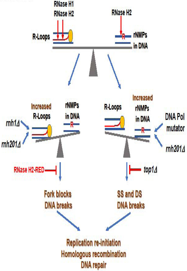
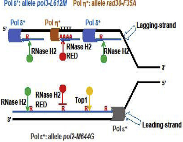
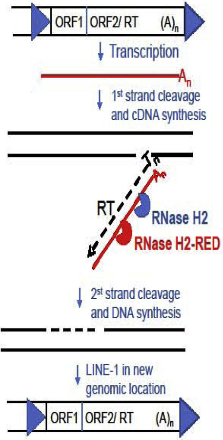
Eukaryotic RNases H2 have dual functions in initiating the removal of ribonucleoside monophosphates (rNMPs) incorporated by DNA polymerases during DNA synthesis and in cleaving the RNA moiety of RNA/DNA hybrids formed during transcription and retrotransposition. The other major cellular RNase H, RNase H1, shares the hybrid processing activity, but not all substrates. After RNase H2 incision at the rNMPs in DNA the Ribonucleotide Excision Repair (RER) pathway completes the removal, restoring dsDNA. The development of the RNase H2-RED (Ribonucleotide Excision Defective) mutant enzyme, which can process RNA/DNA hybrids but is unable to cleave rNMPs embedded in DNA has unlinked the two activities and illuminated the roles of RNase H2 in cellular metabolism. Studies mostly in Saccharomyces cerevisiae, have shown both activities of RNase H2 are necessary to maintain genome integrity and that RNase H1 and H2 have overlapping as well as distinct RNA/DNA hybrid substrates. In mouse RNase H2-RED confirmed that rNMPs in DNA during embryogenesis induce lethality in a p53-dependent DNA damage response. In mammalian cell cultures, RNase H2-RED helped identifying DNA lesions produced by Top1 cleavage at rNMPs and led to determine that RNase H2 participates in the retrotransposition of LINE-1 elements. In this review, we summarize the studies and conclusions reached by utilization of RNase H2-RED enzyme in different model systems.


Conflict of interest statement

Conflict of Interest: The authors declare that there are no conflicts of interest

Figures

Figure 1.
Figure 1.. Recognition of rNMP in dsDNA by RNase H2.
(A). Surface representation of Tm-RNase H2 with the catalytic center in yellow. The nucleic acid is shown in cartoon with DNA in blue and the single ribonucleotide in red. The close-up is a view of the interaction of the 2’-OH (pink sphere) with adjacent residues. The nucleotides are numbered relative to the scissile phosphate, which is indicate with an arrow. Cartoon adapted from Rychlik et al., 2010 (Rychlik et al., 2010). (B). Models of Bst-RNase H3 (in green) and Tm-RNase H2 (in orange) active sites interactions with substrates. Residues interacting with DpRpD and possible hydrogen bonds (dotted lines) are shown. The nucleotides are number as in A. Four catalytic residues (DEDD for Tm-RNase H2 and DEDE for Bst-RNase H3) are indicated in numerical order. Cartoon adapted from Chon et al., 2013 (Chon et al., 2013). (C). Sequence alignment of the active site and the region around the crucial Tyr residue is shown for human RNase H2A (Hu2A), S. cerevisiae Rnh201 (Sc2A), T. maritima RNase H2 (Tma2), B. stearothermophilus RNase H3 (Bst3), Streptococcus pneumoniae RNase H3 (Spn3) and Thermovibrio ammonificans HB-1 RNase h3 (Tam3). Amino acids important for catalysis are highlighted in grey. Residues interacting with the 2’-OH are boxed (Y in orange for Tma2 and D in green for Bst3).
Figure 2.
Figure 2.. R-Loops and rNMPs are kept in check by RNase H1 and RNase H2.
The coordinate work of RNase H1 and RNase H2 limits the presence of R-loops (top left, DNA in blue, RNA in red. The RNA is extruding from the RNA polymerase represented as a yellow oval) and rNMPs in DNA (top right, DNA in blue, single rNMP represented by a red “R”) to preserve genome stability (top). In the absence of RNase H1 and RNase H2, R-loops accumulate and block replication creating breaks and DNA damage, outweighing the defect that might cause rNMPs in DNA in the absence of RNase H2. The RNase H2-RED mutant would process these hybrids preserving genome integrity (middle left). When RNase H2 is missing in combination with replicases mutants that include rNMPs at higher frequency than the WT counterparts, especially leading strand Pol ε-M644G, the high density of unrepaired rNMPs in DNA would result in SSB and DSB in DNA upon Top1 processing, while R-loops would not accumulate significantly because RNase H1 is active. Deletion of TOP1 would prevent DNA damage (middle right). DNA damage created by increased R-loops or increased rNMPs in DNA induces replication re-initiation, HR and DNA repair to maintain genome integrity (bottom).
Figure 3.
Figure 3.. Removal of rNMPs inserted by DNA polymerases.
Representation of the replication fork, with template strand in black and newly synthesized strands in blue. The leading strand Pol ε (represented as a grey cylinder) processively copies the template strand. Allele pol2-M644G codes for a mutant enzyme that incorporates rNMPs at higher frequency than the WT Pol ε. These newly embedded rNMPs in DNA are nicked by WT RNase H2 (green circle), but not by the mutant RNase H2-RED (red circle), to initiate the RER process. In the absence of RNase H2, Top1 (yellow circle) can incise at the 3’-end of the rNMP inducing DNA breaks and mutagenesis. The bulk of lagging strand synthesis is carried out by Pol δ (blue cylinder) in OFs that need processing and maturation. Allele pol3-L612M codes for a mutant with higher propensity than WT Pol δ to incorporate rNMPs in DNA. RNase H2, but not RNase H2-RED, can initiate the removal of single rNMPs embedded in lagging strand DNA. In regions difficult to replicate, such as homo-polymeric tracks, especially runs of Ts in lagging-strand DNA template, the translesion polymerase Pol η (orange cylinder) replaces Pol δ and copies DNA with lower fidelity. The allele rad30-F35A codes for a Pol η mutant protein that incorporates rNMPs in DNA at a higher frequency than the WT Pol η. RNase H2-RED would incise in tracks of polyribonucleotide in DNA incorporated by the mutant Pol η to initiate their removal.
Figure 4.
Figure 4.. RNase H2 participates in the LINE-1 retrotransposition.
Retrotransposition competent LINE-1 elements consist of a promoter in their 5’-untranslated region (blue arrow in the top left) followed by ORF1, which codes for an RNA-binding protein and ORF2/RT which codes for a protein with both endonuclease and reverse transcriptase activity (Goodier, 2016). At the 3’-end of the element is a short 3’-UTR followed by a poly-A tail and a short repeat created during the insertion (blue arrow at the top right). The process of retrotransposition starts by transcription of the LINE-1 element into a mRNA (red line) including a poly-A tail (An). Then, LINE-1 mRNA is reverse transcribed, and the new cDNA is integrated in a new genomic location by the concerted work of ORF2/RT. The endonuclease recognizes and cleaves the bottom strand of a target sequence containing a run of Ts, which are used as primer for the RT to copy the LINE-1 mRNA and make a cDNA (black dotted line in middle panel), resulting in an RNA/DNA hybrid covalently attached to the genome. The following step requires the removal of the mRNA template and because LINE-1 does not code for an RNase H, it uses the cellular RNase H2 (blue). The RNase H2-RED mutant (red) can also participate in this hydrolysis. Subsequently, in a process not completely understood, after removal of the RNA, the top strand is cleaved and the newly cDNA is used as template for top strand synthesis, resulting in a new LINE-1 insertion.

Similar articles

Cited by

References

    1. Abeshouse A, Ahn J, Akbani R, Ally A, Amin S, Andry Christopher D., Annala M, Aprikian A, Armenia J, Arora A, et al. (2015). The Molecular Taxonomy of Primary Prostate Cancer. Cell 163, 1011–1025. - PMC - PubMed
    1. Aden K, Bartsch K, Dahl J, Reijns MAM, Esser D, Sheibani-Tezerji R, Sinha A, Wottawa F, Ito G, Mishra N, et al. (2019). Epithelial RNase H2 Maintains Genome Integrity and Prevents Intestinal Tumorigenesis in Mice. Gastroenterology 156, 145–159.e119. - PMC - PubMed
    1. Ait Saada A, Lambert SAE, and Carr AM (2018). Preserving replication fork integrity and competence via the homologous recombination pathway. DNA Repair 71, 135–147. - PMC - PubMed
    1. Allen-Soltero S, Martinez SL, Putnam CD, and Kolodner RD (2014). A saccharomyces cerevisiae RNase H2 interaction network functions to suppress genome instability. Mol Cell Biol 34, 1521–1534. - PMC - PubMed
    1. Alvino GM, Collingwood D, Murphy JM, Delrow J, Brewer BJ, and Raghuraman MK (2007). Replication in Hydroxyurea: It’s a Matter of Time. Mol Cell Biol 27, 6396–6406. - PMC - PubMed

Publication types