Review
. 2019 Nov 8:18:e20190061.
doi: 10.1590/1677-5449.190061.

Pelvic congestion syndrome and embolization of pelvic varicose veins

Affiliations
Review

Pelvic congestion syndrome and embolization of pelvic varicose veins

Mateus Picada Corrêa et al. J Vasc Bras. .

Abstract

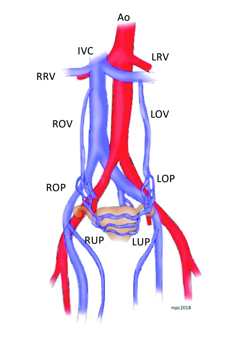
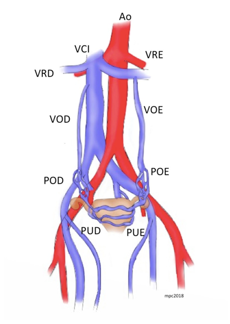
Pelvic congestion syndrome (PGS) is defined as chronic pelvic pain for more than 6 months associated with perineal and vulvar varicose veins caused by reflux or obstruction in gonadal, gluteal, or parauterine veins. PGS accounts for 16-31% of cases of chronic pelvic pain, and is usually diagnosed in the third and fourth decades of life. Interest in this condition among vascular surgeons has been increasing over recent years because of its association with venous insufficiency of the lower limbs. Despite its significant prevalence, PGS is still poorly diagnosed in both gynecology and angiology offices. Therefore, in this article we review the etiology and diagnosis of this condition and the outcomes of the different types of treatment available.

Resumo: A síndrome da congestão pélvica (SCP) é definida como dor pélvica crônica há mais de 6 meses associada a varizes perineais ou vulvares, resultantes do refluxo ou obstrução das veias gonadais, glúteas ou periuterinas. A SCP é responsável por 16-31% dos casos de dor pélvica crônica, sendo diagnosticada sobretudo na terceira e quarta décadas de vida. Nos últimos anos, houve um interesse maior nessa patologia por parte dos cirurgiões vasculares devido à sua associação com insuficiência venosa de membros inferiores. Apesar de prevalente, a SCP ainda é pouco diagnosticada tanto nos consultórios ginecológicos quanto nos de angiologistas. Portanto, neste artigo revisaremos a etiologia e o diagnóstico desta patologia e os resultados dos diversos tipos de tratamentos disponíveis.

Keywords: embolization; varicose veins; venous insufficiency.


Conflict of interest statement

Conflicts of interest: No conflicts of interest declared concerning the publication of this article.

Figures

Figure 1
Figure 1. Schematic illustration of the anatomy of the pelvic veins. Ao = aorta; IVC = inferior vena cava; = LRV left renal vein; RRV = right renal vein; LOV= left ovarian vein; ROV= right ovarian vein; ROP = right ovarian plexus; LOP= left ovarian plexus; RUP = right uterine plexus; LUP = left uterine plexus.
Figure 2
Figure 2. Axial angiotomography slice in venous phase showing several varicose parauterine veins of varying diameters, as large as 11 mm.
Figure 3
Figure 3. Pelvic phlebography during Valsalva maneuver, showing large varicose vessels. There is contrast reflux to the left common femoral vein (arrow) and the right parauterine complex (broken arrow).
Figure 4
Figure 4. Reconstruction of left gonadal phlebography showing increased diameter and reflux to parauterine veins (A). After injection of polidocanol foam, the pelvic veins can no longer be seen (B), and the MPA2 catheter tip is maintained at the distal portion of the iliac bone, with the aim of preventing retrograde flow of foam to the gonadal vein (arrow). After embolization of the left gonadal vein with six 0.035” coils, the left ovarian vein is completely excluded (broken arrow (C)).
Figura 1
Figura 1. Representação esquemática da anatomia das veias pélvicas. Ao = aorta; VCI = veia cava inferior; VRE = veia renal esquerda; VRD = veia renal direita; VOE = veia ovariana esquerda; VOD = veia ovariana direita; POD = plexo ovariano direito; POE = plexo ovariano esquerdo; PUD = plexo uterino direito; PUE = plexo uterino esquerdo.
Figura 2
Figura 2. Corte axial de uma angiotomografia em fase venosa demonstrando diversas varizes parauterinas de diversos diâmetros, alcançando até 11 mm.
Figura 3
Figura 3. Flebografia pélvica em Valsalva demonstrando grandes vasos varicosos. Há refluxo de contraste para a veia femoral comum esquerda (seta) e para o plexo parauterino direito (seta pontilhada).
Figura 4
Figura 4. Reconstrução de flebografia de gonadal esquerda demonstrando aumento de diâmetro e refluxo para veias parauterinas (A). Após injeção de espuma de polidocanol, as veias pélvicas não são mais visualizadas (B), e a ponta do cateter MPA2 mantém-se na parte distal do osso ilíaco com o intuito de evitar o fluxo retrógrado da espuma até a veia gonadal (seta). Após a embolização da veia gonadal esquerda com seis molas 0.035”, há completa exclusão da veia ovariana esquerda (seta pontilhada (C)).

References

    1. Taylor HC., Jr Vascular congestion and hyperemia; their effect on function and structure in the female reproductive organs; the clinical aspects of the congestion-fibrosis syndrome. Am J Obstet Gynecol. 1949;57(4):637–653. doi: 10.1016/0002-9378(49)90704-8. - DOI - PubMed
    1. Meissner MH, Gibson K. Clinical outcome after treatment of pelvic congestion syndrome: sense and nonsense. Phlebology. 2015;30(1) Suppl:73–80. doi: 10.1177/0268355514568067. - DOI - PubMed
    1. O’Brien MT, Gillespie DL. Diagnosis and treatment of the pelvic congestion syndrome. J Vasc Surg Venous Lymphat Disord. 2015;3(1):96–106. doi: 10.1016/j.jvsv.2014.05.007. - DOI - PubMed
    1. Asciutto G, Asciutto KC, Mumme A, Geier B. Pelvic venous incompetence: reflux patterns and treatment results. Eur J Vasc Endovasc Surg. 2009;38(3):381–386. doi: 10.1016/j.ejvs.2009.05.023. - DOI - PubMed
    1. Lechter A, Lopez G, Martinez C, Camacho J. Anatomy of the gonadal veins: a reappraisal. Surgery. 1991;109(6):735–739. - PubMed

LinkOut - more resources