

Extracellular vesicles and coagulation in blood from healthy humans revisited
- PMID: 31762964
- PMCID: PMC6853244
- DOI: 10.1080/20013078.2019.1688936
Extracellular vesicles and coagulation in blood from healthy humans revisited
Abstract
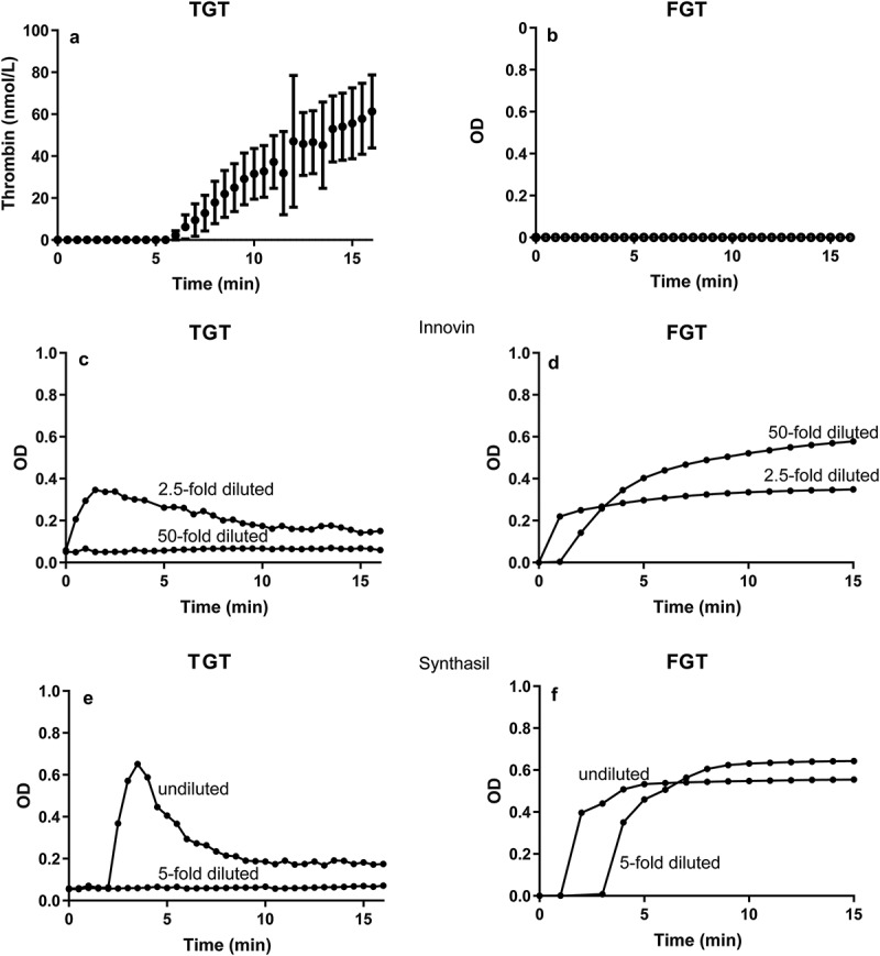
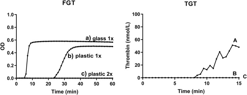
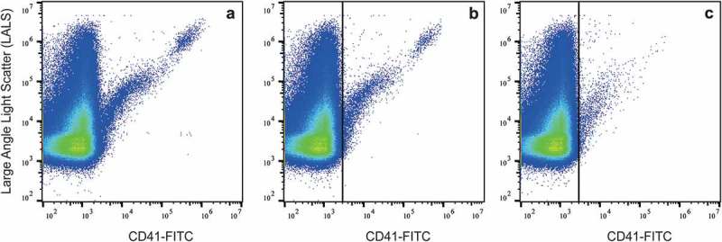
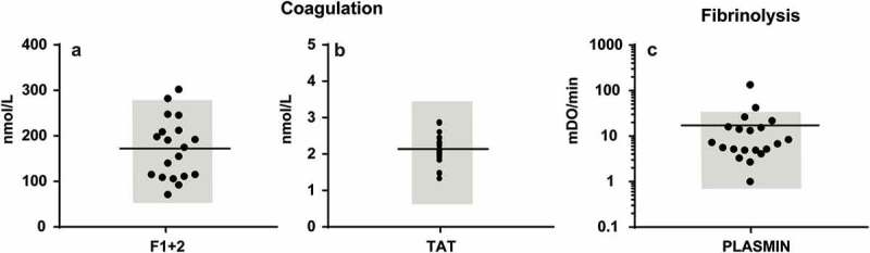
Background: In 2001, we studied the presence and coagulant properties of "microparticles" in the blood of healthy humans. Since then, multiple improvements in detection, isolation and functional characterization of the now called "extracellular vesicles" (EVs) have been made, and shortcomings were identified. Aim: To revisit the presence and function of EVs in blood from healthy humans. Methods: Blood was collected from 20 healthy donors. EV-containing plasma was prepared according to new guidelines, and plasma was diluted to prevent swarm detection. Single EVs were measured by flow cytometry with known sensitivity of fluorescence and light scatter. The haemostatic properties of EVs were measured by thrombin-, fibrin-, and plasmin generation. Plasma concentrations of thrombin-antithrombin complexes and prothrombin fragment 1 + 2 were measured to assess the coagulation status in vivo. Results: Compared to 2001, the total concentrations of detected EVs increased from 190- to 264-fold. In contrast to 2001, however, EVs are non-coagulant which we show can be attributed to improvements in blood collection and plasma preparation. No relation is present between the plasma concentrations of EVs and either TAT or F1 + 2. Finally, we show that EVs support plasmin generation. Discussion: Improvements in blood collection, plasma preparation and detection of EVs reveal that results from earlier studies have to be interpreted with care. Compared to 2001, higher concentrations of EVs are detected in blood of healthy humans which promote fibrinolysis rather than coagulation.
Keywords: Coagulation; exosomes; extracellular vesicles; fibrinolysis; flow cytometry; microparticles.
© 2019 The Author(s). Published by Informa UK Limited, trading as Taylor & Francis Group.
Figures
References
-
- Berckmans RJ, Nieuwland R, Boing AN, et al. Cell-derived microparticles circulate in healthy humans and support low grade thrombin generation. Thromb Haemost. 2001;85:639–10. - PubMed
-
- Arraud N, Linares R, Tan S, et al. Extracellular vesicles from blood plasma: determination of their morphology, size, phenotype and concentration. J Thromb Haemost. 2014;12:614–627. - PubMed
-
- Coumans FAW, Brisson AR, Buzas EI, et al. Methodological guidelines to study extracellular vesicles. Circ Res. 2017;120:1632–1648. - PubMed
-
- van der Pol E, Sturk A, van Leeuwen T, et al. group I-S-VW. Standardization of extracellular vesicle measurements by flow cytometry through vesicle diameter approximation. J Thromb Haemost. 2018;16:1236–1245. - PubMed
LinkOut - more resources
Full Text Sources
