Review
. 2019 Aug 16;10(34):7835-7851.
doi: 10.1039/c9sc03368h. eCollection 2019 Sep 14.

Biomedical applications of copper-free click chemistry: in vitro, in vivo, and ex vivo

Affiliations
Review

Biomedical applications of copper-free click chemistry: in vitro, in vivo, and ex vivo

Eunha Kim et al. Chem Sci. .

Abstract

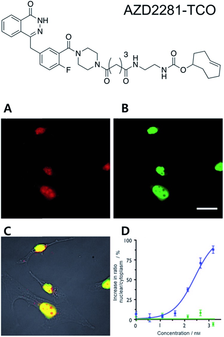
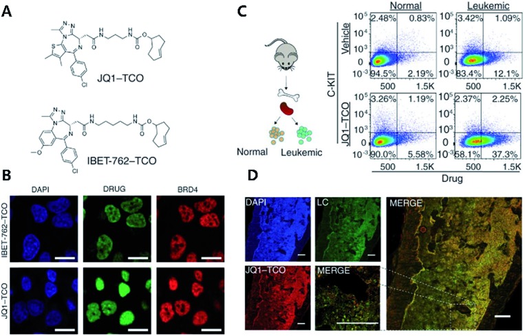
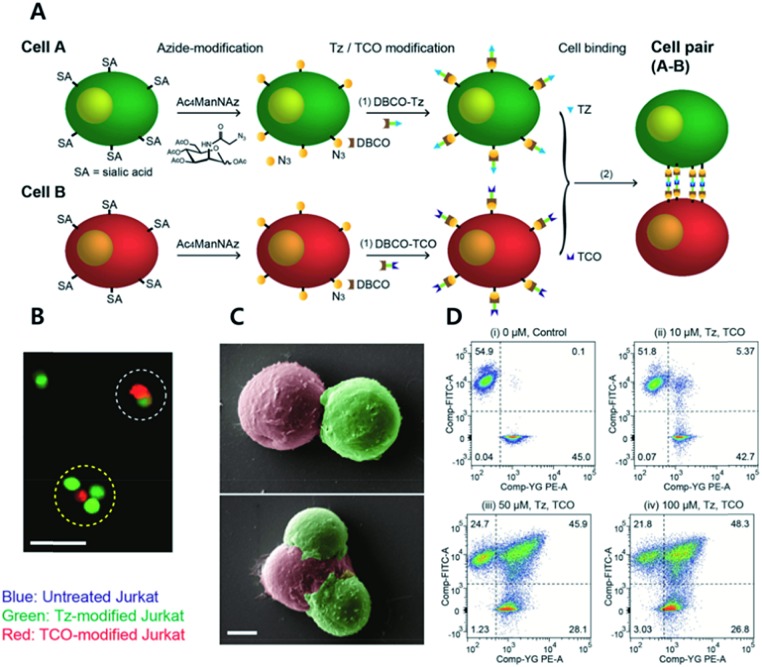
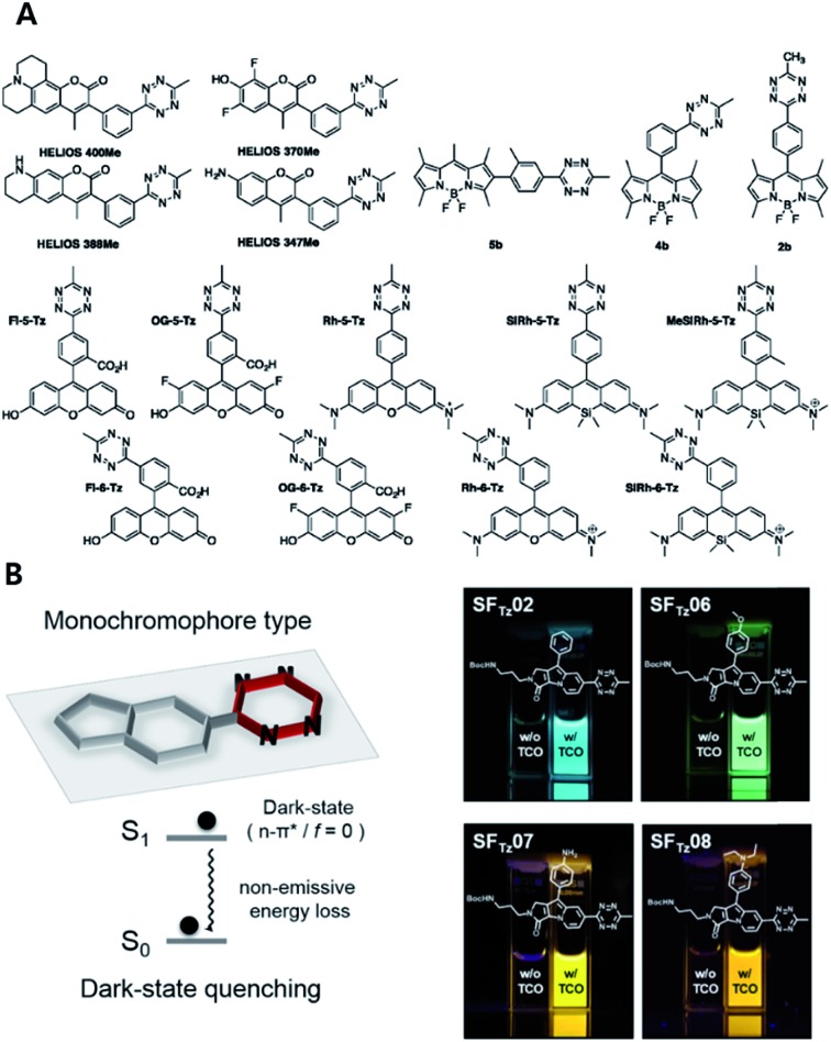
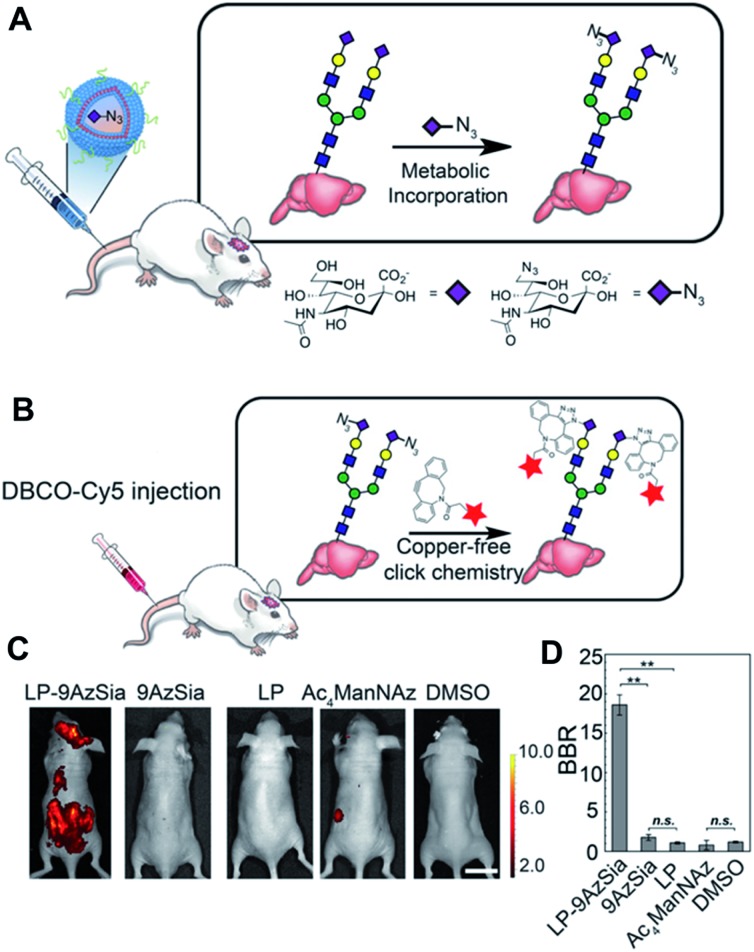
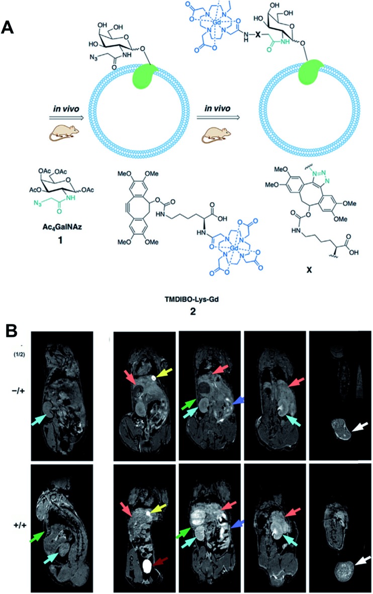
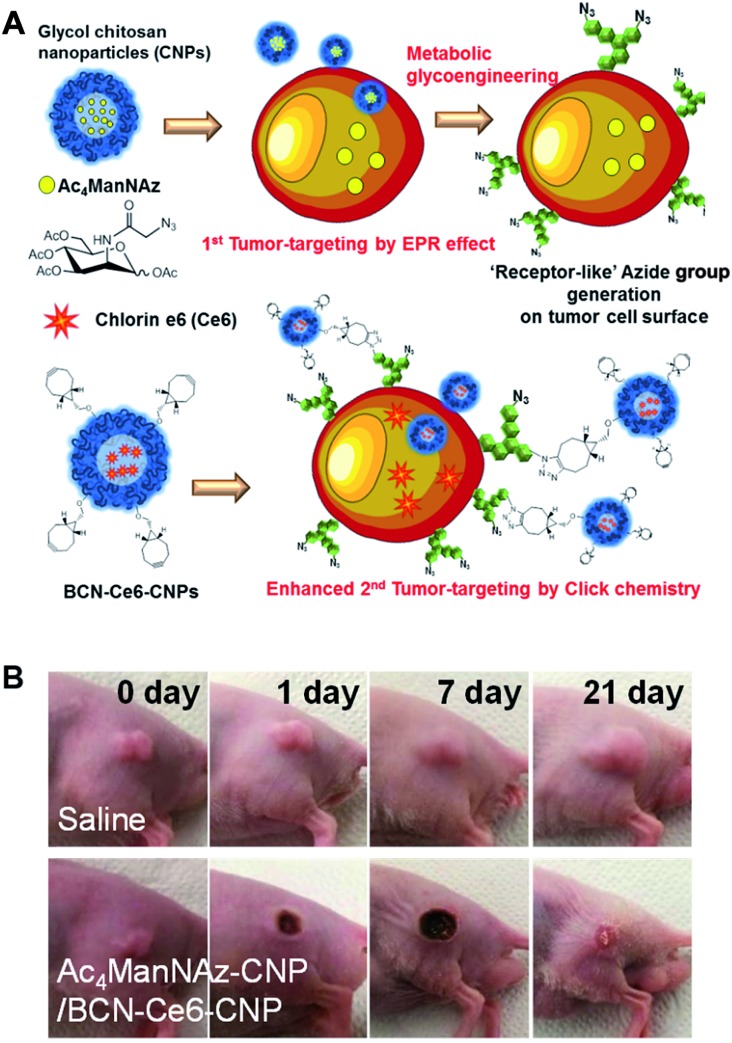
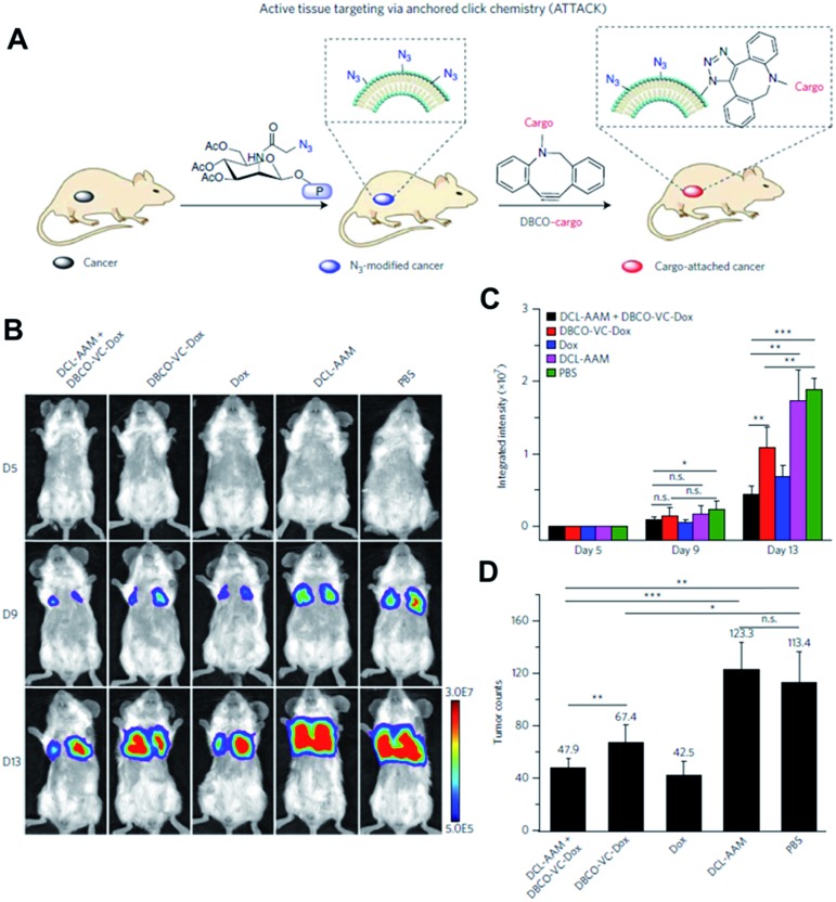
Recently, click chemistry has provided important advances in biomedical research fields. Particularly, copper-free click chemistry including strain-promoted azide-alkyne cycloaddition (SPAAC) and inverse-electron-demand Diels-Alder (iEDDA) reactions enable fast and specific chemical conjugation under aqueous conditions without the need for toxic catalysts. Click chemistry has resulted in a change of paradigm, showing that artificial chemical reactions can occur on cell surfaces, in cell cytosol, or within the body, which is not easy with most other chemical reactions. Click chemistry in vitro allows specific labelling of cellular target proteins and studying of drug target engagement with drug surrogates in live cells. Furthermore, cellular membrane lipids and proteins could be selectively labelled with click chemistry in vitro and cells could be adhered together using click chemistry. Click chemistry in vivo enables efficient and effective molecular imaging and drug delivery for diagnosis and therapy. Click chemistry ex vivo can be used to develop molecular tools to understand tissue development, diagnosis of diseases, and therapeutic monitoring. Overall, the results from research to date suggest that click chemistry has emerged as a valuable tool in biomedical fields as well as in organic chemistry.


Figures

Fig. 1
Fig. 1. Clickable drug surrogates for imaging of PARP1 protein in vitro. (A) Copper-free click reaction between AZD2281-TCO and Texas Red-Tz in MDA-MB436 cells. (B) Immunofluorescence staining of PARP1 protein with green fluorescent monoclonal anti-PARP antibody. (C) A composite overlay on phase contrast. (D) Increase in the ratio of nuclear/cytoplasmic fluorescence signal for AZD2291-TCO/Texas Red-Tz without (circle) and with (square) the blocking reagent AZD2281. Scale bar: 20 μm. Reproduced from ref. 35 with permission from John Wiley and Sons, copyright 2010.
Fig. 2
Fig. 2. Clickable drug surrogates for mechanism study in vitro. (A) Chemical structures of clickable BET inhibitor analogs, JQ1-TCO and IBET-762-TCO. (B) Fluorescence imaging of HeLa cells incubated with IBET-762-TCO or JQ1-TCO. Nuclei were stained with 4′,6-diamidino-2-phenylindole (DAPI), drug surrogates were labelled with Cy5-tetrazine, and BRD4 was stained with an anti-BRD4 antibody. Scale bar: 20 μm. (C) Schematic illustration of the procedure to detect clickable small molecules in vivo and flow cytometry analysis of drug levels within normal hematopoietic cells and leukemia cells. (D) Confocal microscopy of individual leukemia cells containing drug surrogates in mouse femur tissue treated with 100 mg kg–1 of JQ1-TCO. Leukemia cells (LCs) are identified by the Venus reporter. Scale bar: 187 μm. Reproduced from ref. 43 with permission from The American Association for the Advancement of Science, copyright 2017.
Fig. 3
Fig. 3. Click chemistry for labelling cellular membrane protein. (A) In vitro bioorthogonal labelling of apoptotic cells with an S-allyl handle by iEDDA copper-free click chemistry. (B) SDS-PAGE of AnxV containing an S-allyl handle labeled with Tz-Cy3 or Tz-Rhod. Fluorescence scanning of gels imaged (right) before staining with Coomassie blue (left). (C) In vitro fluorescence imaging of apoptotic cells with AnxV containing a chemical handle for the iEDDA reaction. Apoptotic cells were specifically labeled with AnxV S-allyl Cys incubation and followed by treatment with fluorogenic Tz-Cy3. Scale bar: 100 μm. Reproduced from ref. 66 with permission from John Wiley and Sons, copyright 2016.
Fig. 4
Fig. 4. Click chemistry for artificial cell attachment. (A) Schematic illustration of the artificial cellular gluing method via metabolic glycoengineering and copper-free click chemistry. (B) Fluorescence images and (C) SEM images of glued cells visualized doublet to quartet glued cells. Scale bar: 50 μm in (B) and 5 μm in (C). (D) Flow cytometry data of glued cells confirmed dose-dependent population increase of glued cells. Reproduced from ref. 73 with permission from John Wiley and Sons, copyright 2015.
Fig. 5
Fig. 5. Tetrazine-based turn on probe development. (A) Chemical structures of fluorogenic tetrazine fluorophores (FLTz). (B) Monochromophore type FLTz allows wavelength independent fluorescence quenching efficiency via nonradiative energy decay due to the lowest lysing dark state at S1 presumably originated from a non-radiative n → π* transition. Reproduced from ref. 86 with permission from American Chemical Society, copyright 2017.
Fig. 6
Fig. 6. In vivo fluorescence imaging of sialoglycans in mouse brain by copper-free click chemistry. (A) Azide group labelling of sialoglycans in mouse brain by 9-azido sialic acid-loaded liposome and metabolic glycoengineering. Liposomes were injected intravenously to mice and delivered 9-azido sialic acid to brain cells across BBB. Then, azide groups were generated in brain cells by metabolic glycoengineering. (B) SPAAC in vivo between DBCO-Cy5 and sialoglycans in mouse brain. Intravenously injected DBCO-Cy5 could label newly synthesized brain sialoglycans. (C) In vivo fluorescence images showing sialoglycans targeted by SPAAC. 9-Azido sialic acid-loaded liposomes were administered daily to mice for 7 days and DBCO-Cy5 was injected at day 8. (D) Brain signal-to-background ratio (BBR) of (C). **P < 0.01; n.s., not significant (one-way ANOVA). Reproduced from ref. 51 with permission from the National Academy of Sciences of the United States of America (NAS), copyright 2016.
Fig. 7
Fig. 7. In vivo MR imaging of LL2 tumor glycans by copper-free click chemistry. (A) Two step strategy including i.p. injection of Ac4GalNAz and i.v. injection of TMBIDO-modified gadolinium into the mice. Azido-labeled glycoproteins were labeled with TMBIDO-modified gadolinium by SPAAC for MR imaging. (B) In vivo T1-MR images of LL2 tumor glycans labeled by SPAAC. Mice were injected with solvent vehicles (upper) or Ac4GalNAz (lower), and TMBIDO-modified gadolinium was injected into both mice. Images were displayed from the ventral towards the dorsal side. Tumor (white arrows), kidney (cyan), liver (orange), gut (purple), and spleen (green). Reproduced from ref. 53 with permission from John Wiley and Sons, copyright 2016.
Fig. 8
Fig. 8. In vivo tumor-targeted delivery of photosensitizers and photodynamic therapy by copper-free click chemistry. (A) Azide group labelling of tumor cells by Ac4ManNAz-loaded nanoparticles and second tumor targeting by DBCO-modified nanoparticles containing photosensitizers. Both nanoparticles were injected into mice sequentially, and tumor-targeting was enhanced by SPAAC between azide groups and DBCO. (B) In vivo results of photodynamic therapy after copper-free click chemistry-based tumor-targeting. Ac4ManNAz-loaded nanoparticles and DBCO-modified nanoparticles containing photosensitizers were injected sequentially, and laser was irradiated over the tumor site. Reproduced from ref. 27 with permission from the American Chemical Society, copyright 2014.
Fig. 9
Fig. 9. In vivo tumor-targeted delivery of doxorubicin and chemotherapy by copper-free click chemistry. (A) Tumor tissue labeling by a cathepsin-responsive precursor and targeted delivery of DBCO-modified cargo by SPAAC. The cathepsin-responsive precursor generated azide groups on tumor cells specifically, and they were targeted by DBCO-modified cargo. (B) In vivo luminescence images showing enhanced targeting of doxorubicin by SPAAC and improved therapeutic efficacy. First, the cathepsin-responsive precursor was injected into luciferase-engineered 4T1 tumor-bearing BALB/c mice. Then, DBCO-peptide-doxorubicin was injected into the mice and tumor growth was monitored by luminescence imaging. (C) Quantitative analysis of luminescence signals in (B). (D) Tumor counts in mice after click chemistry-based chemotherapy. All the numerical data are presented as mean ± s.e.m. and analyzed by one-way ANOVA (Fisher; 0.01 < *P ≤ 0.05; **P ≤ 0.01; ***P ≤ 0.001). Reproduced from ref. 56 with permission from Springer Nature, copyright 2017.
Fig. 10
Fig. 10. Protein profiling on microvesicles ex vivo by copper-free click chemistry. (A) Schematic illustration of labelling of extravesicular markers with copper-free click chemistry to maximize magnetic nanoparticle binding to target markers for μNMR measurement. (B) Microfluidic system for on-chip analysis of markers on microvesicles (MVs). (C) Sensitive detection of MV numbers from three different cell lines with μNMR assay. (D) Comparable sensitivity between μNMR measurement and fluorescence ELISA for expression level analysis of EGFR and EGFRvIII markers on MVs. (E) Comparison of the detection sensitivity of MVs in the sample (WB: western blotting, FC: flow cytometry, NTA: nanoparticle tracking analysis). Reproduced from ref. 60 with permission from Springer Nature, copyright 2012.
Fig. 11
Fig. 11. iEDDA type signal amplification strategy for bioorthogonal diagnostic sensing ex vivo. (A) Schematic illustration of bioorthogonal signal amplification strategy for biomarker analysis with copper-free click chemistry. (B) Expression level profiling of four different markers (EGFR, EpCAM, HER2, and MUC1) at human clinical ascites from pancreatic cancer. (C) The DMR system offers better sensitivity than the flow cytometry method for MUC1 detection in clinical samples. Reproduced from ref. 61 with permission from American Chemical Society, copyright 2012.
Fig. 12
Fig. 12. Copper-free click chemistry for protein conformational change study. (A) Schematic illustration of double-site specific labelling strategy with click chemistry. (B) Fluorescence scanning images of SDS-PAGE result of CaM labeled with BODIPY-TMR-X (10) or/and BODIPY-FL (9) via iEDDA reaction. (C) FRET signal changes between 10 and 9 according to concentrations of urea. (D) Relative donor fluorescence changes according to concentrations of Ca2+. Reproduced from ref. 63 with permission from Springer Nature, copyright 2014.
None
Eunha Kim
None
Heebeom Koo

References

    1. Kolb H. C., Finn M. G., Sharpless K. B. Angew. Chem., Int. Ed. 2001;40:2004–2021. - PubMed
    1. Jewett J. C., Bertozzi C. R. Chem. Soc. Rev. 2010;39:1272–1279. - PMC - PubMed
    1. Boyce M., Carrico I. S., Ganguli A. S., Yu S.-H., Hangauer M. J., Hubbard S. C., Kohler J. J., Bertozzi C. R. Proc. Natl. Acad. Sci. U. S. A. 2011;108:3141–3146. - PMC - PubMed
    1. Boyce M., Bertozzi C. R. Nat. Methods. 2011;8:638–642. - PMC - PubMed
    1. Cañeque T., Müller S., Rodriguez R. Nat. Rev. Chem. 2018;2:202–215.