. 2019 Sep 12;6(22):1901690.
doi: 10.1002/advs.201901690. eCollection 2019 Nov.

Janus Nanobullets Combine Photodynamic Therapy and Magnetic Hyperthermia to Potentiate Synergetic Anti-Metastatic Immunotherapy

Affiliations

Janus Nanobullets Combine Photodynamic Therapy and Magnetic Hyperthermia to Potentiate Synergetic Anti-Metastatic Immunotherapy

Zheng Wang et al. Adv Sci (Weinh). .

Abstract

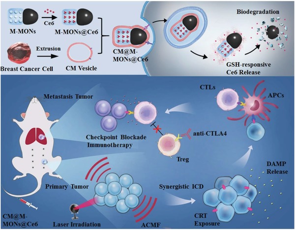
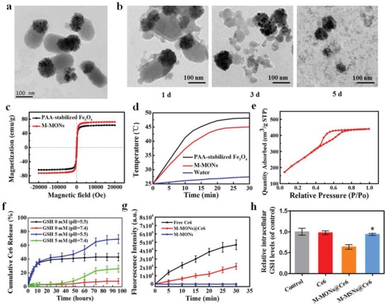
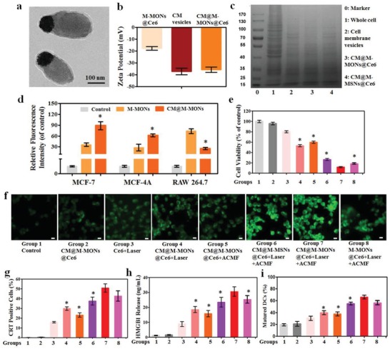
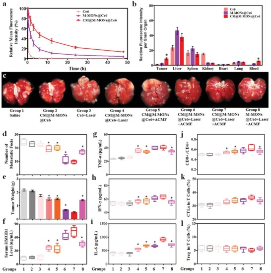
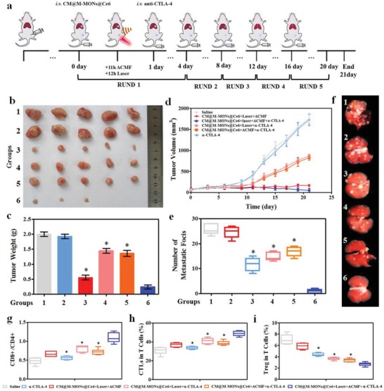
Photodynamic therapy (PDT) is clinically promising in destructing primary tumors but ineffective against distant metastases. This study reports the use of immunogenic nanoparticles mediated combination of PDT and magnetic hyperthermia to synergistically augment the anti-metastatic efficacy of immunotherapy. Janus nanobullets integrating chlorine e6 (Ce6) loaded, disulfide-bridged mesoporous organosilica bodies with magnetic heads (M-MONs@Ce6) are tailored for redox/pH-triggered photosensitizer release accompanying their matrix degradation. Cancer cell membrane cloaking enables favorable tumor-targeted accumulation and prolonged blood circulation time of M-MONs@Ce6. The combination of PDT and magnetic hyperthermia has a strong synergy anticancer activity and simultaneously elicits a sequence of immunogenic cell death, resulting in synergistically tumor-specific immune responses. When combined with anti-CTLA-4 antibody, the biomimetic and biodegradable nanoparticle enables the notable eradication of primary and deeply metastatic tumors with low systematic toxicity, thus potentially advancing the development of combined hyperthermia, PDT, and checkpoint blockade immunotherapy to combat cancer metastasis.

Keywords: Janus nanoparticles; cancer metastasis; checkpoint blockade immunotherapy; magnetic hyperthermia; photodynamic therapy.


Conflict of interest statement

The authors declare no conflict of interest.

Figures

Scheme 1
Scheme 1
A schematic of the synthetic procedure for the cancer cell membrane‐cloaked Ce6‐loaded Janus magnetic mesoporous organosilica nanoparticles (CM@M‐MON@Ce6) and their application for combined PDT and magnetic hyperthermia to further potentiate a CTLA‐4 blockade to enhance synergistic antitumor immunity in combating cancer metastasis.
Figure 1
Figure 1
Characterization of M‐MON@Ce6. a) TEM images of M‐MONs. b) M‐MONs were immersed in 5 × 10⁻³ m GSH solution for 1, 3, and 5 d. c) The magnetization curve, d) temperature–time curves, and e) N2 sorption isotherms of M‐MONs. f) Drug release profiles of Ce6@M‐MONs in 0 and 5 × 10⁻³ m GSH (pH = 7.4 and 5.5). g) Time‐dependent SOSG fluorescence in Ce6 and M‐MON@Ce6 solutions. h) Intracellular GSH levels of MCF‐7 cells after treatment with M‐MON@Ce6 for 12 h. The data are presented as the mean ± S.D. (n = 3). *p < 0.05 versus the M‐MON@Ce6 group.
Figure 2
Figure 2
Combined PDT and magnetic hyperthermia by CM@M‐MON@Ce6 in vitro. a) TEM images, b) zeta potential, and c) SDS‐PAGE protein analysis of CM@M‐MON@Ce6. d) The relative fluorescence intensity of MCF‐7, MCF‐10A, and RAW264.7 cells after incubation with CM@FITC‐M‐MONs for 6 h. The data are presented as the mean ± S.D. (n = 3). *p < 0.05 compared with the M‐MON group. e–h) MCF‐7 cells incubated with CM@M‐MON@Ce6 (12.5 µg mL−1) for 2 h, followed by a 20 min exposure to an ACMF or/and 5 min of exposure to laser irradiation with a 20 min exposure to an ACMF. e) Cell viability after 24 h of exposure. f) Intracellular reactive oxygen species (ROS) fluorescence images after 6 h of exposure; the scale bars indicate 10 µm. g) The percentage of CRT‐positive cells and h) the amount of released HMGB1 after 24 h of exposure. i) The percent of mature DCs (CD11c+CD80+CD86+) in BMDCs after co‐incubation with different treated MCF‐7 cells for 24 h. The data are presented as the mean ± S.D. (n = 3). *p < 0.05 compared with the CM@M‐MON@Ce6+Laser+ACMF group. Group description of (e), (g), (h), and (i) was the same as illustrated in (f).
Figure 3
Figure 3
Anti‐tumor effects and immune responses after combined PDT and magnetic hyperthermia with CM@M‐MON@Ce6. a) The blood circulation time, and b) biodistribution of CM@M‐MON@Ce6 in MCF‐7 tumor‐bearing mice. c) Representative images of lung tissues with observable metastatic nodules. d) The number of pulmonary metastatic nodules and e) primary tumor weights of 4T1 tumor‐bearing mice from each group over 21 d. After 5 d of combined PDT and magnetic hyperthermia, serum and primary tumor tissue were collected for the analysis of f) HMGB1, g) TNF‐α, h) IFN‐γ, and i) IL‐6 levels in serum and for the analysis of j) the ratios of CD8+T cells/CD4+T cells, k) CTL content, and l) Treg content in the primary tumor tissues. The data are presented as the mean ± S.D. (n = 5, *p < 0.05 compared with the CM@M‐MON@Ce6+Laser+ACMF group).
Figure 4
Figure 4
The synergistic effects of CM@M‐MON@Ce6‐mediated PDT and magnetic hyperthermia in combination with anti‐CTLA4 checkpoint blockade. a) A schematic of CM@M‐MON@Ce6‐mediated PDT and magnetic hyperthermia and anti‐CTLA4 checkpoint blockade combined therapy. 4T1 tumor‐bearing mice were randomly divided into the following eight groups: saline (group 1), α‐CTLA‐4 (group 2), CM@M‐MON@Ce6+Laser+ACMF (group 3), CM@M‐MON@Ce6+Laser+α‐CTLA‐4 (group 4), CM@M‐MON@Ce6+ACMF+α‐CTLA‐4 (group 5), and CM@M‐MON@Ce6+Laser+ACMF+α‐CTLA‐4 (group 6). b) Representative tumor images, c) tumor weights, d) tumor volumes, e) number of pulmonary metastatic nodules, and f) representative images of lung tissues with observable metastatic nodules of 4T1 tumor‐bearing mice from each group over 21 d. After 9 d of combined PDT, magnetic hyperthermia, and immune checkpoint therapy, serum and primary tumor tissue were collected for the analysis of g) the ratios of CD8+T cells/CD4+T cells, h) CTL content, and i) Treg content in the metastatic tumor tissues. The data are presented as the mean ± S.D. (n = 5, *p < 0.05 compared with the CM@M‐MON@Ce6+Laser+ACMF group).

References

    1. Riggi N., Aguet M., I. Stamenkovic, I , Annu. Rev. Pathol.: Mech. Dis. 2018, 13, 117. - PubMed
    1. a) Agostinis P., Berg K., Cengel K. A., Foster T. H., Girotti A. W., Gollnick S. O., Hahn S. M., Hamblin M. R., Juzeniene A., Kessel D., Ca‐Cancer J. Clin. 2011, 61, 250; - PMC - PubMed
    2. b) Dolmans D. E., Fukumura D., Jain R. K., Nat. Rev. Cancer 2003, 3, 380; - PubMed
    3. c) Fan W., Huang P., Chen X., Chem. Soc. Rev. 2016, 45, 6488; - PubMed
    4. d) Sun W., Li S., Häupler B., Liu J., Jin S., Steffen W., Schubert U. S., Butt H. J., Liang X. J., Wu S., Adv. Mater. 2017, 29, 1603702. - PubMed
    1. Banerjee S., MacRobert A., Mosse C., Periera B., Bown S., Keshtgar M., Breast 2017, 31, 105. - PubMed
    1. a) Beik J., Abed Z., Ghoreishi F. S., Hosseini‐Nami S., Mehrzadi S., Shakeri‐Zadeh A., Kamrava S. K., J. Controlled Release 2016, 235, 205; - PubMed
    2. b) Mallory M., Gogineni E., Jones G. C., Greer L., Simone C. B. II, Crit. Rev. Oncol. Hematol. 2016, 97, 560. - PubMed
    1. a) Sun X., Xing L., Clifton Ling C., Li G. C., Int. J. Hyperthermia 2010, 26, 224; - PubMed
    2. b) Bezu L., Gomes‐da‐Silva L. C., Dewitte H., Breckpot K., Fucikova J., Spisek R., Galluzzi L., Kepp O., Kroemer G., Int. J. Hyperthermia 2010, 26, 224.

LinkOut - more resources