Ethnic Variation of Sinonasal Anatomy on CT Scan and Volumetric Analysis
- PMID: 31763314
- PMCID: PMC6848680
- DOI: 10.1007/s12070-019-01600-6
Ethnic Variation of Sinonasal Anatomy on CT Scan and Volumetric Analysis
Abstract
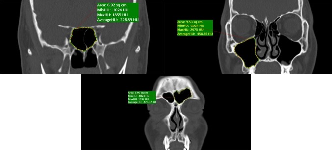
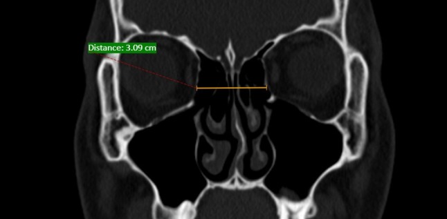
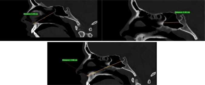
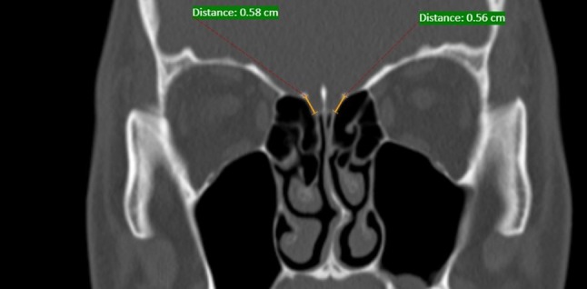
To determine Ethnic differences in the frequency of the relatively common anatomical variants along with difference in anatomy of sinonasal region with surgical importance. A study was conducted to determine the frequency of anatomical variants, volumes of paranasal sinuses using computed tomography and to identify any difference between Group A consisting of people of Indian subcontinent and Group B consisting of people from north east Asian region. Volumetric analysis done using cumulative of area multiplied by slice thickness. The results were compared using Chi square test, p value < 0.05 was considered statistically significant. Among the common and uncommon anatomical variants (Agger nasi, pneumatized uncinate, concha bullosa etc.) there was no significant difference between the two groups. In both the groups Keros Type 1 was the most common type of ethmoid roof seen. On volumetric analysis sphenoid sinus volume was found to be higher in Indians without mongoloid features. Hence it's ideal that in this era of endoscopic sinus surgery we tailor make approaches to address individual anatomical variation.
Keywords: Anatomic variation; CT-scan; Ethnic; Paranasal sinuses; Volumetric analysis.
© Association of Otolaryngologists of India 2019.
Conflict of interest statement
Conflict of interestThe authors declare that they have no conflict of interest.
Figures


Similar articles
-
Ethnic variation in sinonasal anatomy on CT-scanning.Rhinology. 2005 Sep;43(3):210-4. Rhinology. 2005. PMID: 16218515
-
An analysis of the anatomic variations of the paranasal sinuses and ethmoid roof using computed tomography.Eurasian J Med. 2013 Jun;45(2):115-25. doi: 10.5152/eajm.2013.23. Eurasian J Med. 2013. PMID: 25610263 Free PMC article.
-
Prevalence of Anatomical Variations of the Sinonasal Region and their Relationship with Chronic Rhinosinusitis.Kathmandu Univ Med J (KUMJ). 2016 Oct.-Dec.;14(56):342-346. Kathmandu Univ Med J (KUMJ). 2016. PMID: 29336423
-
Anatomical Variations of the Nasal Cavities and Paranasal Sinuses: A Systematic Review.Cureus. 2021 Jan 15;13(1):e12727. doi: 10.7759/cureus.12727. Cureus. 2021. PMID: 33614330 Free PMC article. Review.
-
Remarkable anatomic variations in paranasal sinus region and their clinical importance.Eur J Radiol. 2004 Jun;50(3):296-302. doi: 10.1016/j.ejrad.2003.08.012. Eur J Radiol. 2004. PMID: 15145491 Review.
Cited by
-
Prevalence of the anatomical variations of concha bullosa and its relation with sinusitis among Saudi population: a computed tomography scan study.Anat Cell Biol. 2021 Jun 30;54(2):193-201. doi: 10.5115/acb.20.247. Anat Cell Biol. 2021. PMID: 33896798 Free PMC article.
-
A CT-Scan review of anatomical variants of sinonasal region and its correlation with symptoms of sinusitis (nasal obstruction, facial pain and rhinorrhea).Pak J Med Sci. 2021 Jan-Feb;37(1):195-200. doi: 10.12669/pjms.37.1.3260. Pak J Med Sci. 2021. PMID: 33437276 Free PMC article.
-
Anatomical Variants of Internal Carotid Artery-Results from a Retrospective Study.Medicina (Kaunas). 2023 May 31;59(6):1057. doi: 10.3390/medicina59061057. Medicina (Kaunas). 2023. PMID: 37374261 Free PMC article.
-
Computed tomography assessment of maxillary sinus hypoplasia and associated anatomical variations: a systematic review and meta-analysis of global evidence.Oral Radiol. 2024 Apr;40(2):124-137. doi: 10.1007/s11282-023-00726-2. Epub 2023 Dec 11. Oral Radiol. 2024. PMID: 38079051
-
Anatomical Variations in Nasal Cavities and Paranasal Sinuses on Computed Tomography in Chronic Rhinosinusitis: Implications for Diagnosis and Treatment.Indian J Otolaryngol Head Neck Surg. 2025 Jan;77(1):120-126. doi: 10.1007/s12070-024-05123-7. Epub 2024 Oct 24. Indian J Otolaryngol Head Neck Surg. 2025. PMID: 40066395
References
LinkOut - more resources
Full Text Sources