. 2019 Nov 2:11:100227.
doi: 10.1016/j.bonr.2019.100227. eCollection 2019 Dec.

Characterizing how probiotic Lactobacillus reuteri 6475 and lactobacillic acid mediate suppression of osteoclast differentiation

Affiliations

Characterizing how probiotic Lactobacillus reuteri 6475 and lactobacillic acid mediate suppression of osteoclast differentiation

Darin Quach et al. Bone Rep. .

Abstract

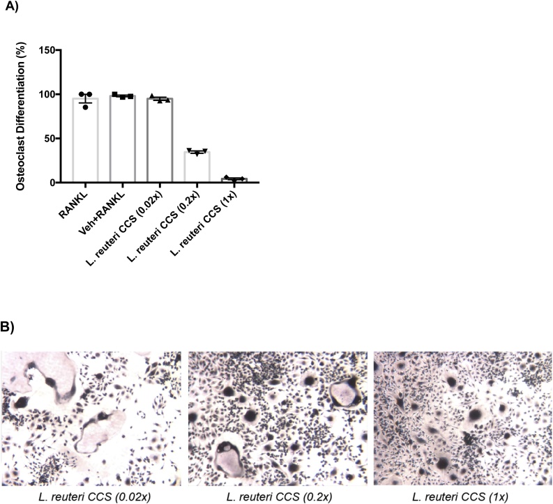
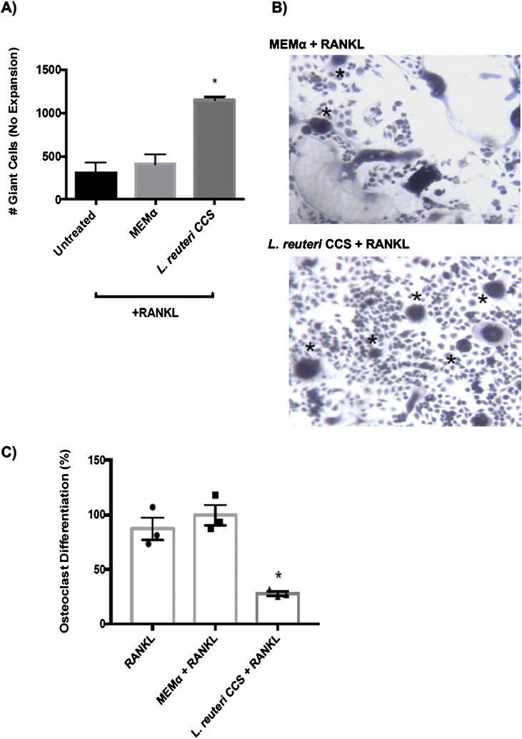
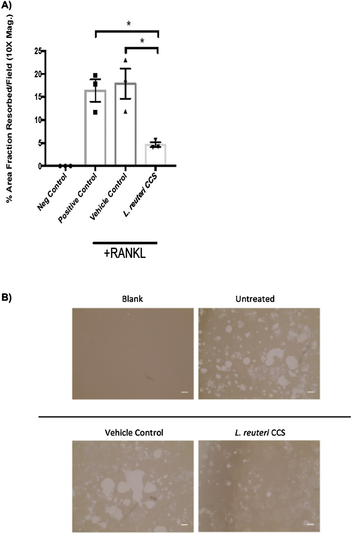
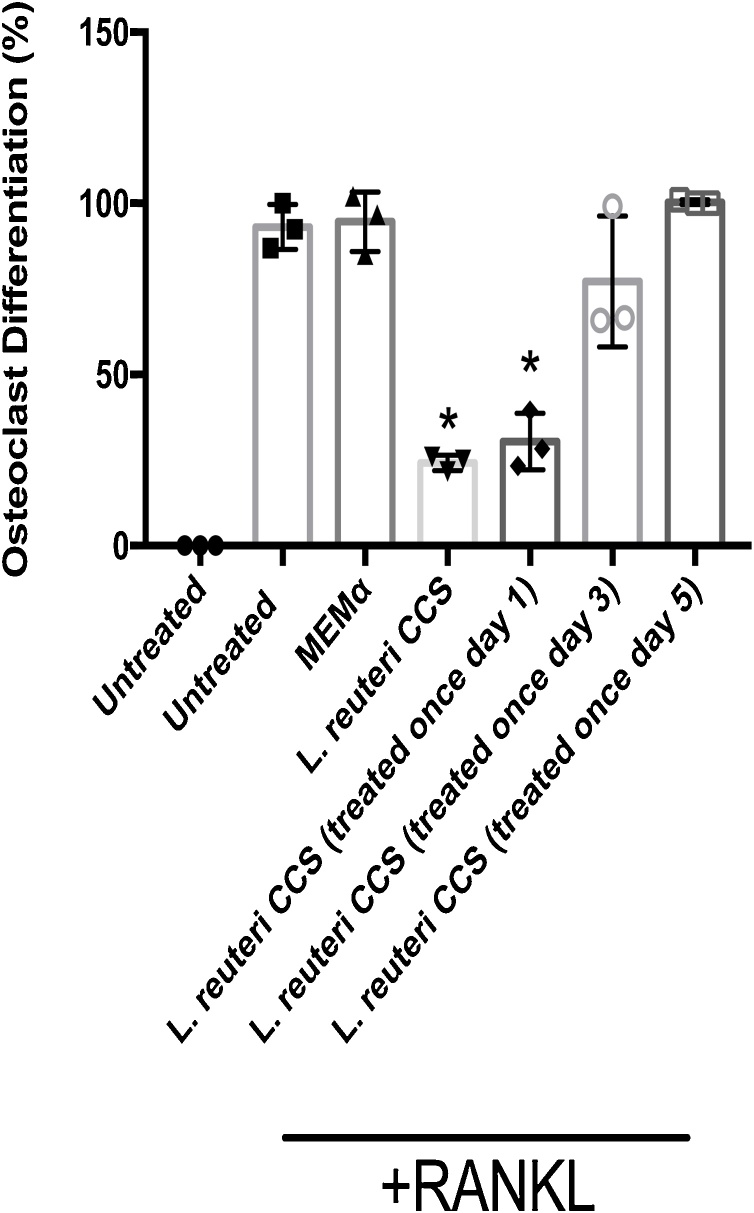
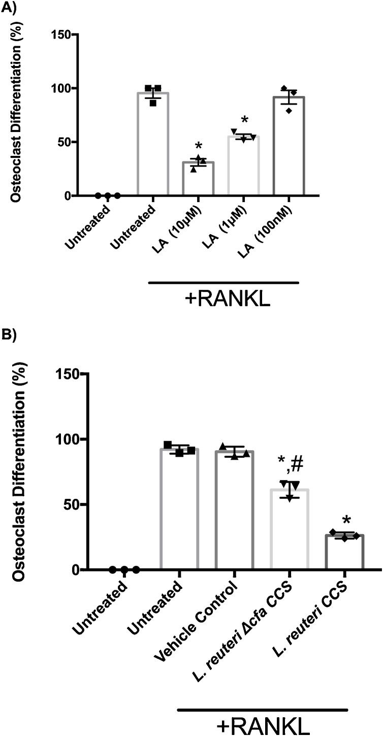
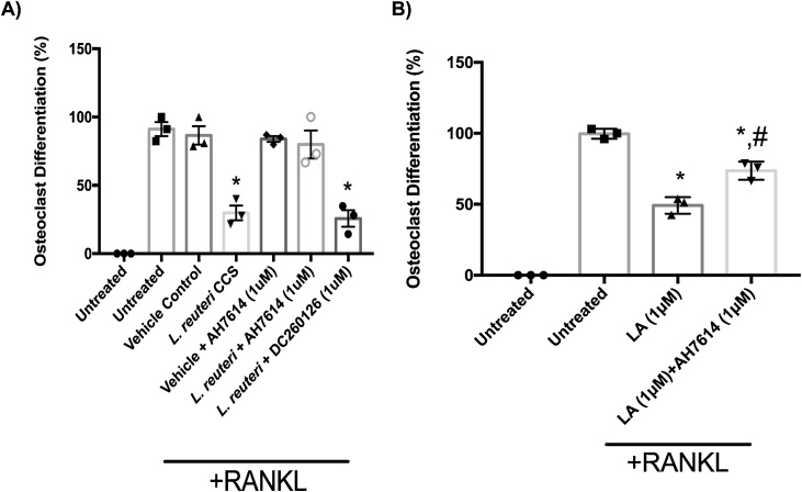
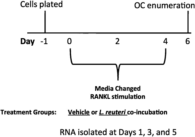
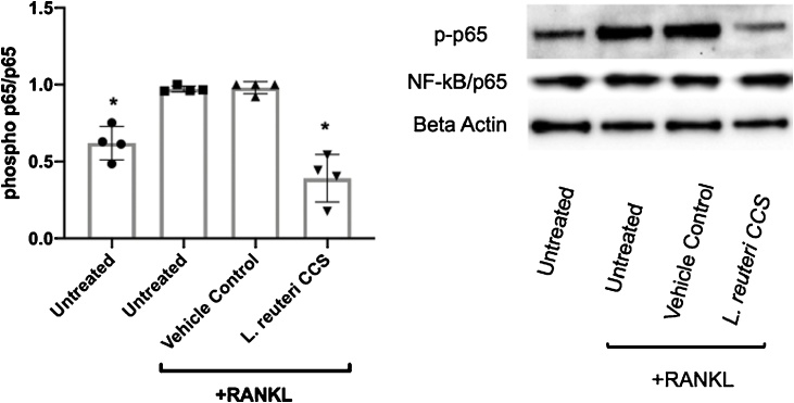
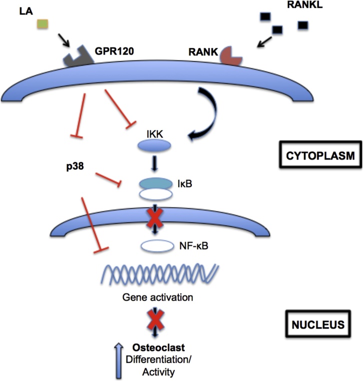
Osteoporosis is a disease that impacts over 200 million people worldwide. Taking into consideration the side effects stemming from medications used to treat this illness, investigators have increased their efforts to develop novel therapeutics for osteoporosis. In a previous study, we demonstrated that ovariectomy-induced bone loss in mice was prevented by treatment with the probiotic bacterium Lactobacillus reuteri 6475 (L. reuteri), an effect that correlated with reduced osteoclastogenesis in the bone marrow of L. reuteri treated mice. We also demonstrated that L. reuteri directly inhibited osteoclastogenesis in vitro. To better understand how L. reuteri impacts osteoclast formation, we used additional in vitro analyses to identify that conditioned supernatant from L. reuteri inhibited osteoclastogenesis at the intermediate stage of fused polykaryons. To elucidate the effect of L. reuteri treatment on host cell physiology, we performed RNAseq at multiple time points during in vitro osteoclastogenesis and established that L. reuteri downregulated several KEGG pathways including osteoclast differentiation as well as TNF-α, NF-κB, and MAP kinase signaling. These results were consistent with Western Blot data demonstrating that NF-κB and p38 activation were decreased by L. reuteri treatment. We further identified that lactobacillic acid (LA), a cyclopropane fatty acid produced by L. reuteri, contributed significantly to the suppression of osteoclastogenesis. Additionally, we demonstrated that L. reuteri is signaling through the long chain fatty acid receptor, GPR120, to impact osteoclastogenesis. Overall, these studies provide both bacterial and host mechanisms by which L. reuteri impacts osteoclastogenesis and suggest that long chain fatty acid receptors could be targets for preventing osteoclastogenesis.

Keywords: Bone; Lactobacillic acid; Osteoclast; Osteoporosis; Probiotic.


Conflict of interest statement

The authors report that they have no competing interests.

Figures

Fig. 1
Fig. 1
Dose-dependent inhibition of osteoclast formation byL.reuteri. Osteoclast differentiation was induced with the addition of 100 ng/mL RANKL. Giant multinucleated cells that stained positive for TRAP and with ≥ 3 nuclei were considered osteoclasts (A). Light microscopy images (B) were taken and demonstrate an increase in osteoclast formation with decreasing levels of L. reuteri CCS used. This was performed three times in total and the reported result is a representative experiment with associated standard deviation, *p < 0.05 compared to untreated and MEM-α (vehicle control) conditions as determined by one-way ANOVA.
Fig. 2
Fig. 2
Progression of osteoclastogenesis over time and accumulation of polykaryons. RAW264.7 cells were stimulated for osteoclast differentiation with RANKL (100 ng/ml) and treated with a vehicle (MEM-α) or L. reuteri CCS. (A, B) Fused polykaryons (asterisks) were present in both conditions but L. reuteri treatment led to an accumulation of them by day 7. (C) After a 10 day washout period, osteoclast differentiation was still inhibited in cells treated by L. reuteri CCS at day 17. *p < 0.05 compared to untreated and MEM-α (vehicle control) conditions as determined by one-way ANOVA. n = 3.
Fig. 3
Fig. 3
Suppression of mineral resorption byL.reuteri. RAW264.7 cells were plated for on Osteoassay plates for 1 day and then stimulated concurrently with RANKL (100 ng/ml) and either the vehicle control (MEM-α) or L. reuteri CCS. Fresh medium was replenished every 2 days. The analysis was performed using the Image J software package to measure densitometry. (A) L. reuteri CCS significantly decreased the amount of absorption from calcium and phosphate coated plates. (B) The microscopy images also support that less absorption had taken place in the presence of L. reuteri CCS. Three biological replicates are depicted with associated standard error of mean, *p < 0.05 compared to untreated and MEM-α (vehicle control) conditions as determined by one-way ANOVA. Scale bars signify 100 μm increments. n = 3.
Fig. 4
Fig. 4
Impact ofL.reuteri CCS on osteoclast differentiation at different time points. RAW264.7 cells were plated and osteoclast differentiation was induced with the addition of 100 ng/mL RANKL. L. reuteri CCS was added normally when the media was replenished (days 1, 3, and 5) or just once on day 1, 3, or 5. After 7 days, the number of giant multinucleated (≥ 3 nuclei) cells staining positive for TRAP were quantified. Treatment by L. reuteri CCS on day 1 was sufficient to suppress osteoclastogenesis. Three biological replicates are depicted with associated standard error of mean, *p < 0.05 compared to untreated and MEM-α (vehicle control) conditions as determined by one-way ANOVA.
Fig. 5
Fig. 5
Lactobacillic acid (LA) involved with the suppression of osteoclastogenesis. RAW264.7 cells were plated and osteoclast differentiation was induced with the addition of 100 ng/mL RANKL. (A) Dose-dependent suppression of osteoclastogenesis was observed with LA at the concentrations of 10 μM, 1 μM, and 100 nM. (B) A mutant unable to produce LA in the L. reuteri genetic background was not as effective as the WT strain in suppressing osteoclastogenesis. Three biological replicates are depicted with associated standard error of mean, *p < 0.05 compared to untreated and MEM-α (vehicle control) conditions, #p < 0.05 compared to L. reuteri as determined by one-way ANOVA.
Fig. 6
Fig. 6
Suppression of osteoclastogenesis byL.reuteri was mediated through GPR120 signaling. RAW264.7 cells were plated. After 24 h, the cells were pretreated with 1 μM of AH7614 for 1 h. Then, osteoclast differentiation was induced with the addition of 100 ng/mL RANKL. Cell differentiation medium (containing RANKL and the inhibitor) was replenished every 2 days. (A) The presence of the inhibitor attenuated the suppression of osteoclastogenesis by L. reuteri. (B) Pharmacological inhibition of GPR120 decreased the ability of LA to suppress osteoclast formation. Three biological replicates are depicted with associated standard error of mean, *p < 0.05 compared to untreated and MEM-α (vehicle control), #p <0.05 compared to LA conditions as determined by one-way ANOVA.
Fig. 7
Fig. 7
Experimental layout for gene expression analysis. RAW264.7 cells were plated for 1 day and then stimulated concurrently with RANKL (100 ng/ml) and either the vehicle control (MEM-α) or L. reuteri CCS. RNA was extracted at days 1, 3, and 5.
Fig. 8
Fig. 8
Effect ofL.reuteri on RANKL-induced NF-κB/p65 phosphorylation. RAW264.7 cells were concurrently treated with RANKL (100 ng/ml) and MEM-α or L. reuteri CCS for 60 min. Total intracellular contents were prepared and an equal amount of protein was analyzed. Western blot analysis using antibodies to the phosphorylated NF-κB/p65 subunit was performed. Band intensity was measured by densitometry using the ImageJ software package. Results were normalized to levels of total p65. Four biological replicates are depicted with associated standard error of mean, *p < 0.05 compared to untreated and MEM-α (vehicle control) conditions as determined by one-way ANOVA.
Fig. 9
Fig. 9
Impact ofL.reuteri on MAPK signaling. RAW264.7 cells were concurrently treated with RANKL (100 ng/ml) and MEM-α or L. reuteri CCS for 60 min. Total intracellular contents were prepared and an equal amount of protein was analyzed. Western blot analysis using antibodies to the p38, phosphorylated p38, ERK, phosphorylated ERK, JNK, phosphorylated JNK. Band intensity was measured by densitometry using the ImageJ software package. Results were normalized to levels of total p38. Three biological replicates are depicted with associated standard error of mean, *p < 0.05 compared to untreated and MEM-α (vehicle control) conditions as determined by one-way ANOVA.
Fig. 10
Fig. 10
Working model of osteoclastogenesis suppression byL.reuteri and LA. RAW264.7 cells are stimulated for osteoclastogenesis by RANKL. L. reuteri and LA activated GPR120 or a receptor yet to be identified to suppress osteoclastogenesis. GPR120 receptor antagonism partially inhibited the ability of L. reuteri and LA to suppress osteoclastogenesis. MAPK and NF-κB signaling has been shown to be downstream of GPR120 signaling and we demonstrated that L. reuteri impacts several arms of these pathways.
None

References

    1. Aharon R., Bar-Shavit Z. Involvement of aquaporin 9 in osteoclast differentiation. J. Biol. Chem. 2006;281:19305–19309. - PubMed
    1. Ahn S.H., Park S.-Y., Baek J.-E., Lee S.-Y., Baek W.-Y., Lee S.-Y., Lee Y.-S., Ju Yoo H., Kim H., Lee S.H., Im D.-S., Lee S.-K., Kim B.-J., Koh J.-M. Free fatty acid receptor 4 (GPR120) stimulates bone formation and suppresses bone resorption in the presence of elevated n -3 fatty acid levels. Endocrinology. 2016;4 - PubMed
    1. Alam H., Gu B., Lee M.G. Histone methylation modifiers in cellular signaling pathways. Cell. Mol. Life Sci. 2015;72:4577–4592. - PMC - PubMed
    1. Anders S., McCarthy D.J., Chen Y., Okoniewski M., Smyth G.K., Huber W., Robinson M.D. Count-based differential expression analysis of RNA sequencing data using R and Bioconductor. Nat. Protoc. 2013;8:1765–1786. doi:nprot.2013.099[pii]\r10.1038/nprot.2013.099. - PubMed
    1. Arthur J.S., Ley S.C. Mitogen-activated protein kinases in innate immunity. Nat. Rev. Immunol. 2013;13:679–692. - PubMed