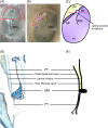
Developmental aspects of the tympanic membrane: Shedding light on function and disease
- PMID: 31763764
- PMCID: PMC7154630
- DOI: 10.1002/dvg.23348
Developmental aspects of the tympanic membrane: Shedding light on function and disease
Abstract
The ear drum, or tympanic membrane (TM), is a key component in the intricate relay that transmits air-borne sound to our fluid-filled inner ear. Despite early belief that the mammalian ear drum evolved as a transformation of a reptilian drum, newer fossil data suggests a parallel and independent evolution of this structure in mammals. The term "drum" belies what is in fact a complex three-dimensional structure formed from multiple embryonic cell lineages. Intriguingly, disease affects the ear drum differently in its different parts, with the superior and posterior parts being much more frequently affected. This suggests a key role for the developmental details of TM formation in its final form and function, both in homeostasis and regeneration. Here we review recent studies in rodent models and humans that are beginning to address large knowledge gaps in TM cell dynamics from a developmental biologist's point of view. We outline the biological and clinical uncertainties that remain, with a view to guiding the indispensable contribution that developmental biology will be able to make to better understanding the TM.
Keywords: development; middle ear; tympanic membrane.
© 2019 The Authors. Genesis published by Wiley Periodicals, Inc.
Conflict of interest statement
The authors have no conflicts of interest to declare.
Figures


Similar articles
-
Major evolutionary transitions and innovations: the tympanic middle ear.Philos Trans R Soc Lond B Biol Sci. 2017 Feb 5;372(1713):20150483. doi: 10.1098/rstb.2015.0483. Philos Trans R Soc Lond B Biol Sci. 2017. PMID: 27994124 Free PMC article. Review.
-
Deep-time origin of tympanic hearing in crown reptiles.Curr Biol. 2024 Nov 18;34(22):5334-5340.e5. doi: 10.1016/j.cub.2024.09.041. Epub 2024 Oct 10. Curr Biol. 2024. PMID: 39393352
-
Can you hear me now? Understanding vertebrate middle ear development.Front Biosci (Landmark Ed). 2011 Jan 1;16(5):1675-92. doi: 10.2741/3813. Front Biosci (Landmark Ed). 2011. PMID: 21196256 Free PMC article. Review.
-
Developmental mechanisms of the tympanic membrane in mammals and non-mammalian amniotes.Congenit Anom (Kyoto). 2016 Jan;56(1):12-7. doi: 10.1111/cga.12132. Congenit Anom (Kyoto). 2016. PMID: 26754466 Review.
-
Ear drum mobility and middle ear volume measured with tympanometry.Scand Audiol. 1984;13(3):147-50. doi: 10.3109/01050398409043053. Scand Audiol. 1984. PMID: 6494799
Cited by
-
In vivo microstructural investigation of the human tympanic membrane by endoscopic polarization-sensitive optical coherence tomography.J Biomed Opt. 2023 Dec;28(12):121203. doi: 10.1117/1.JBO.28.12.121203. Epub 2023 Mar 29. J Biomed Opt. 2023. PMID: 37007626 Free PMC article.
-
Effect of Growth Factor-Loaded Acellular Dermal Matrix/MSCs on Regeneration of Chronic Tympanic Membrane Perforations in Rats.J Clin Med. 2021 Apr 6;10(7):1541. doi: 10.3390/jcm10071541. J Clin Med. 2021. PMID: 33917576 Free PMC article.
-
The continued importance of comparative auditory research to modern scientific discovery.Hear Res. 2023 Jun;433:108766. doi: 10.1016/j.heares.2023.108766. Epub 2023 Apr 6. Hear Res. 2023. PMID: 37084504 Free PMC article. Review.
-
Collagen Matrix to Restore the Tympanic Membrane: Developing a Novel Platform to Treat Perforations.Polymers (Basel). 2024 Jan 15;16(2):248. doi: 10.3390/polym16020248. Polymers (Basel). 2024. PMID: 38257047 Free PMC article.
-
Thymosin beta-4 - A potential tool in healing middle ear lesions in adult mammals.Int Immunopharmacol. 2023 Mar;116:109830. doi: 10.1016/j.intimp.2023.109830. Epub 2023 Feb 14. Int Immunopharmacol. 2023. PMID: 38706788 Free PMC article.
References
-
- Ankamreddy, H. , Min, H. , Kim, J. Y. , Yang, X. , Cho, E.‐S. , Kim, U.‐K. , & Bok, J. (2019). Region‐specific endodermal signals direct neural crest cells to form the three middle ear ossicles. Development (Epub ahead of print). - PubMed
Publication types
MeSH terms
Grants and funding
LinkOut - more resources
Full Text Sources

