. 2019 Dec;127(1):e91.
doi: 10.1002/cpim.91.

Isolation and Analysis of Tumor-Derived Exosomes

Affiliations

Isolation and Analysis of Tumor-Derived Exosomes

Nils Ludwig et al. Curr Protoc Immunol. 2019 Dec.

Abstract

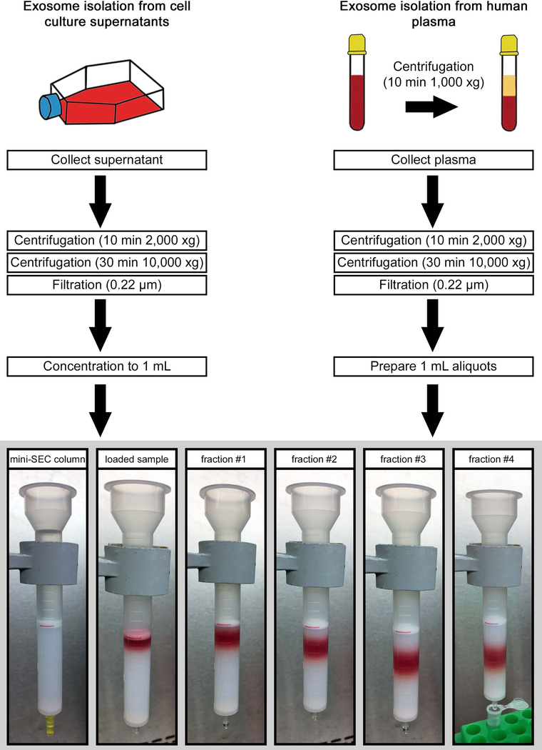
A method for isolation of exosomes from tumor cell supernatants or cancer patients' plasma is presented. Tumor-derived exosomes (TEX) are defined as a subset of extracellular vesicles (EVs) sized at 30 to 150 nm and originating from multivesicular bodies (MVBs). The method utilizes size exclusion chromatography (SEC) for recovery of exosomes from cell-line supernatants or cancer patients' plasma. The recovered exosomes are morphologically intact, aggregate-free, and functionally competent. Their molecular content parallels that of the parent tumor cells and they carry various immunoregulatory ligands known to modulate functions of immune cells. All exosomes isolated from tumor cell lines are TEX, while those isolated from plasma of cancer patients have to be fractionated into TEX and non-TEX. Mini-SEC allows for exosome isolation and recovery in quantities sufficient for molecular profiling, functional studies, and, in the case of plasma, further fractionation into TEX and non-TEX. The mini-SEC method can also be used for comparative studies of the exosome content in serial specimens of cancer patients' body fluids. © 2019 by John Wiley & Sons, Inc.

Keywords: TEX; exosome isolation; exosomes; size exclusion chromatography.


Figures

Fig. 1:
Fig. 1:
Illustration of the SEC principle. (A) The column bed consists of Sepharose beads. Very small molecules (blue) enter pores in the gel, equilibrating between the gel and the moving buffer, and so travel slowly and are eluted later. Medium sized molecules (green) enter some pores in the gel, equilibrating between the gel and the moving buffer. Large molecules (red) enter few pores in the gel, and so travel rapidly and are eluted sooner. (B) Schematic illustration of the mini-SEC column showing larger molecules being eluted faster followed by medium sized and smaller molecules which enter the pores of the Sepharose beads. (C) Graph describing the time-dependent elution of small, medium sized and large molecules.
Fig. 2:
Fig. 2:
Schema and experimental set up for isolation of exosomes from supernatants or plasma. The red line is added for visual orientation and marks the 10mL mark of the column which equates 6cm of column height. Column is filled with 10 ml of Sepharose 2B with an additional 30µm filter added on top. 1 mL aliquot of pre-cleared and concentrated supernatant is loaded, followed by elution with PBS. 1 mL fractions are collected. For this size/diameter column, fraction #4 is enriched in exosomes and is collected in a microcentrifuge tube.
Fig. 3:
Fig. 3:
Characterization of TEX derived from cell culture supernatants. (A) The yield of exosomes in fraction #4 was measured by using a BCA protein assay (See Supporting Protocol 1). Total exosomal protein (µg) in fraction #4 was found to be higher for exosomes derived from cancer cell lines (grey bars) compared to exosomes derived from normal cells (white bars). Values are expressed as means ± SEM from at least three independent experiments. (B) TEM image of isolated and negatively-stained PCI-13-derived exosomes. (C) Size distribution of PCI-13-derived exosomes measured by qNano according to Supporting Protocol 1. (D) The angiogenesis antibody arrays (Supporting Protocol 1) show comparative protein analysis of UMSCC47 cell lysate (200µg protein; upper array) and exosomes produced by UMSCC47 cells (200µg protein; lower array) (E) Quantification of the arrays shown in D using ImageJ. (F) TEX-induced apoptosis of CD8+ Jurkat cells. Data from experiments in which CD8+ Jurkat cells were co-incubated with increasing protein levels of TEX isolated from supernatants of U251 cells (See Supporting Protocol 2). Values are expressed as means ± SEM. (Figures 3D and E originally appeared in Ludwig N, Yerneni SS, Razzo BM, Whiteside TL. Exosomes from HNSCC Promote Angiogenesis through Reprogramming of Endothelial Cells. Mol Cancer Res. 2018;16(11):1798–808.)
Fig. 4:
Fig. 4:
Characterization of TEX derived from plasma of cancer patients. (A) TEM of fractions #3, #4, and #5 recovered after miniSEC of plasma obtained from a patient with AML. (B) Protein contents in fractions recovered after SEC. An aliquot (50μL) of each fraction was separated by SDS-PAGE gels, stained with Coomassie blue and analyzed by WBs. CD9 is used as an exosome marker. (C) Western blot analysis of exosomes isolated from plasma of 4 AML patients (Pt1 – Pt4) and a normal donor (NC). Each lane was loaded with 10μg exosome protein (D) Proliferation of CFSE+ CD4+ T cells is inhibited by exosomes isolated from plasma of normal donors or HNSCC patients. (E) Apoptosis in CD8+ Jurkat cells is induced by exosomes of normal donors and HNSCC patients. Figures 4A, B, C and D printed by permission of Springer Nature: Springer ebook Acute Myeloid Leukemia: Methods and Protocols, Methods in Molecular Biology by Paolo Fortina et al. (eds.) in the chapter “Isolation of Biologically Active Exosomes from Plasma of Patients with Cancer” by Chang-Sook Hong, Sonja Funk, and Theresa L. Whiteside, copyright 2017.
Fig. 5:
Fig. 5:
Immune capture of CD3(+) exosomes and detection of the exosome cargo by on-bead flow cytometry. (A) After exosome isolation from plasma via mini-SEC, immune capture with anti-CD3 biotinylated antibody on beads can be used to separate CD3(+) from uncaptured CD3(–) exosomes. (B) Surface markers carried by CD3(+) exosomes can be detected by on-bead flow cytometry. Non-captured CD3(–) exosomes can be captured on beads using biotinylated ant-CD63 antibody for detection. (Figures 5A and B adapted from Theodoraki, M., Hoffmann, T. K. and Whiteside, T. L. (2018), Separation of plasma-derived exosomes into CD3(+) and CD3(-) fractions allows for association of immune cell and tumor cell markers with disease activity in HNSCC patients. Clin Exp Immunol, 192: 271–283.).

References

    1. Böing AN, van der Pol E, Grootemaat AE, Coumans FAW, Sturk A, and Nieuwland R 2014. Single-step isolation of extracellular vesicles by size-exclusion chromatography. Journal of Extracellular Vesicles 1:1–11. - PMC - PubMed
    1. Boriachek K, Islam MN, Möller A, Salomon C, Nguyen NT, Hossain MSA, Yamauchi Y, and Shiddiky MJA 2018. Biological Functions and Current Advances in Isolation and Detection Strategies for Exosome Nanovesicles. Small 14:1–21. - PubMed
    1. Chen C, Skog J, Hsu C, Lessard R, Balaj L, Wurdinger T, Carter B, Breakefield X, Toner M, and Irimia D 2010. Microfluidic isolation and transcriptome analysis of serum microvesicles. Lab Chip 10:505–511. - PMC - PubMed
    1. Chen T, Guo J, Yang M, Zhu X, and Cao X 2011. Chemokine-Containing Exosomes Are Released from Heat-Stressed Tumor Cells via Lipid Raft-Dependent Pathway and Act as Efficient Tumor Vaccine. The Journal of Immunology 186:2219–2228. - PubMed
    1. Gardiner C, Vizio D. Di, Sahoo S, Théry C, Witwer KW, Wauben M, and Hill AF 2016. Techniques used for the isolation and characterization of extracellular vesicles: Results of a worldwide survey. Journal of Extracellular Vesicles 5:1–6. - PMC - PubMed

Publication types

LinkOut - more resources