Randomized Controlled Trial
. 2019 Dec;35(4):e39-e45.
doi: 10.1097/YCT.0000000000000597.

Central-Positive Complexes: A Novel Characterization of Ictal Markers Induced During Electroconvulsive Therapy

Affiliations
Randomized Controlled Trial

Central-Positive Complexes: A Novel Characterization of Ictal Markers Induced During Electroconvulsive Therapy

R Edward Hogan et al. J ECT. 2019 Dec.

Abstract

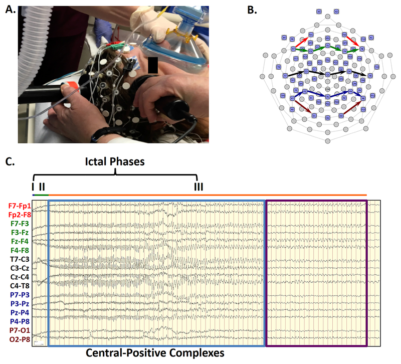
Objectives: Electroencephalography (EEG) allows monitoring of generalized seizures induced during electroconvulsive therapy (ECT). Scalp EEG recordings show different phases of electroencephalographic ictal activity during ECT seizures, documenting a pattern of seizures that may vary within and across individuals. In this case series, we used 64-electrode high-density EEG recording to detect topographic electroencephalographic changes not typically evident with conventional limited montages commonly used during ECT.

Methods: The EEG recordings were acquired from 5 participants (24 ECT sessions) during index courses for treatment-resistant depression. Using previously proposed staging criteria, the ictal EEG and simultaneously acquired video were interpreted by an expert reviewer blinded to study treatment parameters.

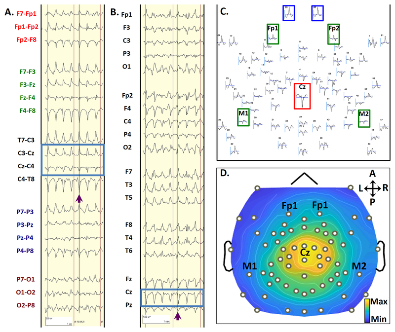
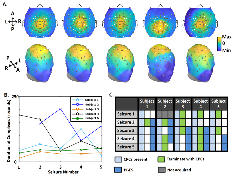
Results: The EEG recordings of all seizures showed generalized, high-amplitude, central-positive complexes (CPCs), which emerged at the beginning of phase III (polyspike and slow wave activity), with median duration of 47 seconds (interquartile range, 77 seconds), ranging from 14 to 203 seconds. Although individuals showed variability in frequency and amplitude of CPCs, CPCs typically evolved from 4.0 to 1.5 Hz in frequency and decreased in amplitude as the seizure progressed. Elaborating on previously described phases of ECT-induced electrographic seizures, we describe variability in morphology at seizure termination. Initiation of CPCs typically corresponded with clonic movements, but often terminated after motor signs ceased.

Conclusions: Generalized, high-amplitude, CPCs during ECT are a previously uncharacterized ictal waveform during ECT, which may have important scientific and clinical value. These complexes offer a specific marker for correlating clinical outcomes in ECT and greater understanding of generalized tonic-clonic seizures.


Conflict of interest statement

Conflicts of interest: The study was funded by the James S. McDonnell Foundation (US), Recipient: Michael S. Avidan, M.B.B.Ch. There are no other relevant conflicts of interest.

Figures

FIGURE 1:
FIGURE 1:. Ictal Phases Following ECT Charge Delivery.
1A. Modified 64-channel EEG sensor net allows high-density EEG recording without interference with ECT stimulation electrode placement. 1B. The sensor net diagram shows the localization of the standard 10–20 electrode locations. Arrows indicate the locations of the recording chains of the bipolar transverse montage in FIGURE 1C. 1C. High-amplitude complexes are demonstrated in a bipolar transverse montage. The electrodes contributing to each EEG channel in a chain are connected by color-coded arrows on the electrode map. Intervals for the different ictal phases are depicted (blue, very short Phase I; green, Phase II; orange, Phase III). As each hash mark represents one second, clearly-formed, rhythmic complexes (blue box) arise by the 8th second of the record, with the highest differential amplitudes over the midline chain (black). The polarity of the complexes shifts from relatively negative polarity (upward deflection) to relatively positive (downward deflection) following the chain from the left (T7) to the right (T8), identifying localization at the vertex electrode (Cz). These complexes were initially at a frequency of 4.0 Hz but gradually decreased to 2.0 Hz. At second 50, these complexes abruptly stop, while the seizure continues for another 21 seconds of Phase III (purple box). Signals have been bandpass filtered from 1–70 Hz.
FIGURE 2:
FIGURE 2:. Topology of CPCs.
A typical CPC from seizure 2 in subject 1, in a bipolar transverse montage (2A) and an average referenced montage (2B). Purple lines and arrows highlight a single CPC, which occurred 13 seconds after seizure onset during Phase III of Seizure 2. Blue boxes highlight the phase reversal in bipolar montages (2A) and electrodes with maximal amplitude referenced to the average signal (2B). The scalp topography layout for this single CPC is shown in 2C. The topographic plot layout shows all 64 electrodes in relative space as they are distributed over the head, using an average reference. The standard electrode numbers for 64-channel high-density recordings are marked above each waveform. This complex showed a maximal positive voltage over the vertex (Cz, red inset) electrode and maximal negative voltage over sub-ocular electrodes 62 and 63 (blue insets). Electrode locations approximating those for conventional frontomastoid EEG are inset in green. Duration of display (represented by the horizontal gray bar below each waveform) is 300 ms. 2D shows a topographic voltage map of the peak amplitude of the CPC over a 2-ms time window, using an average reference. This voltage map displays the maximal positive voltages over the vertex and surrounding regions (red), and maximal negative voltages over the sub-ocular electrodes (blue). Black dots represent electrode locations, with selected 10–20 locations annotated. Signals have been bandpass filtered from 1–70 Hz.
FIGURE 3.
FIGURE 3.. Consistency of CPCs.
3A. Topograms showing distribution of EEG power across the scalp during a CPC from one seizure for each participant over a 2 ms window. Each complex was taken during the first 5 seconds of Phase III in seizure 2, which was the first seizure after the dose-charge titration. The topology of CPCs remains consistent across all patients. 3B. Duration of CPCs in all seizures of the 5 subjects. The duration of CPCs for all seizures varied across patients, but showed relative consistency in two study participants (Subjects 3 and 5). 3C. The table shows characteristics of seizures, with three columns for each subject. The first column shows that CPCs were present during Phase III of all 24 seizures (light blue). The second column shows seizures that terminated with CPCs (green), and those that did not (white). The third column marks seizures with postictal generalized EEG suppression (PGES, dark blue), and those without PGES (white).

References

    1. Lisanby SH. Electroconvulsive therapy for depression. N Engl J Med 2007;357:1939–45. - PubMed
    1. Weiner RD. Indications for use of electroconvulsive therapy In: Weiner RD, ed. The Practice of Electroconvulsive Therapy: Recommendations for Treatment, Training, and Privileging (A Task Force Report of the American Psychiatric Association). Washington, D.C.: American Psychiatric Publishing, Inc.; 2000:5–26.
    1. Abrams R Electroconvulsive Therapy. 4th edition New York, NY: Oxford University Press; 2002.
    1. Krystal AD, Weiner RD. EEG correlates of the response to ECT: a possible antidepressant role of brain-derived neurotrophic factor. J ECT 1999;15:27–38. - PubMed
    1. Mayur P. Ictal electroencephalographic characteristics during electroconvulsive therapy: a review of determination and clinical relevance. J ECT 2006;22:213–7. - PubMed

Publication types