. 2019 Nov 25:8:e48118.
doi: 10.7554/eLife.48118.

Novel charged sodium and calcium channel inhibitor active against neurogenic inflammation

Affiliations

Novel charged sodium and calcium channel inhibitor active against neurogenic inflammation

Seungkyu Lee et al. Elife. .

Abstract

Voltage-dependent sodium and calcium channels in pain-initiating nociceptor neurons are attractive targets for new analgesics. We made a permanently charged cationic derivative of an N-type calcium channel-inhibitor. Unlike cationic derivatives of local anesthetic sodium channel blockers like QX-314, this cationic compound inhibited N-type calcium channels more effectively with extracellular than intracellular application. Surprisingly, the compound is also a highly effective sodium channel inhibitor when applied extracellularly, producing more potent inhibition than lidocaine or bupivacaine. The charged inhibitor produced potent and long-lasting analgesia in mouse models of incisional wound and inflammatory pain, inhibited release of the neuropeptide calcitonin gene-related peptide (CGRP) from dorsal root ganglion neurons, and reduced inflammation in a mouse model of allergic asthma, which has a strong neurogenic component. The results show that some cationic molecules applied extracellularly can powerfully inhibit both sodium channels and calcium channels, thereby blocking both nociceptor excitability and pro-inflammatory peptide release.

Keywords: Cav2.2; Nav1.7; asthma; calcitonin gene-related peptide; dorsal root ganglion; inflammatory peptide; mouse; neuroscience.


Conflict of interest statement

SL, JL, CW, BB is named as an inventor on a patent application (U.S. Patent Office No. 62/769,420) related to this work, SJ, ST, HZ, MK, NA, MP, PL, TJ, MP, LH, AJ No competing interests declared

Figures

Figure 1.
Figure 1.. State dependent inhibition of Cav2.2 channels by the neutral compound NNCB-2.
(A) Time course of inhibition of Cav2.2 current by external NNCB-2 applied at concentrations from 3 to 100 µM. Current carried by 5 mM Ba2+ was evoked by 50-millisecond steps from −70 to +10 mV delivered every 3 s. Data points show mean SEM (n = 4). Inset: Structure of NNCB-2 (3-Cyclopentylmethylsulfanyl-2-(3,3-dimethyl-butylamino)-N-(4-methoxy-benzyl)-propionamide). (B) Inhibition by 10 µM NNCB-2 of Cav2.2 current with current evoked from a holding potential of −70 mV (top) or −100 mV (bottom). (C) Test for reversibility of NNCB-2 following exposure for 1 min, using 50 ms test pulses to +10 mV from a holding potential of either −70 mV (n = 3) or −100 mV (n = 7). Source data 1.
Figure 2.
Figure 2.. State dependent inhibition of Cav2.2 channels by the charged compound CNCB-2.
(A) Time course of inhibition of Cav2.2 current by external CNCB-2 applied at concentrations from 10 to 300 µM. Current carried by 5 mM Ba2+ current was evoked by 50-millisecond steps from −70 to +10 mV delivered every 3 s. Data points show mean ± SEM (n = 4). Inset: Structure of CNCB-2 ([2-Cyclopentylmethylsulfanyl-1-(4-methoxy-benzylcarbamoyl)-ethyl]−(3,3-dimethyl-butyl)-dimethyl-ammonium, chloride salt). (B) Inhibition by 30 µM CNCB-2 of Cav2.2 current with current evoked from a holding potential of −70 mV (top) or −100 mV (bottom). (C) Minimal reversal of CNCB-2 inhibition by washing following exposure for 1 min, using 50 ms test pulses to +10 mV from a holding potential of either −70 mV (n = 9) or −100 mV (n = 7). Source data can be found in Source data 1.
Figure 3.
Figure 3.. Test of whether Cav2.2 inhibition by NNCB-2 and CNCB-2 requires channel opening and comparison of internal and external application of CNCB-2 .
(A) Calcium channel inhibition by NNCB-2 or CNCB-2 does not require channel opening. Ba2+ current was elicited in a whole-cell recording by 50-millisecond steps from −70 to + 10 mV delivered every 10 s. Five pulses were applied in control solution, then NNCB-2 (n = 4) or CNCB-2 (n = 4 to 5) was applied for 3 min with no activation of channels and then depolarizing pulses were resumed for 50 s (in the continuing presence of drug). Then, the cell was perfused with drug-free solution for 3 min without stimulation and then depolarizing pulses were resumed for 50 s to assay recovery. (B) Slow inhibition of N-type Ca2+ channels by intracellularly applied CNCB-2. 300 µM CNCB-2 applied in the pipette solution (green triangles) produced inhibition that developed relatively slowly over several minutes (green triangles, mean ± SEM, n = 3). The time course of inhibition was similar to that of 100 µM CNCB-2 applied externally (black circles, mean ± SEM, n = 4) and slower than 300 µM CNCB-2 applied externally (red circles, mean ± SEM, n = 4); data points for external application from Figure 2. Source data can be found in Source data 1.
Figure 4.
Figure 4.. Dose-dependent inhibition of native N-type Ca2+ channels in mouse sympathetic neurons by CNCB-2 applied extracellularly.
(A) Time course of inhibition by 10 µM CNCB-2. Current carried by 5 mM Ba2+ was evoked by 50 ms steps from −70 to +10 mV delivered every 3 s. The external solution contained 5 μM nimodipine to block L-type calcium channels and 1 μM TTX to block sodium channels. Inset: Current before and after application of CNCB-2. Tail currents in control trace are truncated. (B) Current remaining in CNCB-2 applied at 3, 10, or 30 µM (mean ± SEM, n = 5 for 3 and 10 μM CNCB-2, n = 6 for 30 µM CNCB-2). Experiments performed at 37°C. Source data can be found in Source data 1.
Figure 5.
Figure 5.. CNCB-2 applied extracellularly inhibits Nav1.7 sodium channels.
(A) Time course of inhibition by 3 µM CNCB-2. Current was evoked by 10 ms steps from −70 to 0 mV delivered every 5 s. Mean ± SEM, n = 5. (B) Inhibition by 3 µM CNCB-2 at different holding potentials. Each holding potential was established for 2 s before a test pulse to 0 mV. (C) Collected results showing inhibition by 3 µM CNCB-2 as a function of holding potential, with current normalized to largest current in control (from a holding potential of −120 mV). Filled symbols show mean ± SEM, n = 5. Solid lines are drawn according to the Boltzmann equation 1/(1 + exp((V - Vh)/k)), where V is holding voltage, Vh is voltage of half-maximal availability, and k is the slope factor. Control: Vh = −63 mV, k = 6.5 mV; 3 µM CNCB-2: Vh = −71 mV, k = 7.2 mV. Dashed red line: fit for CNCB-2 data scaled to 1. Experiments performed at 37°C. Source data can be found in Source data 1.
Figure 6.
Figure 6.. Use-dependent inhibition of Nav1.7 sodium channels by externally-applied CNCB-2 compared to lidocaine and bupivacaine.
(A) Inhibition of Nav1.7 current by 3 µM CNCB-2 applied with stimulation at 0.1 Hz (20-msec depolarizations from −100 mV to 0 mV) for two minutes followed by two minutes of stimulation at 10 Hz, a return to 0.1 Hz stimulation and wash-out of compound. Black symbols: same pulse protocol in the absence of drug, recorded before application of drug. (B) Same protocol with 30 µM lidocaine. (C) Same protocol with 10 µM bupivacaine. (D) Collected results showing current at the end of 2 min of 10 Hz stimulation relative to that before application of drug. Mean ± SEM, n = 12 for no drug, n = 5 for 3 µM CNCB-2, n = 6 for 30 µM lidocaine, n = 5 for 10 µM bupivacaine. Experiments performed at 37°C. Source data can be found in Source data 1.
Figure 7.
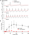
Figure 7.. Inhibition of action potential generation in mouse DRG neurons by CNCB-2.
(A) Action potential firing evoked by injections of 40 pA, 150 pA, and 200 pA in a small-diameter mouse DRG neuron before and after 5 min exposure to 3 µM CNCB-2. Resting potential in CNCB-2 was adjusted to match the resting potential in control (near −67 mV) by injection of −20 pA. (B) Collected results showing the number of action potentials evoked by 1 s current injections of increasing sizes before and after 5 min exposure to 3 µM CNCB-2. Resting potentials in CNCB-2 were adjusted to match the resting potential in control by injection of steady holding current. Mean ± SEM, n = 4. Experiments at 37°C. Source data can be found in Source data 1.
Figure 8.
Figure 8.. Inhibition of TTX-resistant sodium current in mouse DRG neurons by CNCB-2.
(A) Time-course of inhibition of TTX-resistant sodium current in a small mouse DRG neuron by 3 µM CNCB-2. TTX-resistant sodium current was isolated by solutions containing 300 nM TTX. Current was evoked by a 10-msec step depolarization from −80 mV to −10 mV delivered once every 5 s. Inset: currents before and after CNCB-2. (B) Collected results showing current after application of 3 µM CNCB-2 (n = 6) or 10 µM CNCB-2 (n = 4) for long enough to reach steady-state (10–15 min for 3 µM CNCB-2 and 5–10 min for 10 µM CNCB-2). Experiments at 37°C. Source data can be found in Source data 1.
Figure 9.
Figure 9.. Effect of CNCB-2 in mouse pain models.
(A) Inhibition of paw withdrawal in paw incision model of post-operative pain. Threshold for response to von Frey filaments tested 1, 4 and 8 hr after injection of CNCB-2 into an injured paw 24 hr after the incisional injury (5 mm incision). Mean ± SEM for 50% withdrawal threshold (n = 8 for each group). (B) Inhibition by CNCB-2 of paw withdrawal in the zymosan model of inflammatory pain. Either 10 μL of CNCB-2 (30 mM) or vehicle (5% DMSO in normal saline) was injected in hind paw. One hour later the same paw was injected subcutaneously with 20 μL of zymosan (5 mg/ml in saline) and tested 4 hr after the zymosan injection by the von Frey test (Mean ± SEM for 50% withdrawal threshold, n = 5 for each group). (C) Direct analgesic effect of CNCB-2: inhibition of paw withdrawal by 10 μL of CNCB-2 (30 mM) injected into uninjured paws (n = 8 for each group). Source data can be found in Source data 1.
Figure 10.
Figure 10.. Inhibition of CGRP release and neurogenic lung inflammation by NNCB-2 and CNCB-2.
(A) Effect of 30 µM NNCB-2 or 30 µM CNCB-2 on release of CGRP induced by application of 50 mM KCl to DRG cultures (p=0.15 for 30 µM NNCB-2 (n = 4) compared to 50 mM KCl with no drug (n = 4), p=0.028 for 30 µM CNCB-2 (n = 8) compared to 50 mM KCl with no drug (n = 4); one-tailed t-test for non-homoscedastic data). (B) Effect of 100 µM NNCB-2 or 30 µM CNCB-2 on inflammation-associated immune cells in broncheoalveolar lavage fluid in lungs from mice with ovalbumin-induced lung inflammation. p=0.019 for CD45+ cells from ovalbumin-exposed mice application of 100 µM NNCB-2 (n = 8) compared to vehicle (n = 8); p=0.014 for CD45+ cells from ovalbumin-exposed mice with application of 30 µM CNCB-2 (n = 7) compared to vehicle (n = 8). p=0.031 for CD3+ cells from ovalbumin-exposed mice with application of 100 µM NNCB-2 (n = 8) compared to vehicle (n = 8); p=0.028 for CD3+ cells from ovalbumin-exposed mice with application of 30 µM CNCB-2 (n = 7) compared to vehicle (n = 8). p=0.10 for macrophages from ovalbumin-exposed mice with application of 100 µM NNCB-2 (n = 8) compared to vehicle (n = 8); p=0.11 for macrophages from ovalbumin-exposed mice with application of 30 µM CNCB-2 (n = 7) compared to vehicle (n = 8). p=0.008 for eosinophils from ovalbumin-exposed mice with application of 100 µM NNCB-2 (n = 8) compared to vehicle (n = 8); p=0.005 for eosinophils from ovalbumin-exposed mice with application of 30 µM CNCB-2 (n = 7) compared to vehicle (n = 8); one-tailed t-test for non-homoscedastic data. Source data can be found in Source data 1.

References

    1. Abbadie C, McManus OB, Sun SY, Bugianesi RM, Dai G, Haedo RJ, Herrington JB, Kaczorowski GJ, Smith MM, Swensen AM, Warren VA, Williams B, Arneric SP, Eduljee C, Snutch TP, Tringham EW, Jochnowitz N, Liang A, Euan MacIntyre D, McGowan E, Mistry S, White VV, Hoyt SB, London C, Lyons KA, Bunting PB, Volksdorf S, Duffy JL. Analgesic effects of a substituted N-triazole oxindole (TROX-1), a state-dependent, voltage-gated calcium channel 2 blocker. Journal of Pharmacology and Experimental Therapeutics. 2010;334:545–555. doi: 10.1124/jpet.110.166363. - DOI - PubMed
    1. Abildgaard JT, Chung AS, Tokish JM, Hattrup SJ. Clinical efficacy of liposomal bupivacaine: a systematic review of prospective, randomized controlled trials in orthopaedic surgery. JBJS Reviews. 2019;7:e8. doi: 10.2106/JBJS.RVW.18.00192. - DOI - PubMed
    1. Ahuja S, Mukund S, Deng L, Khakh K, Chang E, Ho H, Shriver S, Young C, Lin S, Johnson JP, Wu P, Li J, Coons M, Tam C, Brillantes B, Sampang H, Mortara K, Bowman KK, Clark KR, Estevez A, Xie Z, Verschoof H, Grimwood M, Dehnhardt C, Andrez JC, Focken T, Sutherlin DP, Safina BS, Starovasnik MA, Ortwine DF, Franke Y, Cohen CJ, Hackos DH, Koth CM, Payandeh J. Structural basis of Nav1.7 inhibition by an isoform-selective small-molecule antagonist. Science. 2015;350:aac5464. doi: 10.1126/science.aac5464. - DOI - PubMed
    1. Amrutkar DV, Ploug KB, Olesen J, Jansen-Olesen I. Role for voltage gated calcium channels in calcitonin gene-related peptide release in the rat trigeminovascular system. Neuroscience. 2011;172:510–517. doi: 10.1016/j.neuroscience.2010.10.032. - DOI - PubMed
    1. Aubdool AA, Brain SD. Neurovascular aspects of skin neurogenic inflammation. Journal of Investigative Dermatology Symposium Proceedings. 2011;15:33–39. doi: 10.1038/jidsymp.2011.8. - DOI - PubMed

Publication types

MeSH terms