. 2019 Nov 25;14(11):e0225422.
doi: 10.1371/journal.pone.0225422. eCollection 2019.

Insulin-like growth factor (IGF)-II- mediated fibrosis in pathogenic lung conditions

Affiliations

Insulin-like growth factor (IGF)-II- mediated fibrosis in pathogenic lung conditions

Sara M Garrett et al. PLoS One. .

Abstract

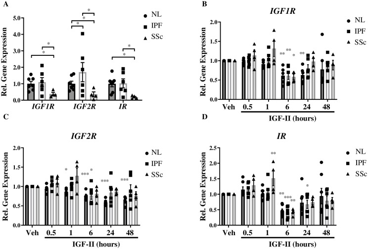
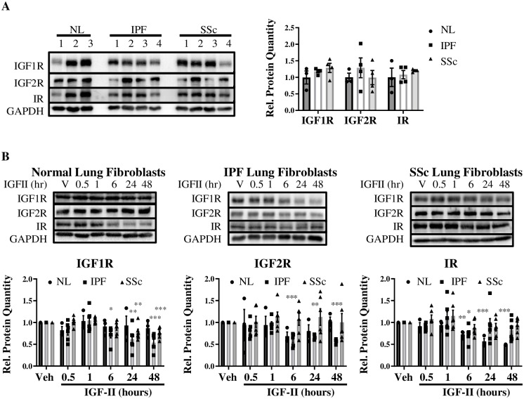
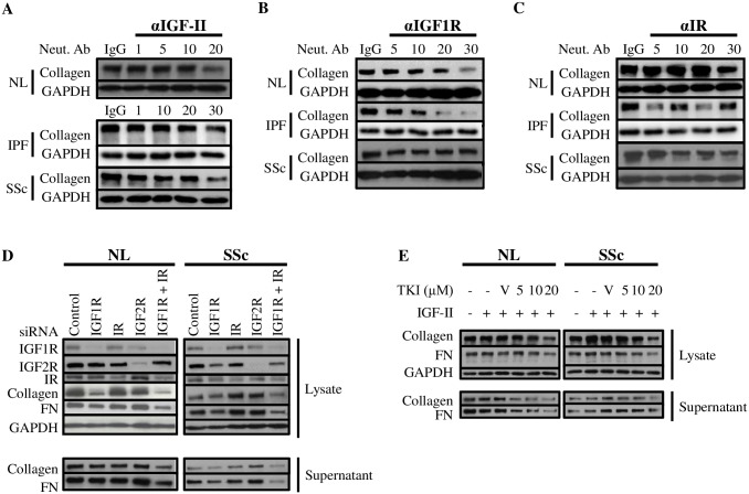
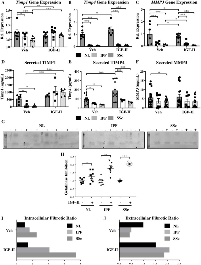
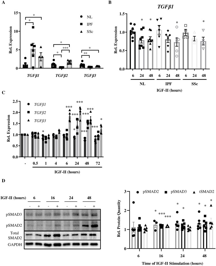
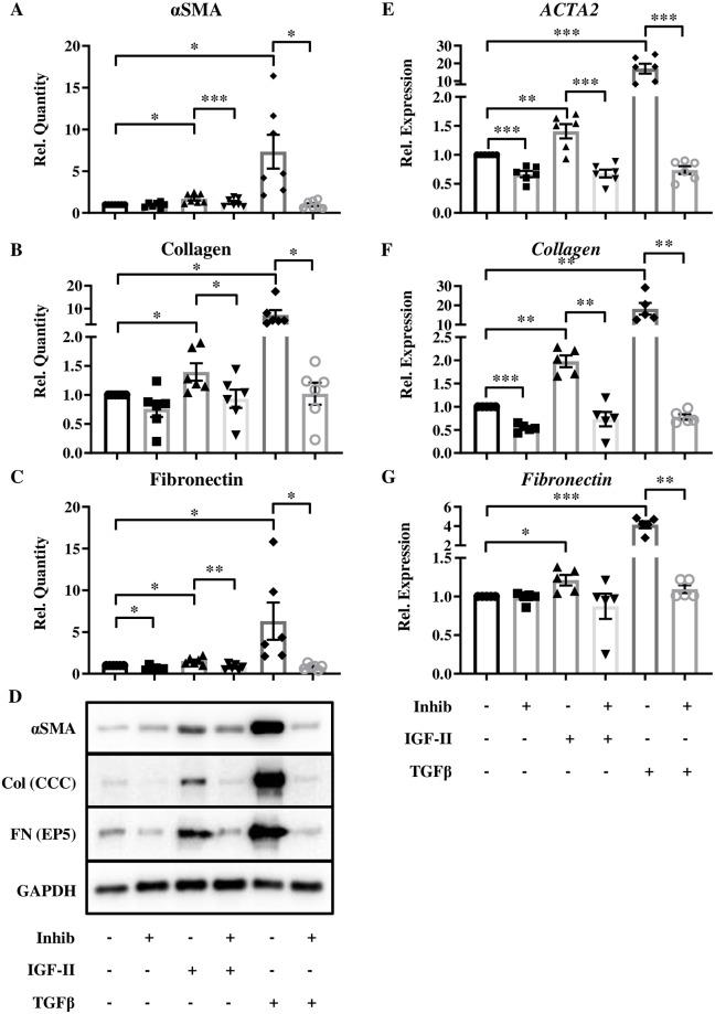
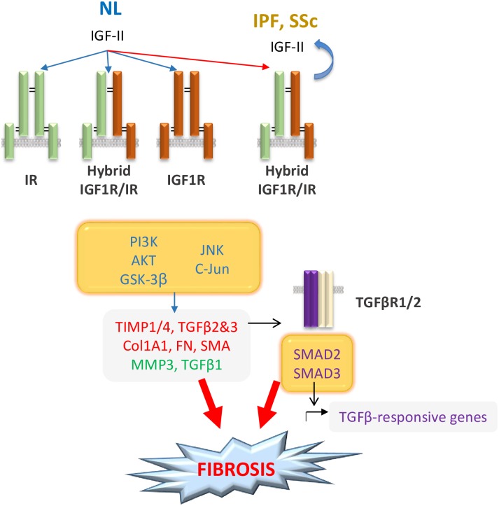
Type 2 insulin-like growth factor (IGF-II) levels are increased in fibrosing lung diseases such as idiopathic pulmonary fibrosis (IPF) and scleroderma/systemic sclerosis-associated pulmonary fibrosis (SSc). Our goal was to investigate the contribution of IGF receptors to IGF-II-mediated fibrosis in these diseases and identify other potential mechanisms key to the fibrotic process. Cognate receptor gene and protein expression were analyzed with qRT-PCR and immunoblot in primary fibroblasts derived from lung tissues of normal donors (NL) and patients with IPF or SSc. Compared to NL, steady-state receptor gene expression was decreased in SSc but not in IPF. IGF-II stimulation differentially decreased receptor mRNA and protein levels in NL, IPF, and SSc fibroblasts. Neutralizing antibody, siRNA, and receptor inhibition targeting endogenous IGF-II and its primary receptors, type 1 IGF receptor (IGF1R), IGF2R, and insulin receptor (IR) resulted in loss of the IGF-II response. IGF-II tipped the TIMP:MMP balance, promoting a fibrotic environment both intracellularly and extracellularly. Differentiation of fibroblasts into myofibroblasts by IGF-II was blocked with a TGFβ1 receptor inhibitor. IGF-II also increased TGFβ2 and TGFβ3 expression, with subsequent activation of canonical SMAD2/3 signaling. Therefore, IGF-II promoted fibrosis through IGF1R, IR, and IGF1R/IR, differentiated fibroblasts into myofibroblasts, decreased protease production and extracellular matrix degradation, and stimulated expression of two TGFβ isoforms, suggesting that IGF-II exerts pro-fibrotic effects via multiple mechanisms.


Conflict of interest statement

The authors have declared that no competing interests exist.

Figures

Fig 1
Fig 1. Steady-state and IGF-II-stimulated changes in receptor gene expression.
Basal (A) and IGF-II (200 ng/mL)-mediated gene expression of IGF1R (B), IGF2R (C), and IR (D) receptors in Normal Lung (NL), IPF, and SSc fibroblasts normalized to GAPDH housekeeping gene. N = 3–6. *p<0.05, **p<0.01, ***p<0.001 by paired 2-tailed Student’s T Test (A) and 1-way ANOVA with Dunnett’s multiple comparison test compared to vehicle (PBS, B-D).
Fig 2
Fig 2. Steady-state and IGF-II-stimulated changes in receptor protein expression.
Basal (A) and IGF-II (200 ng/mL)-stimulated protein expression of IGF1R, IGF2R, and IR receptors (B) in Normal Lung (NL), IPF, and SSc fibroblasts normalized to GAPDH housekeeping protein. Quantification of protein expression was graphed as histograms with mean +/- standard error of the mean and representative immunoblots included. N = 3–6. *p<0.05, **p<0.01, ***p<0.001 by paired 2-tailed Student’s T Test (A) and 1-way ANOVA with Dunnett’s multiple comparison test compared to vehicle (PBS, B).
Fig 3
Fig 3. Blockade of endogenous IGF-II or its receptors abrogates ECM component production.
NL, IPF, and SSc lung fibroblasts were treated with neutralizing antibodies to endogenous IGF-II (A), IGF1R (B), IR (C), or appropriate isotype control (respective highest antibody concentration, 20–30 μg/mL) for 1 hr, followed by stimulation with IGF-II (200 ng/mL) for 48 hr and lysates were probed for Collagen and GAPDH by immunoblot. D: Lung fibroblasts were subjected to knockdown of IGF1R, IR, IGF2R, dual knockdown of IGF1R + IR, or scrambled siRNA in the presence of IGF-II (200 ng/mL) and probed for protein expression of Collagen, Fibronectin (FN), and GAPDH in lysates and supernatants. E: Lung fibroblasts were treated with increasing concentrations of IGF1R tyrosine kinase inhibitor (TKI, Tyrphostin AG 538) in the presence of 200 ng/mL IGF-II and probed for protein expression of Collagen, Fibronectin, and GAPDH in lysates and supernatants. Representative immunoblots from at least three experiments.
Fig 4
Fig 4. IGF-II promotes an intracellular and an extracellular fibrotic environment.
Transcript levels of TIMP1 (A), TIMP4 (B), and MMP3 (C) normalized to GAPDH housekeeping gene by qRT-PCR and secreted TIMP1 (D), TIMP4 (E), and MMP3 (F) levels in supernatant by ELISA in NL, IPF, and SSc fibroblasts treated with vehicle (PBS) or IGF-II (200 ng/mL) for 48 hr. N = 5–10. *p<0.05, **p<0.01, ***p<0.001 by 1-way ANOVA with Dunnett’s multiple comparison test. G: Reverse gelatin zymography of supernatants from fibroblasts treated with vehicle (-) or IGF-II (200 ng/mL, +) for 48 hr and quantification of relative gelatinase inhibition (H). Individual samples are represented with mean +/- standard error of the mean as indicated. N = 5–7. *p<0.05, **p<0.01, and ***p<0.001 by paired 2-tailed Student’s T test. Fibrotic ratios of relative TIMP1 & TIMP4 to relative MMP3 transcript (I, intracellular) and secreted protein (J, extracellular) levels in NL, IPF, and SSc fibroblasts. Ratio was calculated by taking the average of relative TIMP1 and relative TIMP4, then dividing by relative MMP3. High fibrotic ratio indicates increased ECM deposition and/or decreased ECM breakdown.
Fig 5
Fig 5. IGF-II stimulates TGFβ isoform expression.
A: Steady-state expression of TGFB1, TGFB2, and TGFB3 in NL, IPF, and SSc fibroblasts. B: TGFB1 gene expression in NL, IPF, and SSc fibroblasts over time with IGF-II stimulation (200 ng/mL). C: Gene expression of TGFB1, TGFB2, and TGFB3 in NL fibroblasts treated with IGF-II (200 ng/mL) or vehicle (PBS). D: Representative images and quantification of SMAD2/3 activation in NL fibroblasts over time. N = 3–8. Histograms show data normalized to GAPDH presented as mean +/- standard error of the mean with significance by 1-way ANOVA with Dunnett’s multiple comparison test indicated as *p<0.05, **p<0.01, and ***p<0.001. (-): vehicle (PBS), (+): IGF-II (200 ng/mL).
Fig 6
Fig 6. IGF-II contributes to transdifferentiation of NL fibroblasts into myofibroblasts.
Expression of αSMA (A), Collagen (B), and Fibronectin (C) proteins with representative images (D) and αSMA/ACTA2 (E), Collagen (F), and Fibronectin (G) transcripts in NL after IGF-II (200 ng/mL) stimulation for 96 hr (protein) or 72 hr (transcript) following 1 hr pre-treatment with TGFBR1 inhibitor (SB431542). N = 6–7. Histograms are mean +/- standard error of the mean with significance by 1-way ANOVA with Dunnett’s multiple comparison test indicated as *p<0.05, **p<0.01, and ***p<0.001.
Fig 7
Fig 7. IGF-II mediates lung fibrosis through multiple mechanisms.
IGF-II signals through IR, IGF1R, or hybrid receptors (IGF1R/IR), preferentially switching to the hybrid receptor species in IPF and SSc. Signaling via PI3K/AKT/GSK3β and JNK/c-Jun [12] results in upregulation of ACTA2, TIMP1/4, Collagen, Fibronectin, & TGFβ2/3 and down-regulation of MMP3 & TGFβ1. Stimulation by IGF-II also causes TGFβ2/3 upregulation and canonical SMAD2/3 activation. Through differential gene expression and receptor species preferences, IGF-II induces formation of myofibroblasts and contributes to ECM accumulation, leading to fibrosis.

References

    1. Raghu G, Chen SY, Hou Q, Yeh WS, Collard HR. Incidence and prevalence of idiopathic pulmonary fibrosis in US adults 18–64 years old. Eur Respir J. 2016;48(1):179–86. 10.1183/13993003.01653-2015 . - DOI - PubMed
    1. Herzog EL, Mathur A, Tager AM, Feghali-Bostwick C, Schneider F, Varga J. Review: interstitial lung disease associated with systemic sclerosis and idiopathic pulmonary fibrosis: how similar and distinct? Arthritis Rheumatol. 2014;66(8):1967–78. 10.1002/art.38702 . - DOI - PMC - PubMed
    1. Martyanov V, Kim GJ, Hayes W, Du S, Ganguly BJ, Sy O, et al. Novel lung imaging biomarkers and skin gene expression subsetting in dasatinib treatment of systemic sclerosis-associated interstitial lung disease. PLoS One. 2017;12(11):e0187580 10.1371/journal.pone.0187580 . - DOI - PMC - PubMed
    1. Hsu E, Shi H, Jordan RM, Lyons-Weiler J, Pilewski JM, Feghali-Bostwick CA. Lung tissues in patients with systemic sclerosis have gene expression patterns unique to pulmonary fibrosis and pulmonary hypertension. Arthritis Rheum. 2011;63(3):783–94. 10.1002/art.30159 . - DOI - PMC - PubMed
    1. Garrett SM, Baker Frost D, Feghali-Bostwick C. The mighty fibroblast and its utility in scleroderma research. J Scleroderma Relat Disord. 2017;2(2):69–134. 10.5301/jsrd.5000240 . - DOI - PMC - PubMed

Publication types

MeSH terms

Substances