Review
. 2019 Nov 21;11(12):1836.
doi: 10.3390/cancers11121836.

Cancer Cell Membrane-Coated Nanoparticles for Cancer Management

Affiliations
Review

Cancer Cell Membrane-Coated Nanoparticles for Cancer Management

Jenna C Harris et al. Cancers (Basel). .

Abstract

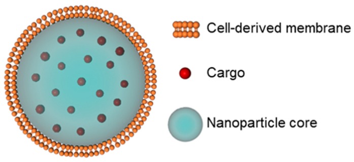
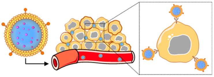
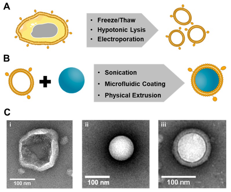
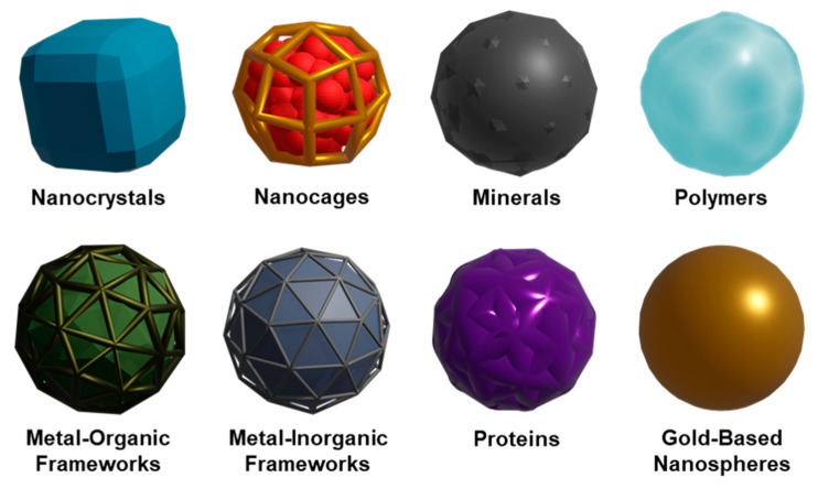
Cancer is a global health problem in need of transformative treatment solutions for improved patient outcomes. Many conventional treatments prove ineffective and produce undesirable side effects because they are incapable of targeting only cancer cells within tumors and metastases post administration. There is a desperate need for targeted therapies that can maximize treatment success and minimize toxicity. Nanoparticles (NPs) with tunable physicochemical properties have potential to meet the need for high precision cancer therapies. At the forefront of nanomedicine is biomimetic nanotechnology, which hides NPs from the immune system and provides superior targeting capabilities by cloaking NPs in cell-derived membranes. Cancer cell membranes expressing "markers of self" and "self-recognition molecules" can be removed from cancer cells and wrapped around a variety of NPs, providing homotypic targeting and circumventing the challenge of synthetically replicating natural cell surfaces. Compared to unwrapped NPs, cancer cell membrane-wrapped NPs (CCNPs) provide reduced accumulation in healthy tissues and higher accumulation in tumors and metastases. The unique biointerfacing capabilities of CCNPs enable their use as targeted nanovehicles for enhanced drug delivery, localized phototherapy, intensified imaging, or more potent immunotherapy. This review summarizes the state-of-the-art in CCNP technology and provides insight to the path forward for clinical implementation.

Keywords: biomimetic; cancer; drug delivery; imaging; immunotherapy; membrane-wrapped; nanocarrier; photodynamic therapy; photothermal therapy; targeted delivery.


Conflict of interest statement

The authors declare no conflict of interest. The funders had no role in the design of the study; in the collection, analyses, or interpretation of data; in the writing of the manuscript, or in the decision to publish the results.

Figures

Figure 1
Figure 1
Scheme depicting the components of a representative membrane-wrapped nanoparticle.
Figure 2
Figure 2
Scheme depicting the delivery of cancer cell membrane-wrapped nanoparticles (CCNPs) to tumors. Upon systemic administration, CCNPs exhibit long circulation due to the presence of “markers of self” on the membrane surface that minimize immune recognition. Additionally, CCNP membranes contain “self-recognition” molecules that allow the NPs to bind homotypic tumor cells after escaping from tumor vessels.
Figure 3
Figure 3
Illustration of the synthesis of membrane-wrapped nanoparticles. (A) Cell membranes can be extracted from their source cells by applying one of three methods. (B) Membranes can be wrapped around different types of nanoparticles using one of the three membrane–core fusion methods. (C) Transmission electron microscopy images of a (i) 4T1 breast cancer cell membrane vesicle, (ii) bare poly(lactic-co-glycolic acid) (PLGA) nanoparticle, and (iii) 4T1 cancer-cell membrane-wrapped PLGA nanoparticle prepared by the authors using the hypotonic lysis method depicted in (A) and the physical extrusion method depicted in (B).
Figure 4
Figure 4
Summary of various nanoparticle formulations that have been wrapped with cell-derived membranes to enable cancer treatment and imaging.
Figure 5
Figure 5
Demonstration of homotypic tumor targeting by CCNPs. (A) Illustration of experimental design for data shown in (B). Mice bearing human squamous carcinoma (UM-SCC-7) tumors were treated with doxorubicin (DOX) alone or with DOX and magnetic iron oxide nanoparticles that were wrapped with membranes derived from three different sources (COS7 monkey kidney cells, HeLa cervical cancer cells, or homotypic UM-SCC-7 squamous carcinoma cells). (B) In vivo fluorescence images of mice bearing UM-SCC-7 tumors 24 hours post-injection with membrane-wrapped nanoparticles prepared with (a) UM-SCC-7, (b) COS7, or (c) HeLa membranes as described in A, or post-injection with (d) DOX at an equivalent DOX dosage. The highest tumor accumulation is observed for homotypic membrane-wrapped nanoparticles. (C) Illustration of the dual tumor-bearing mouse model in which one flank harbored a hepatocellular carcinoma (H22) tumor and the other harbored a UM-SCC-7 tumor. The animals were injected with membrane-wrapped NPs designed to homotypically target one tumor or the other. Twelve hours post-injection, in vivo fluorescence images and ex vivo images of tumors were acquired. Both types of membrane-wrapped nanoparticles evaluated exhibited preferential accumulation in homotypic tumors (matched to the source membrane) versus heterotypic tumors with membrane mismatch. Reprinted (adapted) with permission from Reference [44]: Zhu, J.Y.; Zheng, D.W.; Zhang, M.K.; et al. Nano Lett. 2016, 16, 5895–5901. Copyright (2016) American Chemical Society.

References

    1. Jemal A., Siegel R., Ward E., Murray T., Xu J., Thun M.J. Cancer Statistics, 2019. CA. Cancer J. Clin. 2019;57:43–66. doi: 10.3322/canjclin.57.1.43. - DOI - PubMed
    1. Lang T., Yin Q., Li Y. Progress of Cell-Derived Biomimetic Drug Delivery Systems for Cancer Therapy. Adv. Ther. 2018;1:1800053. doi: 10.1002/adtp.201800053. - DOI
    1. Steichen S.D., Caldorera-Moore M., Peppas N.A. A review of current nanoparticle and targeting moieties for the delivery of cancer therapeutics. Eur. J. Pharm. Sci. 2013;48:416–427. doi: 10.1016/j.ejps.2012.12.006. - DOI - PMC - PubMed
    1. Sun T., Zhang Y.S., Pang B., Hyun D.C., Yang M., Xia Y. Engineered nanoparticles for drug delivery in cancer therapy. Angew. Chem. Int. Ed. 2014;53:12320–12364. - PubMed
    1. Albanese A., Tang P.S., Chan W.C.W. The Effect of Nanoparticle Size, Shape, and Surface Chemistry on Biological Systems. Annu. Rev. Biomed. Eng. 2012 doi: 10.1146/annurev-bioeng-071811-150124. - DOI - PubMed

LinkOut - more resources