Review
. 2019 Nov 21;8(12):533.
doi: 10.3390/plants8120533.

Structure-Function Analysis Reveals the Singularity of Plant Mitochondrial DNA Replication Components: A Mosaic and Redundant System

Affiliations
Review

Structure-Function Analysis Reveals the Singularity of Plant Mitochondrial DNA Replication Components: A Mosaic and Redundant System

Luis Gabriel Brieba. Plants (Basel). .

Abstract

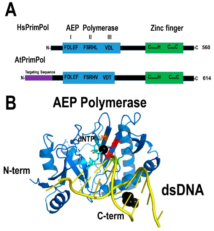
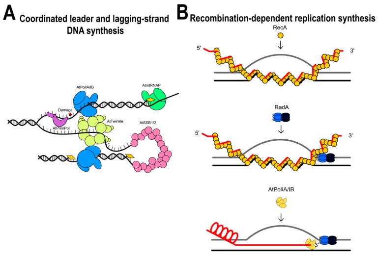
Plants are sessile organisms, and their DNA is particularly exposed to damaging agents. The integrity of plant mitochondrial and plastid genomes is necessary for cell survival. During evolution, plants have evolved mechanisms to replicate their mitochondrial genomes while minimizing the effects of DNA damaging agents. The recombinogenic character of plant mitochondrial DNA, absence of defined origins of replication, and its linear structure suggest that mitochondrial DNA replication is achieved by a recombination-dependent replication mechanism. Here, I review the mitochondrial proteins possibly involved in mitochondrial DNA replication from a structural point of view. A revision of these proteins supports the idea that mitochondrial DNA replication could be replicated by several processes. The analysis indicates that DNA replication in plant mitochondria could be achieved by a recombination-dependent replication mechanism, but also by a replisome in which primers are synthesized by three different enzymes: Mitochondrial RNA polymerase, Primase-Helicase, and Primase-Polymerase. The recombination-dependent replication model and primers synthesized by the Primase-Polymerase may be responsible for the presence of genomic rearrangements in plant mitochondria.

Keywords: DNA replication 1; evolution 2; recombination-dependent replication 4; replisome 3.


Conflict of interest statement

The author declares no conflict of interest.

Figures

Figure 1
Figure 1
AtTwinkle is a homolog of bacteriophage T7 primase-helicase and mitochondrial Twinkle. (A) Schematic representation of the bifunctional T7 primase-helicase in comparison to AtTwinkle and human Twinkle. T7 primase-helicase and AtTwinkle contain the conserved motifs necessary for primase and helicase activities, whereas human Twinkle is inactive as a primase. (B) Homology model of AtTwinkle showing its RNA polymerase domain and helicase modules with basis on the crystal structure of the heptameric T7 primase-helicase [31]. (C) Close view of a monomeric module of the RNAP and helicase of AtTwinkle. (D) Close view of the primase module composed of the zinc binding domain (ZBD) and RNAP domain. The conserved cysteines that coordinate the zinc atom are colored in red and magenta.
Figure 2
Figure 2
Bacteriophage-type plant organellar RNA polymerases. (A) Domain organization of bacteriophage-related RNAP. These enzymes share a C-terminal or polymerization domain that is divided into three subdomains: Fingers, palm, and thumb, and a N-terminal domain involved in promoter opening and RNA binding. The N-terminal domain is colored orange and the subdomains of the fingers, thumb, and palm of blue, green, and red, respectively. mtHsRNAP associates with two accessory subunits (TFB2M and TFAM) to open double-stranded DNA and contains a N-terminal pentatricopeptide repeat (PPR)-domain and a tether helix not present in plant mitochondrial RNAPs. (B) Structural model of the mtAtRNAP compared to bacteriophage T7RNAP and human mtRNAP during transcription initiation [40,53].
Figure 3
Figure 3
Homology model of tetrameric AtmtSSB1. (A) Homology model of AtmtSBB1 illustrating its oligonucleotide/oligosaccharide/binding (OB)-fold and an acid C-terminal tail. (B) An amino acid sequence alignment illustrates that the C-terminal tail of AtmtSSB2 is composed of two aromatic amino acids, whereas AtmtSSB1 is acidic.
Figure 4
Figure 4
Structural conservation of plant and bacterial RecAs. (A) Crystal structure of the bacterial RecA postsynaptic nucleoprotein filament determined by Chen, Yang, and Pavletich [83]. Each of the five RecA monomers is individually colored and labeled with numbers. The search strand is colored in yellow and the complementary strand in red. The crystal structure comprises solely the RecA fold and the C-terminal domain is not present in the initial construct. (B) Domain organization of AtRecA2 and AtRecA3 in comparison to bacterial RecA. AtRecA3 lacks the C-terminal regulatory domain.
Figure 5
Figure 5
Structural organization of AtRecX. (A) Crystal structure of RecX from E. coli (PDB: 3c1d). RecX is composed of three repeats of a three-helix motifs, (B) modular organization of AtRecX in comparison to bacterial RecX. Plant RecX harbor a mitochondrial targeting sequence (MTS) and a N-terminal domain of unkown function. AtRecX share more than 30% amino acid identity with bacterial RecXs.
Figure 6
Figure 6
AtODB1 resembles human Rad52. (A) Structural domain organization of AtODB1 in comparison to human Rad52. AtODB1 lacks the C-terminal domain necessary to interact with RPA and Rad51; (B) crystal structure of the undecameric ring of human Rad52. The undecameric structure is stabilized by alpha-helix 5 that interacts with alpha-helix 1 of the neighbor molecule. Each subunit (residues 1 to 172 is individually colored) and the C-terminal residues (172 to 212) are colored in read. (C) Model of AtODB1 as a undecameric ring lacking alpha-helix 5 of human Rad52.
Figure 7
Figure 7
Plant RadA resembles the bacterial enzyme. (A) Structural organization of AtRadA in comparision to bacterial RadA. AtRadA shares 63% amino acid similarity with RadA from S. pneumoniae and complete amino acid identity in the catalytic amino acids. Bacterial RadA harbor a zinc finger (ZnF), a Rec-A like ATPase domain with a unique KNRFG motif, and a region homologous to the Lon protease. (B) Crystal structure of the Rec-A like ATPase and Lon protease domains of RadA from S. pneumoniae showing its resemblance to a hexameric helicase. The ZnF domain is not present in the crystal structure.
Figure 8
Figure 8
Plants harbor a RecG ortholog. (A) AtRecG presents the same domain organization of bacterial RecG, plus the addition of an N-terminal organellar targeting sequence. (B) RecG remodels halted replication forks by promoting fork regression (chicken foot structure) that is converted to a Holliday junction. (C) Crystal structure of T. maritima RecG illustrating its modular assembly.
Figure 9
Figure 9
Structural comparison between AtPolIB and bacterial DNAPs. (A) Domain organization of both DNAPs. The polymerization domains are colored in black and the 3′-5′ exonuclease domains in orange. The unique amino acid insertions in AtPolIB in comparison to bacterial DNAPs I are depicted in a ball-stick representation and colored in red, green, and cyan. AtPolIs contain an N-terminal DTS and a disorder region not present in the structural model. (B) homology model of AtPolIBs with the crystal structures of the Klenow fragment from E. coli DNAP I. In both models, the dsDNA from Bacillus DNAP I is superimposed.
Figure 10
Figure 10
Structural organization of Whirlies. (A) Crystal structure of AtWhy2 (PDB ID: 4kop) with model ssDNA from Solanum whirly. The crystal structure represents residues 45 to 212. The second lysine of the KGKAAL motif is in a ball-stick representation. The C-terminal 310 helix is in red. (B) Structural organization of AtWhy2. The disordered C-terminal tail is indicated in the diagram.
Figure 11
Figure 11
Structural organization of OSBs. (A) Structural model of AtOSB1 showing its predicted OB-fold and PDF motif domains. (B) Modular organization of mitochondrial OSBs in Arabidopsis. AtOSBs consist of an OB-like fold followed by one to three PDF motifs (54). Although AtOSB1 is depicted as a monomer, AtOSB2 in solution assembles as tetramer.
Figure 12
Figure 12
Structural comparison between HsDNAligI and AtDNligI. (A) AtDNAligI has a shorter N-terminal region. However, the core structure that harbors the DNA binding domain (red) the adenylation domain (cyan) and the OB-fold domain (orange) are conserved between both ligases. (B) Homology modeling of AtDNAlig I with basis on the crystal structure of human DNA ligase I (PDB ID: 1X9N).
Figure 13
Figure 13
AtPrimPol resembles HsPrimPol. (A) Both AtPrimPol and HsPrimPol share a modular organization. AtPrimPol contains an N-terminal sequence for dual organellar targeting. (B) Structural model of the archaeo-eukaryotic primase (AEP) domain of AtPrimPol. The structural model was constructed with basis on the crystal structure of the AEP domain of HsPrimPol.
Figure 14
Figure 14
Putative models for DNA replication in plant mitochondria. (A) Leader and lagging-strand DNA synthesis. (B) Recombination-dependent replication systems in plant mitochondria.

References

    1. Gray M.W. Mitochondrial evolution. Cold Spring Harb. Perspect. Biol. 2012;4:a011403. doi: 10.1101/cshperspect.a011403. - DOI - PMC - PubMed
    1. Smith D.R., Keeling P.J. Mitochondrial and plastid genome architecture: Reoccurring themes, but significant differences at the extremes. Proc. Natl. Acad. Sci. USA. 2015;112:10177–10184. doi: 10.1073/pnas.1422049112. - DOI - PMC - PubMed
    1. Gissi C., Iannelli F., Pesole G. Evolution of the mitochondrial genome of Metazoa as exemplified by comparison of congeneric species. Heredity. 2008;101:301–320. doi: 10.1038/hdy.2008.62. - DOI - PubMed
    1. Boore J.L. Animal mitochondrial genomes. Nucleic Acids Res. 1999;27:1767–1780. doi: 10.1093/nar/27.8.1767. - DOI - PMC - PubMed
    1. Davila J.I., Arrieta-Montiel M.P., Wamboldt Y., Cao J., Hagmann J., Shedge V., Xu Y.Z., Weigel D., Mackenzie S.A. Double-strand break repair processes drive evolution of the mitochondrial genome in Arabidopsis. BMC Biol. 2011;9:64. doi: 10.1186/1741-7007-9-64. - DOI - PMC - PubMed

Grants and funding

LinkOut - more resources