. 2019 Nov 25;50(1):97.
doi: 10.1186/s13567-019-0718-z.

Four types of scrapie in goats differentiated from each other and bovine spongiform encephalopathy by biochemical methods

Affiliations

Four types of scrapie in goats differentiated from each other and bovine spongiform encephalopathy by biochemical methods

Jan P M Langeveld et al. Vet Res. .

Abstract

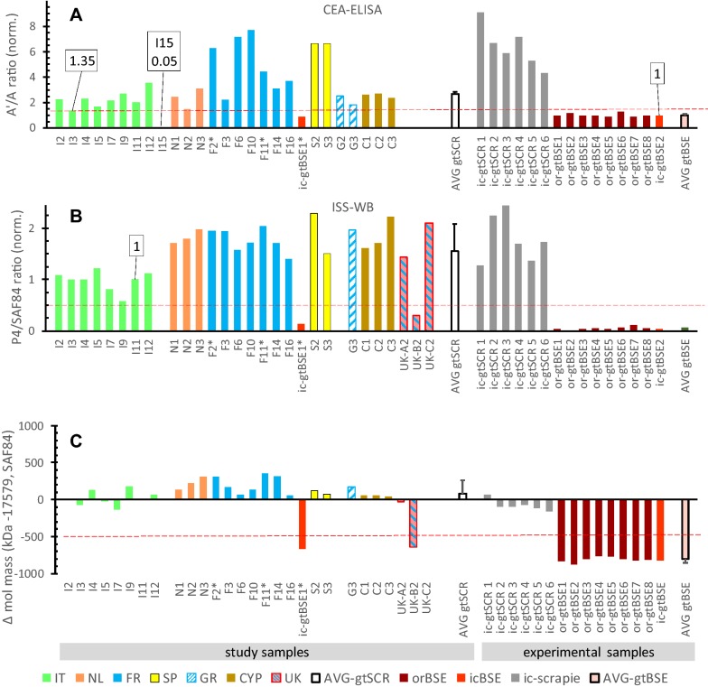
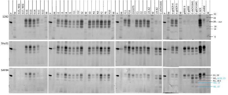
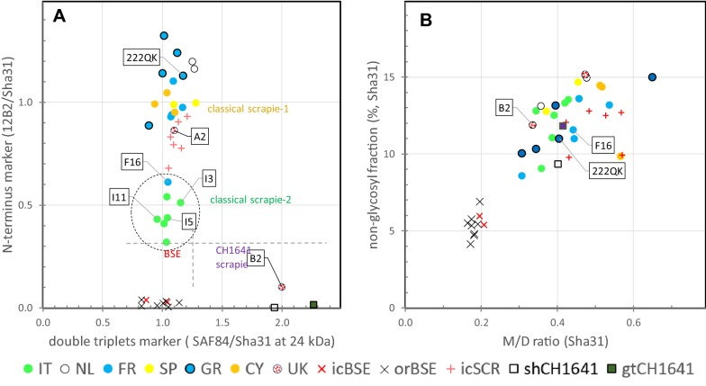
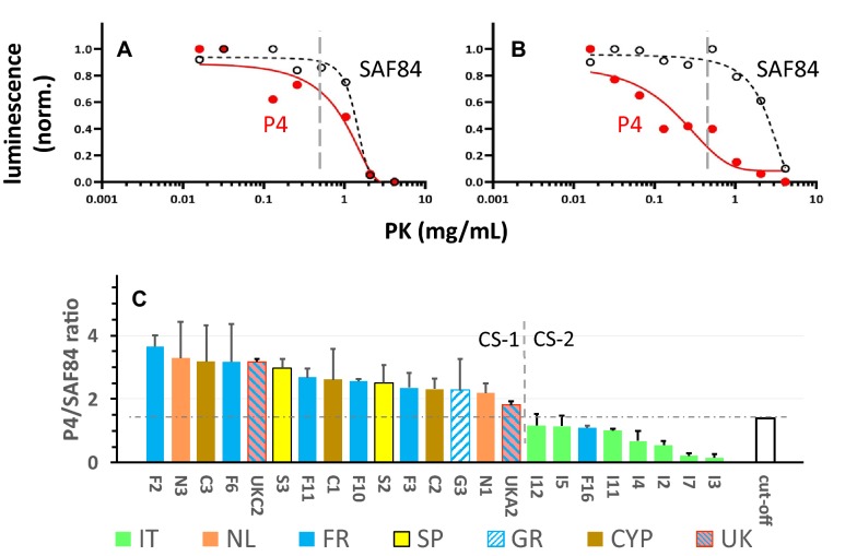
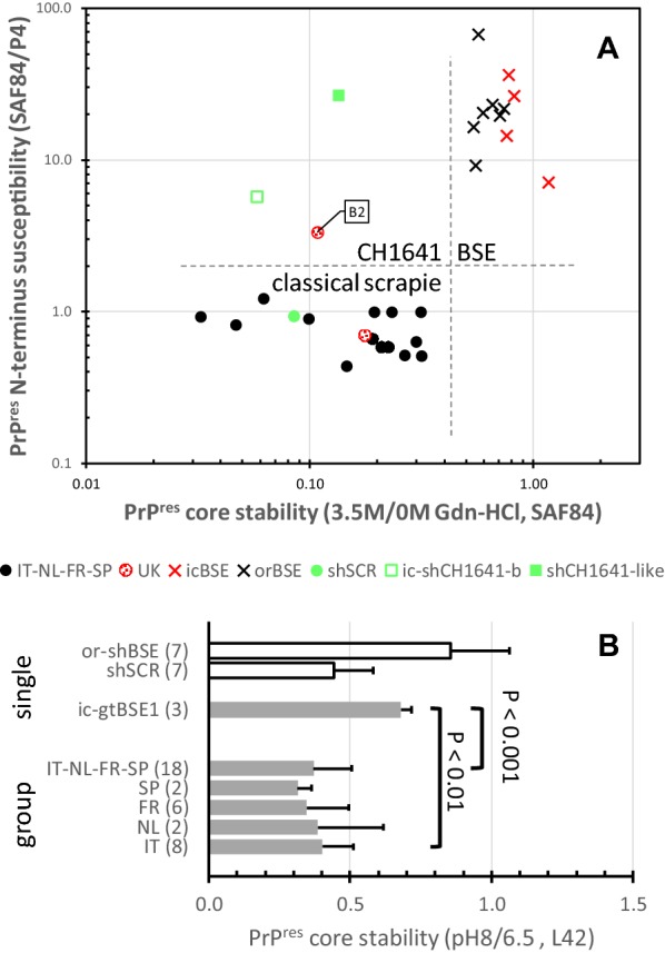
Scrapie in goats has been known since 1942, the archetype of prion diseases in which only prion protein (PrP) in misfolded state (PrPSc) acts as infectious agent with fatal consequence. Emergence of bovine spongiform encephalopathy (BSE) with its zoonotic behaviour and detection in goats enhanced fears that its source was located in small ruminants. However, in goats knowledge on prion strain typing is limited. A European-wide study is presented concerning the biochemical phenotypes of the protease resistant fraction of PrPSc (PrPres) in over thirty brain isolates from transmissible spongiform encephalopathy (TSE) affected goats collected in seven countries. Three different scrapie forms were found: classical scrapie (CS), Nor98/atypical scrapie and one case of CH1641 scrapie. In addition, CS was found in two variants-CS-1 and CS-2 (mainly Italy)-which differed in proteolytic resistance of the PrPres N-terminus. Suitable PrPres markers for discriminating CH1641 from BSE (C-type) appeared to be glycoprofile pattern, presence of two triplets instead of one, and structural (in)stability of its core amino acid region. None of the samples exhibited BSE like features. BSE and these four scrapie types, of which CS-2 is new, can be recognized in goats with combinations of a set of nine biochemical parameters.


Conflict of interest statement

The authors declare that they have no competing interests.

Figures

Figure 1
Figure 1
Discriminatory test results for exclusion of BSE in goat samples. Values below red broken lines indicate that sample concerned is considered BSE suspect. A CEA-ELISA. Values are normalised to sample ic-gtBSE2. B, C ISS-WB results (see Additional file 3). In B are shown N-terminus values relative to the PrPres core as reflected in the P4/SAF84 ratios normalised to the ratio of sample I11. In C is shown the difference in molecular mass in kDa of the N-band between that in a sample and the reference sample (I11). AVG gtSCR and AVG gtBSE represent average and SD of respectively the classical scrapie field samples (except I15 and UKB2) and of all gtBSE samples. Ratios in A and B are inverted compared to original methodology for the logical reason that the bar heights correlate positivley with N terminus values of PrPres.
Figure 2
Figure 2
Triplex-WB of goat study samples from different geographical regions together with TSE controls. Three antibodies used are indicated left. Images are all taken from the same blotting membrane. Lanes P and M, respectively recombinant shPrP and molecular mass standards. Position of molecular mass standards are visible only in the 647 nm (12B2) image and are indicated with kDa figures. Sample identities as in Tables 1 and Additional files 1 and 2 are indicated above the lanes. Only the CH1641 controls and UK-B2 sample exhibit a unique glycoprofile difference between SAF84 and Sha31. In lane C-gtCH1641, the positions of the three bands in triplets PrPres#1 (black, D1, M1, N1) and PrPres#2 (blue, D2, M2, N2) are indicated in the SAF84 panel at the right, including their approximate molecular masses; M1 and D2 have nearly similar molecular masses. Therefore, the signal of SAF84 in the D2 + M1 area is clearly higher than in the D1 area because this co-migration reflects the sum of D2 and M1 with that antibody, while with antibodies having more N-terminus located epitope specificity than SAF84 such as Sha31 the D1-area is higher than the D2 + M1 region.” Tissue equivalents applied vary between 0.5 and 2 mg. Samples were analysed in triplicate WB experiments.
Figure 3
Figure 3
Dot plots of data obtained from Triplex-WB of PrPres as in Figure 2. Each dot represents an individual goat TSE sample analysed in triplicate. Symbols: circles represent field cases and colour the country of origin; other symbols represent experimental samples and control samples from sheep or goat. A Plot with marker for N terminus epitope level on vertical axis versus PrPres double triplets marker on horizontal axis. Horizontal and vertical broken lines indicate the clear separation between CS, CH1641 and BSE. The lower N-terminus marker values of Italian cases and F16 are striking (encircled). B Triplet glycoprofile markers with non-glycosylated (N) fraction on vertical axis and on horizontal axis the ratio between signals in mono-glycosylated (M) and di-glycosylated fraction (D). A difference is obvious between BSE cases (low N and low M/D, due to high D levels in BSE samples) with CS and CH1641 cases.
Figure 4
Figure 4
Proteinase-K (PK) sensitivity of the N-terminal (P4) PrPres epitope in goat classical scrapie (CS) samples. A, B PK digestion curves of respective scrapie goat isolates S2 and I5, showing the differential sensitivities to PK of the epitopes of respectively SAF84 (black) and P4 (red). Vertical lines point to the 1 mg mL−1 PK condition used to study a larger selection of CS cases in C. C Shows the sensitivity of samples to PK at 1 mg mL−1 of the N terminal PrPres P4-epitope relative to that of core epitope SAF84. Vertical dashed line separates the samples between the ones with highest (CS-1) and lowest (CS-2) P4 epitope content consistent with data in Additional file 4. The cut-off between the two groups is reflected by the horizontal dash line and is based on the value of the reference sample I11 plus the average value of the standard deviations of all samples (1 + 0.4 = 1.4).
Figure 5
Figure 5
Stability of PrPres core region after denaturing and basic pretreatment of goat study samples. A The effect of 3.5 M Gdn-HCl treatment on susceptibility to proteolytic degradation was compared to normal condition when analysed by ISS-WB with mAb SAF84. The denaturation yielded high core epitope losses in the CS and CH1641 samples, but not in BSE samples where the PrPres core apparently is highly stable (horizontal axis). The vertical axis shows the susceptibility of the PrPres N-terminus under normal conditions of the ISS-WB process. B The effect of basic pH8 pre-treatment on subsequent proteolytic degradation. Only samples from four countries were tested. The BSE samples appeared the most resistant against proteolytic degradation of the PrPres core. All field samples from Italy, Netherlands, France and Spain together and separately from each country did exhibit a significantly higher susceptibility to Gdn denaturation than goatBSE, as indicated with the P values (Bonferroni–Dunn method).

Similar articles

Cited by

References

    1. Collinge J, Clarke AR. A general model of prion strains and their pathogenicity. Science. 2007;318:930–936. doi: 10.1126/science.1138718. - DOI - PubMed
    1. Prusiner SB. Novel proteinaceous infectious particles cause scrapie. Science. 1982;216:136–144. doi: 10.1126/science.6801762. - DOI - PubMed
    1. Wells GA, Scott AC, Johnson CT, Gunning RF, Hancock RD, Jeffrey M, Dawson M, Bradley R. A novel progressive spongiform encephalopathy in cattle. Vet Rec. 1987;121:419–420. doi: 10.1136/vr.121.18.419. - DOI - PubMed
    1. Wilesmith JW, Wells GA, Cranwell MP, Ryan JB. Bovine spongiform encephalopathy: epidemiological studies. Vet Rec. 1988;123:638–644. - PubMed
    1. Bruce ME, Will RG, Ironside JW, McConnell I, Drummond D, Suttie A, McCardle L, Chree A, Hope J, Birkett C, Cousens S, Fraser H, Bostock CJ. Transmissions to mice indicate that ‘new variant’ CJD is caused by the BSE agent. Nature. 1997;389:498–501. doi: 10.1038/39057. - DOI - PubMed