

Perturbations in imprinted methylation from assisted reproductive technologies but not advanced maternal age in mouse preimplantation embryos
- PMID: 31767035
- PMCID: PMC6878706
- DOI: 10.1186/s13148-019-0751-9
Perturbations in imprinted methylation from assisted reproductive technologies but not advanced maternal age in mouse preimplantation embryos
Abstract
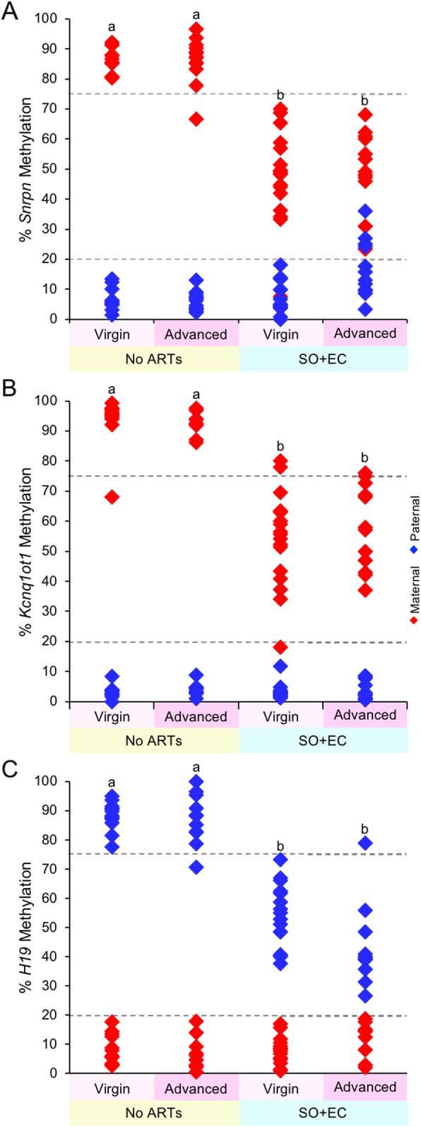
Background: Over the last several decades, the average age of first-time mothers has risen steadily. With increasing maternal age comes a decrease in fertility, which in turn has led to an increase in the use of assisted reproductive technologies by these women. Assisted reproductive technologies (ARTs), including superovulation and embryo culture, have been shown separately to alter imprinted DNA methylation maintenance in blastocysts. However, there has been little investigation on the effects of advanced maternal age, with or without ARTs, on genomic imprinting. We hypothesized that ARTs and advanced maternal age, separately and together, alter imprinted methylation in mouse preimplantation embryos. For this study, we examined imprinted methylation at three genes, Snrpn, Kcnq1ot1, and H19, which in humans are linked to ART-associated methylation errors that lead to imprinting disorders.
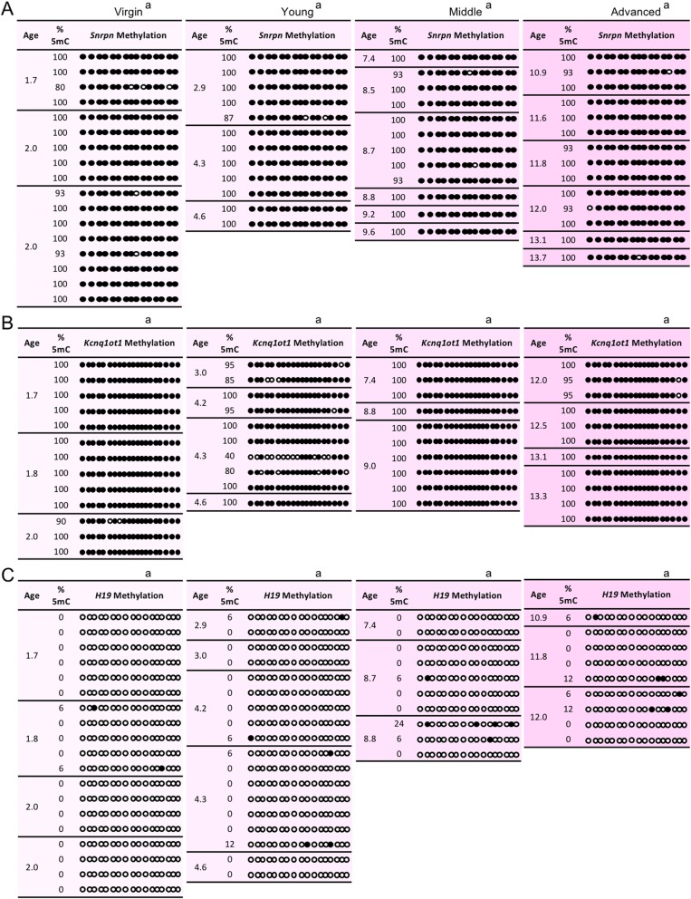
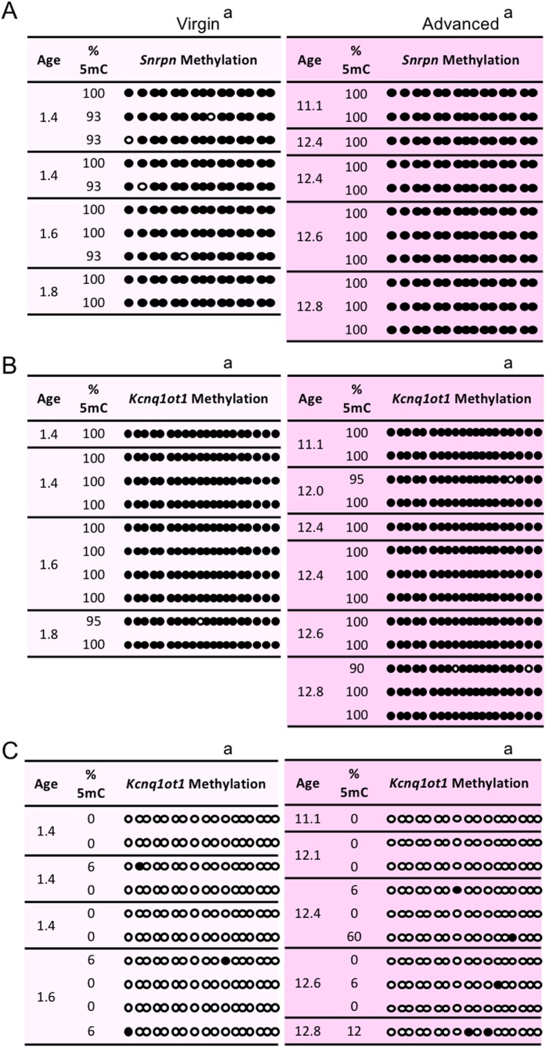
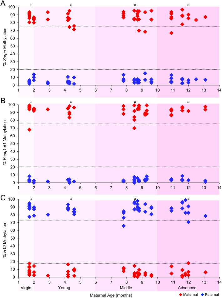
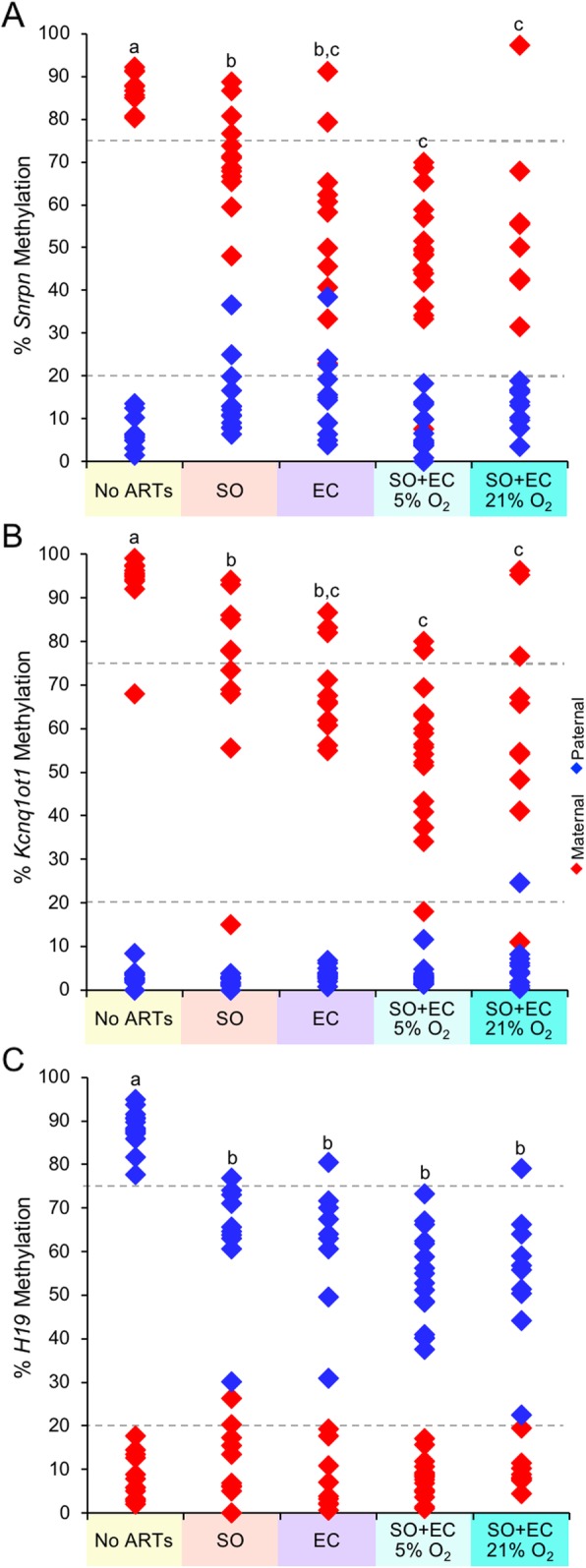
Results: Our data showed that imprinted methylation acquisition in oocytes was unaffected by increasing maternal age. Furthermore, imprinted methylation was normally acquired when advanced maternal age was combined with superovulation. Analysis of blastocyst-stage embryos revealed that imprinted methylation maintenance was also not affected by increasing maternal age. In a comparison of ARTs, we observed that the frequency of blastocysts with imprinted methylation loss was similar between the superovulation only and the embryo culture only groups, while the combination of superovulation and embryo culture resulted in a higher frequency of mouse blastocysts with maternal imprinted methylation perturbations than superovulation alone. Finally, the combination of increasing maternal age with ARTs had no additional effect on the frequency of imprinted methylation errors.
Conclusion: Collectively, increasing maternal age with or without superovulation had no effect of imprinted methylation acquisition at Snrpn, Kcnq1ot1, and H19 in oocytes. Furthermore, during preimplantation development, while ARTs generated perturbations in imprinted methylation maintenance in blastocysts, advanced maternal age did not increase the burden of imprinted methylation errors at Snrpn, Kcnq1ot1, and H19 when combined with ARTs. These results provide cautious optimism that advanced maternal age is not a contributing factor to imprinted methylation errors in embryos produced in the clinic. Furthermore, our data on the effects of ARTs strengthen the need to advance clinical methods to reduce imprinted methylation errors in in vitro-produced embryos.
Keywords: Assisted reproductive technologies; DNA methylation; Fertility; Genomic imprinting; Maternal age; Mouse.
Conflict of interest statement
These authors declare that they have no competing interests.
Figures
Similar articles
-
The loss of imprinted DNA methylation in mouse blastocysts is inflicted to a similar extent by in vitro follicle culture and ovulation induction.Mol Hum Reprod. 2016 Jun;22(6):427-41. doi: 10.1093/molehr/gaw013. Epub 2016 Feb 7. Mol Hum Reprod. 2016. PMID: 26908643
-
Side-by-side comparison of five commercial media systems in a mouse model: suboptimal in vitro culture interferes with imprint maintenance.Biol Reprod. 2010 Dec;83(6):938-50. doi: 10.1095/biolreprod.110.085480. Epub 2010 Aug 11. Biol Reprod. 2010. PMID: 20702853
-
High Frequency of Imprinted Methylation Errors in Human Preimplantation Embryos.Sci Rep. 2015 Dec 2;5:17311. doi: 10.1038/srep17311. Sci Rep. 2015. PMID: 26626153 Free PMC article.
-
Maintenance of Mest imprinted methylation in blastocyst-stage mouse embryos is less stable than other imprinted loci following superovulation or embryo culture.Environ Epigenet. 2017 Aug 29;3(3):dvx015. doi: 10.1093/eep/dvx015. eCollection 2017 Jul. Environ Epigenet. 2017. PMID: 29492315 Free PMC article. Review.
-
Conservation of DNA Methylation Programming Between Mouse and Human Gametes and Preimplantation Embryos.Biol Reprod. 2016 Sep;95(3):61. doi: 10.1095/biolreprod.116.140319. Epub 2016 Jul 27. Biol Reprod. 2016. PMID: 27465133 Review.
Cited by
-
Application of Single-Cell RNA Sequencing in Ovarian Development.Biomolecules. 2022 Dec 27;13(1):47. doi: 10.3390/biom13010047. Biomolecules. 2022. PMID: 36671432 Free PMC article. Review.
-
Early Life Oxidative Stress and Long-Lasting Cardiovascular Effects on Offspring Conceived by Assisted Reproductive Technologies: A Review.Int J Mol Sci. 2020 Jul 22;21(15):5175. doi: 10.3390/ijms21155175. Int J Mol Sci. 2020. PMID: 32707756 Free PMC article. Review.
-
Association between imprinting disorders and assisted reproductive technologies.Epigenomics. 2025 Apr;17(6):397-410. doi: 10.1080/17501911.2025.2471269. Epub 2025 Mar 3. Epigenomics. 2025. PMID: 40033833 Free PMC article. Review.
-
Genome-wide assessment of DNA methylation alterations induced by superovulation, sexual immaturity and in vitro follicle growth in mouse blastocysts.Clin Epigenetics. 2023 Jan 16;15(1):9. doi: 10.1186/s13148-023-01421-z. Clin Epigenetics. 2023. PMID: 36647174 Free PMC article.
-
DNA methylation mechanisms in the maturing and ageing oocyte.Epigenetics Chromatin. 2025 Jun 11;18(1):34. doi: 10.1186/s13072-025-00600-x. Epigenetics Chromatin. 2025. PMID: 40495235 Free PMC article. Review.
References
-
- Ely DM, Hamilton BE. Trends in fertility and mother’s age at first birth among rural and metropolitan counties: United States, 2007-2017. NCHS Data Brief. 2018;(323):1–8. - PubMed
-
- Centers for Disease Control and Prevention ASfRM, Society for Assisted Reproductive Technology. : 2016 Assisted Reproductive Technology National Summary Report 2018.
-
- Chandra A, Copen CE, Stephen EH. Infertility and impaired fecundity in the United States, 1982-2010: data from the National Survey of Family Growth. Natl Health Stat Report. 2013;(67):1–18 11 p following 19. - PubMed
Publication types
MeSH terms
Substances
Grants and funding
LinkOut - more resources
Full Text Sources
Medical
