. 2019 Dec:239:108498.
doi: 10.1016/j.vetmic.2019.108498. Epub 2019 Nov 6.

S100A9 regulates porcine reproductive and respiratory syndrome virus replication by interacting with the viral nucleocapsid protein

Affiliations

S100A9 regulates porcine reproductive and respiratory syndrome virus replication by interacting with the viral nucleocapsid protein

Zhongbao Song et al. Vet Microbiol. 2019 Dec.

Abstract

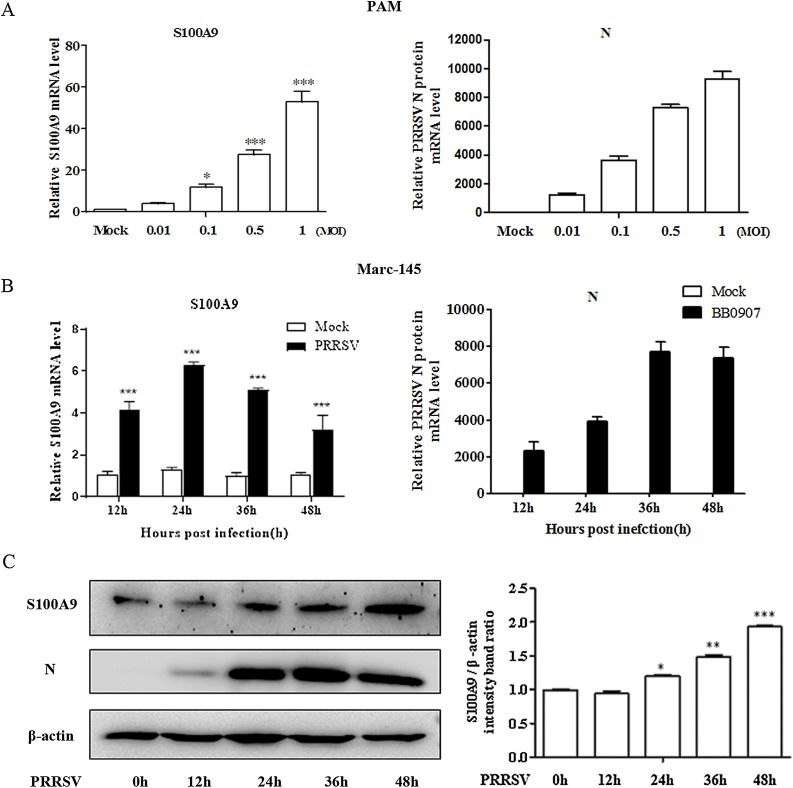
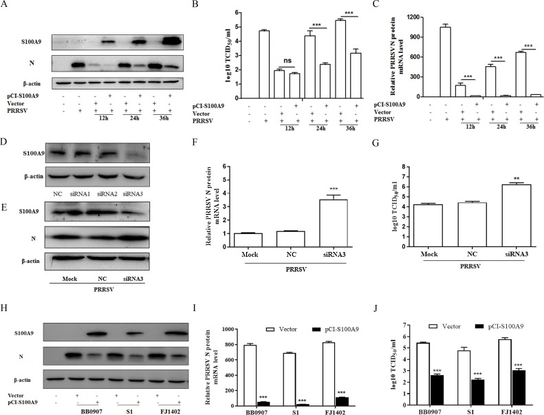
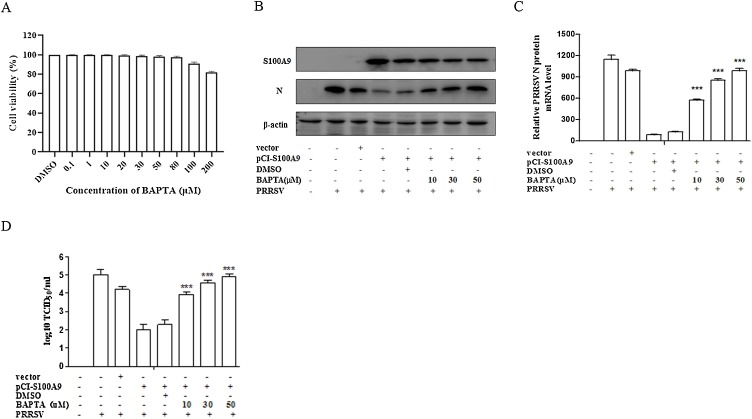
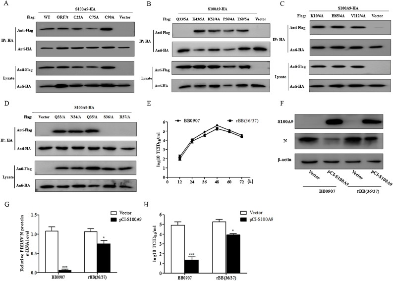
Porcine reproductive and respiratory syndrome virus (PRRSV) has caused huge economic losses to the pig industry worldwide over the last 30 years, yet the associated viral-host interactions remain poorly understood. S100A9 is a damage-associated molecular pattern of the S100 protein family. Here, we found that PRRSV infection stimulated S100A9 expression in porcine alveolar macrophages (PAMs) and Marc-145 cells. S100A9 inhibited PRRSV replication via cellular Ca2+ dependent manner. The viral nucleocapsid (N) protein co-localized with S100A9 in the cytoplasm, and directly interacted at amino acid 78 of S100A9 and amino acids 36-37 of N protein. Moreover, we also found that the mutant S100A9 (E78Q) protein exhibited decreased antiviral activity against PRRSV compared with the parent S100A9. Recombinant PRRSV rBB (36/37) with two mutations in amino acid 36-37 in the N protein exhibited greater replication than the parent PRRSV BB0907 in S100A9-overexpressed PAM and Marc-145 cells. Thus, S100A9 may restrict PRRSV proliferation by interacting with the viral N protein.

Keywords: Inhibit; Nucleocapsid protein; PRRSV; S100A9.


Figures

Fig. 1
Fig. 1
PRRSV infection induces S100A9 in PAM and Marc-145 cells. (A) PAMs were infected with the PRRSV BB0907 strain for 24 h. qRT-PCR was used to quantitate the level of S100A9 and N protein mRNA expression. (B) Marc-145 cells were infected with 0.1 MOI of the BB0907 strain for 12 h, 24 h, 36 h, and 48 h. The qRT-PCR was used to quantitate the level of S100A9 and N protein mRNA expression. (C) Western blot analysis was used to determine the level of S100A9 and N protein expression. All of the results are expressed as the means + standard deviation from three independent experiments.
Fig. 2
Fig. 2
S100A9 inhibits PRRSV replication. (A) Western blot assay for N protein of Marc-145 cells transfected with pCI-S100A9-HA or an empty vector for 24 h, and then infected with the PRRSV BB0907 strain (0.01 MOI), for 24 h, 36 h, and 48 h. (B) The level of N protein mRNA expression by qPCR and (C) PRRSV titer by TCID50 assay. (D) Western blot of Marc-145 cells transfected with three siRNAs targeting monkey S100A9. (E―G) Western blot, qRT-PCR, and TCID50 of Marc-145 cells transfected with siRNA3 for 24 h, then infected with the PRRSV BB0907 strain for 36 h. (H―J) Western blot, qRT-PCR, and TCID50 of Marc-145 cells transfected with pCI-S100A9-HA or control vector for 24 h, then infected with the PRRSV strains BB0907, S1, and FJ1402 for 36 h. All results are expressed as the means + standard deviations from three independent experiments.
Fig. 3
Fig. 3
S100A9 inhibition of PRRSV replication is dependent on intracellular Ca2+. Marc-145 cells were transfected with pCI-S100A9-HA for 6 h, then treated with BAPTA (10 μM, 30 μM, and 50 μM), and infected with PRRSV an MOI of 0.01 for 36 h. Cells were lysed and analyzed by (A) western blot, (B) real time PCR, and (C) TCID50 assay. All the results were confirmed by three independent experiments. Error bars represent the standard deviations of triplicate experiments.
Fig. 4
Fig. 4
S100A9 interacts with the PRRSV N protein. HEK293T cells were co-transfected with pCI-S100A9-HA and plasmids encoding PRRSV proteins (nsp1α, nsp1β, nsp4, nsp5, nsp7, nsp9-12, GP5, M, and N) for 30 h then prepared for immunoprecipitation by (A) an anti-FLAG antibody or (B) anti-HA antibody. (C) Immunofluorescence assay of Marc-145 cells transfected with pCI-S100A9-HA or vector for 24 h, then infected with PRRSV for 30 h. The S100A9 (red) N protein (green) and nuclei (blue). All the results were confirmed by three independent experiments. (For interpretation of the references to colour in this figure legend, the reader is referred to the web version of this article.)
Fig. 5
Fig. 5
The key region of the PRRSV N protein necessary for the interaction with S100A9. (A–D) HEK293 T cells co-transfected for 30 h with pCI-S100A9-HA and pCI-N-FLAG or the pCI-N*-FLAG series of plasmids, the proteins were immunoprecipitated by an anti-FLAG antibody. Whole cell lysates and IP complexes were analyzed by western blot using anti-FLAG and HA antibodies. (E) The growth kinetics of the wild type PRRSV BB0907 strain and recombinant PRRSV rBB(36/37). (F–H) Marc-145 cells transfected with pCI-S100A9-HA or empty vector followed by an incubation with 0.01 MOI of BB0907 or rBB(36/37). A western blot (F), qPCR (G), and TCID50 assay (H) were used to determine the level of viral replication. The data are expressed as the means + standard deviations from three independent experiments.
Fig. 6
Fig. 6
The key region of S100A9 is necessary for the interaction with PRRSV N protein. (A) Western blot of HEK293T cells co-transfected with plasmids expressing pCI-S100A9 or pCI-S100A9(E78Q) and pCI-N-FLAG or empty vector for 30 h. The proteins were immunoprecipitated by an anti-HA antibody. The whole cell lysates and IP complexes were analyzed with an anti-FLAG or HA antibody. (B and C) Marc-145 cells transfected with plasmids expressing mutants of S100A9 for 24 h, then infected with 0.01 MOI of the PRRSV BB0907 strain for 36 h. Viral replication was analyzed by western blot and qPCR. The data are expressed as the means + standard deviations from three independent experiments.
Fig. 7
Fig. 7
S100A9 inhibits PRRSV replication in PAMs. (A) The qRT-PCR of PAMs transfected with siRNA targeting porcine S100A9 or non-specific siRNA. (B and C) Viral replication in PAMs transfected with siRNA-3 or non-specific siRNA for 24 h then infected with 0.01 MOI of PRRSV BB0907, measured by western blot and qPCR. (D–G) Viral replication in PAMs transduced with a lentivirus expressing S100A9 or S100A9(E78Q), (LV-S100A9 and LV-S100A9(E78Q)) and an empty vector (LV-vector). The cells were then infected with 0.01 MOI of PRRSV BB0907 or rBB(36/37), and expression was measured by Western blot and qRT-PCR. The data are expressed as the means + standard deviations from three independent experiments.

Similar articles

Cited by

References

    1. Barr S.D., Smiley J.R., Bushman F.D. The interferon response inhibits HIV particle production by induction of TRIM22. PLoS Pathog. 2008;4 - PMC - PubMed
    1. Beura L.K., Sarkar S.N., Kwon B., Subramaniam S., Jones C., Pattnaik A.K., Osorio F.A. Porcine reproductive and respiratory syndrome virus nonstructural protein 1beta modulates host innate immune response by antagonizing IRF3 activation. J. Virol. 2010;84:1574–1584. - PMC - PubMed
    1. Burkard C., Opriessnig T., Mileham A.J., Stadejek T., Ait-Ali T., Lillico S.G., Whitelaw C., Archibald A.L. Pigs lacking the scavenger receptor cysteine-rich domain 5 of CD163 are resistant to porcine reproductive and respiratory syndrome virus 1 infection. J. Virol. 2018;92 - PMC - PubMed
    1. Chen N., Liu Q., Qiao M., Deng X., Chen X., Sun M. Whole genome characterization of a novel porcine reproductive and respiratory syndrome virus 1 isolate: genetic evidence for recombination between Amervac vaccine and circulating strains in mainland China. Infect. Genet. Evol. 2017;54:308–313. - PubMed
    1. Chen X., Bai J., Liu X., Song Z., Zhang Q., Wang X., Jiang P. Nsp1alpha of porcine reproductive and respiratory syndrome virus strain BB0907 impairs the function of monocyte-derived dendritic cells via the release of soluble CD83. J. Virol. 2018;92 - PMC - PubMed

MeSH terms

LinkOut - more resources