Review
. 2020 Jan;26(1):21-39.
doi: 10.1016/j.molmed.2019.10.007. Epub 2019 Nov 22.

Regulation of Mitochondrial ATP Production: Ca2+ Signaling and Quality Control

Affiliations
Review

Regulation of Mitochondrial ATP Production: Ca2+ Signaling and Quality Control

Liron Boyman et al. Trends Mol Med. 2020 Jan.

Abstract

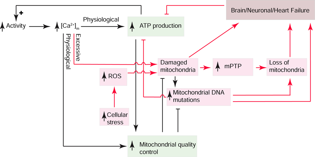
Cardiac ATP production primarily depends on oxidative phosphorylation in mitochondria and is dynamically regulated by Ca2+ levels in the mitochondrial matrix as well as by cytosolic ADP. We discuss mitochondrial Ca2+ signaling and its dysfunction which has recently been linked to cardiac pathologies including arrhythmia and heart failure. Similar dysfunction in other excitable and long-lived cells including neurons is associated with neurodegenerative diseases such as Alzheimer's disease (AD), amyotrophic lateral sclerosis (ALS), and Parkinson's disease (PD). Central to this new understanding is crucial Ca2+ regulation of both mitochondrial quality control and ATP production. Mitochondria-associated membrane (MAM) signaling from the sarcoplasmic reticulum (SR) and the endoplasmic reticulum (ER) to mitochondria is discussed. We propose future research directions that emphasize a need to define quantitatively the physiological roles of MAMs, as well as mitochondrial quality control and ATP production.

Keywords: heart failure; mitochondria-associated membranes; mitochondrial Ca(2+) signaling; mitochondrial quality control; mitophagy; neurodegeneration.


Figures

Figure 1.
Figure 1.. Energy consumption and its dynamic range in different tissues.
A. Daily metabolic rates of major organs and tissues. Shown values are from human subjects. Data is from Wang et al., 2010 [215]. B. Fold changes in oxygen consumption rates of different tissues in different species. Each data point shows the fold change in oxygen consumption determined by distinct studies performed in liver [–20], kidney [21, 22], heart [–32], and skeletal muscle [–36]. All shown measurements were carried out with healthy tissues of wild-type animals under similar, physiologically relevant basal and strenuous activities. The indicated weight (in Kg) is the total body weight.
Figure 2.
Figure 2.. spatial distribution of cardiac mitochondrial Ca2+ signaling components.
A spatial representation of a Ca2+ spark (red gradient) initiated at the Ca2+ release unit (CRU), which is located between the transverse-tubule (TT) and the junctional SR (JSR) membranes. At the peak of a Ca2+ spark, [Ca2+]i briefly (10 ms) bathes the end of a mitochondrion with high [Ca2+]i (5–10 μM). During a [Ca2+]i transient, multiple CRUs release Ca2+, bathing both ends of the mitochondrion with high [Ca2+]i. LCCs, L-type Ca2+ channels; RyR2s (ryanodine receptor type 2); SERCA: sarcoplasmic reticulum and endoplasmic reticulum Ca2+ ATPase; NCX: Na+-Ca2+ exchanger; MCU: mitochondrial Ca2+ uniporter; NCLX: mitochondrial NCX. Adapted from [46].
Figure 3.
Figure 3.. ATP production, Ca2+ movement, and mitochondrial dynamics in the inner (IMM) and outer mitochondrial membranes (OMM) in ventricular myocytes.
A. Top, confocal image of a ventricular cardiomyocyte showing the fluorescence of TMRM in mitochondria. Bottom, zoomed-in view showing a small region of the cell with 4–6 mitochondria. B. Schematic diagram of one of the intermyofibrillar mitochondria. The electron transport chain (ECT) is composed of five complexes: CI CII, CIII, CIV and CV. CI-CIV extrude protons to hyperpolarize IMM to approximately −160 mV. CV (the ATP synthase), uses the energy in ΔΨM to phosphorylate ADP to produce ATP. The adenine nucleotide translocase (ANT) exchanges ADP for ATP across the IMM. The voltage dependent anion channel is permeant to ATP, ADP, Ca2+, etc. The uncoupling proteins (UCPs) on the IMM regulates proton movement from the intermembrane space to the matrix in the production of heat. C. Mitochondrial Ca2+ entry and exit. MCU responsible for Ca2+ entry across the IMM is composed of the MCU, EMRE and MICUR1 subunits and is probably associated with one or more MICU1 (or MICU2 or MICU3) proteins. Ca2+ is extruded from the matrix by the Na+ dependent Ca2+ transporter, NCLX (also called the mitochondrial NCX, Na+-Ca2+ exchanger). D. Proteins involved with mitochondrial dynamics and quality control: OMM: Drp1 which associates with Fis1, Miff, MiD49/51; Parkin and PINK1; translocase of the OMM (Tom) complex, Mfn1, Mfn2. IMM: Opa1, translocase of the IMM (Tim) complex. Note: Tom and Tim proteins form a mitochondrial protein import path.
Figure 4.
Figure 4.. Mitochondrial Associated Membranes (MAMs) and Ca2+ signals.
A. Excitation-contraction coupling (ECC) and MAMs. The sarcoplasmic reticulum (SR) is contiguous with the endoplasmic reticulum (ER) and contribute membranes to MAMs. In cardiac myocytes, [Ca2+]i transient is triggered electrically by action potentials (AP). In ventricular myocytes, there are a very large number of Ca2+ spark sites (~20,000 per cell). Each site includes an array of ~50 SR Ca2+-release channels (ryanodine receptors type 2; RyR2) in the junctional SR that faces a transverse tubule (TT) or the sarcolemma (SL) that become activated upon Ca2+ elevation in the narrow ~15 nm subspace. During cardiac AP, the L-type Ca2+ channels in the TT and SL membranes are depolarization and conduct Ca2+ into the subspace. This high subspace Ca2+ activates the RyR2s enabling Ca2+ release from the SR to produce a Ca2+ spark. B. Three common arrangements of RyR2s in MAMs. Left: A cluster of RyR2s facing away from the mitochondrion, 20 to 100 nm away, similar to those of a Ca2+ spark site. Middle: Isolated RyR2 channel in a MAM facing an OMM at ~15 to 50 nm. These channels are not normally triggered to open. Under quiescent conditions, the opening rate is ~10−4 s−1. Right: Isolated RyR2 in a MAM more than 100 nm away from the mitochondrion. C. Garrote-assisted fission. It has been suggested that mitochondrial fission, normally attributed to the action of Drp1, may be assisted by the encircling action of ER or SR.
Figure 5.
Figure 5.. Signaling elements linking ATP production, mitochondrial quality control and Brain/Neuronal/Heart failure.
Cellular activity controls ATP production and mitochondrial quality control (green boxes) through Ca2+ signaling. Excessive [Ca2+]m and stress-dependent reactive oxygen species (ROS) production (red arrows) appear to damage mitochondria which can lead to manifestations of functional failure of heart and neuronal tissues with positive feedback amplification. Mitochondrial permeability transition pore (mPTP).

References

    1. McCormack JG and Denton RM (1993) The role of intramitochondrial Ca2+ in the regulation of oxidative phosphorylation in mammalian tissues. Biochem Soc Trans 21 ( Pt 3) (3), 793–9. - PubMed
    1. Wescott AP et al. (2019) Voltage-energized Calcium-sensitive ATP Production by Mitochondria. Nature Metabolism In Press. - PMC - PubMed
    1. Boyman L. et al. (2015) The growing importance of mitochondrial calcium in health and disease. Proc Natl Acad Sci U S A 112 (36), 11150–1. - PMC - PubMed
    1. Santulli G. et al. (2015) Mitochondrial calcium overload is a key determinant in heart failure. Proc Natl Acad Sci U S A 112 (36), 11389–94. - PMC - PubMed
    1. Luongo TS et al. (2017) The mitochondrial Na(+)/Ca(2+) exchanger is essential for Ca(2+) homeostasis and viability. Nature 545 (7652), 93–97. - PMC - PubMed

Publication types

MeSH terms

Substances