. 2020 Jun;69(6):1104-1115.
doi: 10.1136/gutjnl-2019-318812. Epub 2019 Nov 25.

Growth differentiation factor 11 attenuates liver fibrosis via expansion of liver progenitor cells

Affiliations

Growth differentiation factor 11 attenuates liver fibrosis via expansion of liver progenitor cells

Zhen Dai et al. Gut. 2020 Jun.

Erratum in

Abstract

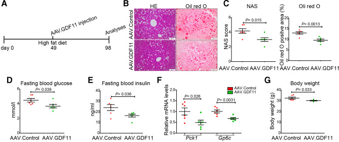
Objective: Liver fibrosis and cirrhosis resulting from chronic liver injury represent a major healthcare burden worldwide. Growth differentiation factor (GDF) 11 has been recently investigated for its role in rejuvenation of ageing organs, but its role in chronic liver diseases has remained unknown. Here, we investigated the expression and function of GDF11 in liver fibrosis, a common feature of most chronic liver diseases.

Design: We analysed the expression of GDF11 in patients with liver fibrosis, in a mouse model of liver fibrosis and in hepatic stellate cells (HSCs) as well as in other liver cell types. The functional relevance of GDF11 in toxin-induced and cholestasis-induced mouse models of liver fibrosis was examined by in vivo modulation of Gdf11 expression using adeno-associated virus (AAV) vectors. The effect of GDF11 on leucine-rich repeat-containing G-protein-coupled receptor 5 (LGR5)+ liver progenitor cells was studied in mouse and human liver organoid culture. Furthermore, in vivo depletion of LGR5+ cells was induced by injecting AAV vectors expressing diptheria toxin A under the transcriptional control of Lgr5 promoter.

Results: We showed that the expression of GDF11 is upregulated in patients with liver fibrosis and in experimentally induced murine liver fibrosis models. Furthermore, we found that therapeutic application of GDF11 mounts a protective response against fibrosis by increasing the number of LGR5+ progenitor cells in the liver.

Conclusion: Collectively, our findings uncover a protective role of GDF11 during liver fibrosis and suggest a potential application of GDF11 for the treatment of chronic liver disease.

Keywords: GDF11 and LGR5; chronic liver disease; fibrosis; hepatic stellate cells; myofibroblasts; stem cells.


Conflict of interest statement

Competing interests: None declared.

Figures

Figure 1
Figure 1
GDF11 is upregulated during liver fibrosis. (A) In situ hybridisation analysis of human GDF11 expression. Scale bars, 100 µm. (B, C) The qPCR-based analysis of human GDF11 mRNA in patients with fibrosis (n=6) and controls (n=6) from Hannover Medical School, Germany (B) and patients with fibrosis (n=26) and controls (n=6) from Haikou Hospital, China (C). (D) In situ hybridisation for mouse Gdf11 in fibrotic and control livers. (E) The qPCR-based analysis of mouse Gdf11 mRNA in fibrotic (n=7) and control (n=7) livers. (F) Western blot for GDF11 in normal and fibrotic mouse livers and its quantification. (G) Measuring the GDF11 content by ELISA in mouse serum. (H) The qPCR analysis of mouse Gdf11 in various liver cells such as hepatocytes (HC), hepatic stellate cells (HSC), Kupffer cells (KC) and liver sinusoidal endothelial cells (LSEC), isolated from fibrotic livers of different mice. Total RNA from kidney was used as a positive control. (I, J) GDF11 and desmin co-immunofluorescence in fibrotic livers of patients and mouse. Scale bars, 100 µm. (K) qPCR analysis for Gdf11 expression in quiescent and HSCs activated by culturing them in the presence of platelet-derived growth factor (PDGF). Experiments were repeated twice for B, C, H and I, and three times for E and F. The values shown in panels G, J and K are the mean of three independent cell isolations. Data are mean±SEM; two-tailed Student’s t-test (panels E, F, G and K) or two-sided Welch’s t-test (panels B and C). CCl4, carbon tetrachloride; DAPI, 4′,6-diamidino-2-phenylindole; DDC, 3,5-diethoxycarbonyl-1,4-dihydrocollidine; GDF11, growth differentiation factor 11; NPC, non-parachamal cells.
Figure 2
Figure 2
Hepatic overexpression of GDF11 attenuates liver fibrosis. (A, G) Schematic overview of experiments analysing the effect of Gdf11 overexpression in CCl4-induced (A) and cholestasis (DDC)-induced (G) mouse models of liver fibrosis (n=8 mice per group of CCl4 and n=8 mice per group of DDC model). (B, H) The qPCR analyses of Gdf11 expression in murine livers after AAV8-GDF11 injection. (C, I) Measurement of the total collagen content by hydroxyproline assay. (D, J) Representative immunohistochemical images of H&E, Sirius Red and desmin staining. Scale bars, 100 µm. (E, K) Quantification of Sirius Red and desmin staining in mice injected with either AAV8-GDF11 or AAV control particles. For each mouse, 6 liver sections were stained in batches and pictures from 12 random fields per section were captured and quantified in a blinded manner using Image-J. (F, I) The qPCR analyses of fibrosis-related genes such as Acta2, p75Ntr, Col1a1 and Col2a1. All experiments shown in this figure were repeated twice. Data are mean±SEM; two-tailed Student’s t-test. AAV8, adeno-associated virus serotype 8; CCl4, carbon tetrachloride; DDC, 3,5-diethoxycarbonyl-1,4-dihydrocollidine; GDF11, growth differentiation factor 11.
Figure 3
Figure 3
GDF11 promotes the expansion of LGR5+ liver progenitor cells (LPCs), which are capable of attenuating liver fibrosis in mice. (A, H) Representative FACS analysis of LGR5+ LPCs in AAV8-GDF11 or AAV control-injected mice in the CCl4 (n=8) (A) and the DDC (n=8) (H) fibrosis model. (B, I) The confocal immunofluorescence images of sorted LGR5+ cells, which were subsequently co-stained with CD133 antibody. Scale bars, 100 µm. (C, J) Schematic overview of the experimental set-up for LGR5+ LPC transplantation in fibrotic mice (n=5 mice per group for both CCl4 and DDC model). The control mice were injected with saline via spleen. (D, K) Measurement of the total collagen content by the hydroxyproline assay in LGR5+ LPC transplanted mice. (E, L) Representative images of H&E, Sirius Red and desmin staining. Scale bars, 100 µm. (F, M) Quantifications of Sirius Red and desmin staining in LGR5+ LPC transplanted and control mice. For each mouse, 6 liver sections were stained in batches and pictures from 12 random fields per section were captured and quantified in a blinded manner using Image-J. (G, N) The qPCR analyses of fibrosis-related genes in LGR5+ LPC transplanted mice. Experiments were repeated twice for A, H and three times for C–G and J–N. Data are mean±SEM; two-tailed Student’s t-test. AAV8, adeno-associated virus serotype 8; CCl4, carbon tetrachloride; DDC, 3,5-diethoxycarbonyl-1,4-dihydrocollidine; FACS, fluorescence-activated cell sorting; GDF11, growth differentiation factor 11; DAPI, 4′,6-diamidino-2-phenylindole; LGR5, leucine-rich repeat-containing G-protein-coupled receptor 5.
Figure 4
Figure 4
GDF11 promotes human liver progenitor cell expansion in ex vivo cultured organoids. (A) Schematic overview of the experimental setup. (B) Serial images showing the growth of human liver organoids. (C) Representative confocal images of human liver organoids stained for KRT19, SOX9, HNF4a. Nuclei were counterstained with DAPI. Scale bars, 100 µm. (D) Analysis of the hepatocyte differentiation by immunofluorescence staining for hepatocyte markers such as albumin (ALB) and major urinary protein (MUP). (E) Representative images of organoids treated with rGDF11 (bottom) and untreated (top) and quantification of organoid numbers at day 4. In each well, all areas with organoids were imaged by serial pictures and quantified in a blinded manner (n=10 wells). (F) qPCR-based analysis of human LGR5, PROM1 and HNF4A mRNA expression in organoids either treated with rGDF11 or untreated. (G) FACS analysis for LGR5+ cells on rGDF11-treated or rGDF11-untreated organoids and dissociated to single cells. (H) The mRNA expression (left) and ELISA for albumin performed with culture medium collected (right) from human liver organoids that were either treated with rGDF11 or remained untreated. (I) Schematic overview of experiments showing human primary hepatic myofibroblasts transfected with GDF11 siRNA and subsequently co-cultured with human liver organoids. (J) qPCR analysis of human GDF11 in human myofibroblasts. (K) Representative images of organoids co-cultured with human myofibroblasts transfected either with siGDF11 (bottom) or scramble (top) and quantification of organoid numbers at day 4. For each well, all areas with organoids were imaged by serial pictures and quantified in a blinded manner (n=10 wells). (L–M) qPCR analysis of human LGR5, PROM1, HNF4A and FACS analysis for LGR5+ cells on human liver organoids co-cultured with human myofibroblasts transfected with siGDF11 (right) or scramble (left). Experiments were repeated twice for B–D, and three times for E–M. Data are mean±SEM; two-tailed Student’s t-test. DAPI, 4′,6-diamidino-2-phenylindole; FACS, fluorescence-activated cell sorting; GDF11, growth differentiation factor 11; LGR5, leucine-rich repeat-containing G-protein-coupled receptor 5.
Figure 5
Figure 5
The depletion of LGR5+ LPCs diminishes GDF11-mediated attenuation of liver fibrosis. (A, G) Schematic overview of the experiments in CCl4 (B–F) and DDC (H–L) fibrosis mouse models (n=5 mice per group for both CCl4 and DDC model). (B, H) Representative FACS analysis of LGR5+ LPCs in AAV.GDF11 and AAV.LGR5.DTA or AAV control-injected mice in the CCl4 (B) and the DDC (H) fibrosis model. (C, I) Measurement of total collagen content by hydroxyproline assay. (D, J) Representative pictures of H&E, Sirius Red and desmin staining. Scale bars, 100 µm. (E, K) Quantifications of Sirius Red and desmin staining shown in panels D and J. For each mouse, 6 liver sections were stained in batches and pictures from 12 random fields per section were captured and quantified in a blinded manner using Image-J. (F, L) The qPCR analyses of fibrosis-related genes such as Acta2, p75Ntr, Col1a1 and Col2a1. All experiments shown in this figure were repeated twice. Data are mean±SEM; two-tailed Student’s t-test. AAV, adeno-associated virus; CCl4, carbon tetrachloride; DDC, 3,5-diethoxycarbonyl-1,4-dihydrocollidine; DTA, diphtheria toxin A; FACS, fluorescence-activated cell sorting; GDF11, growth differentiation factor 11; LGR5, leucine-rich repeat-containing G-protein-coupled receptor 5; LPC, liver progenitor cell.
Figure 6
Figure 6
In vivo modulation of Gdf11 in hepatic myofibroblasts. (A) Preferential targeting of hepatic myofibroblasts is shown by co-staining of GFP and desmin in livers of recipients of AAV.NGF.GFP virus. (B) Schematic overview of the experiments in CCl4-induced fibrosis mouse models (n=4 mice per group). (C) The qPCR analyses of Gdf11 expression in murine livers after AAV.NGF.GDF11 or AAV.NGF.shGDF11 injection. (D) Representative FACS analysis of LGR5+ LPCs in AAV.NGF.GDF11 or AAV.NGF.shGDF11-injected mice in the CCl4 fibrosis model. (E) Measurement of total collagen content by hydroxyproline assay. (F) Representative pictures of H&E, Sirius Red and desmin staining. Scale bars, 100 µm. (G) Quantification of Sirius Red and desmin staining is shown in panel F. For each mouse, 6 liver sections were stained in batches and pictures from 12 random fields per section were captured and quantified in a blinded manner using Image-J. (H) The qPCR analyses of fibrosis-related genes such as Acta2, p75NTR, Col1a1 and Col2a1. Data are mean±SEM; two-tailed Student’s t-test. AAV, adeno-associated virus; CCl4, carbon tetrachloride; DAPI, 4′,6-diamidino-2-phenylindole; GFP, green fluorescent protein; FACS, fluorescence-activated cell sorting; GDF11, growth differentiation factor 11;LGR5, leucine-rich repeat-containing G-protein-coupled receptor 5; LPC, liver progenitor cell.
Figure 7
Figure 7
GDF11 inhibits NASH progression. (A) Schematic overview of the experiments (n=6 mice per group). (B) Representative images of HE and Oil red O-stained liver sections from AAV.control and AAV.GDF11-injected mice fed with high-fat diet (HFD) for 14 weeks. Scale bars, 100 µm. (C) The NAS score and Oil red O-positive area were assessed. For each mouse, 6 liver sections were stained in batches and pictures from 12 random fields per section were captured and quantified in a blinded manner. (D) Fasting glucose concentration in blood. (E) Fasting insulin concentration in blood. (F) The qPCR analysis for gluconeogenic genes Pck1 and G6pc. (G) The body weight of AAV.control and AAV.GDF11-injected mice. Data shown in this figure were repeated twice. AAV, adeno-associated virus; GDF11, growth differentiation factor 11; NAS, Non-alcoholic fatty liver disease Activity Score; NASH, non-alcoholic steatohepatitis.

Comment in

References

    1. Morikawa M, Derynck R, Miyazono K. TGF-β and the TGF-β Family: Context-Dependent Roles in Cell and Tissue Physiology. Cold Spring Harb Perspect Biol 2016;8:a021873 10.1101/cshperspect.a021873 - DOI - PMC - PubMed
    1. McPherron AC, Lawler AM, Lee SJ. Regulation of skeletal muscle mass in mice by a new TGF-beta superfamily member. Nature 1997;387:83–90. 10.1038/387083a0 - DOI - PubMed
    1. McPherron AC, Lawler AM, Lee SJ. Regulation of anterior/posterior patterning of the axial skeleton by growth/differentiation factor 11. Nat Genet 1999;22:260–4. 10.1038/10320 - DOI - PubMed
    1. Loffredo FS, Steinhauser ML, Jay SM, et al. . Growth differentiation factor 11 is a circulating factor that reverses age-related cardiac hypertrophy. Cell 2013;153:828–39. 10.1016/j.cell.2013.04.015 - DOI - PMC - PubMed
    1. Smith SC, Zhang X, Zhang X, et al. . GDF11 does not rescue aging-related pathological hypertrophy. Circ Res 2015;117:926–32. 10.1161/CIRCRESAHA.115.307527 - DOI - PMC - PubMed

Publication types

Substances