. 2020 Jan 10;295(2):435-443.
doi: 10.1074/jbc.AC119.009890. Epub 2019 Nov 25.

Structure of spastin bound to a glutamate-rich peptide implies a hand-over-hand mechanism of substrate translocation

Affiliations

Structure of spastin bound to a glutamate-rich peptide implies a hand-over-hand mechanism of substrate translocation

Han Han et al. J Biol Chem. .

Abstract

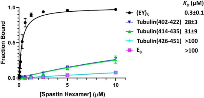
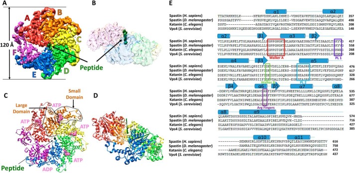
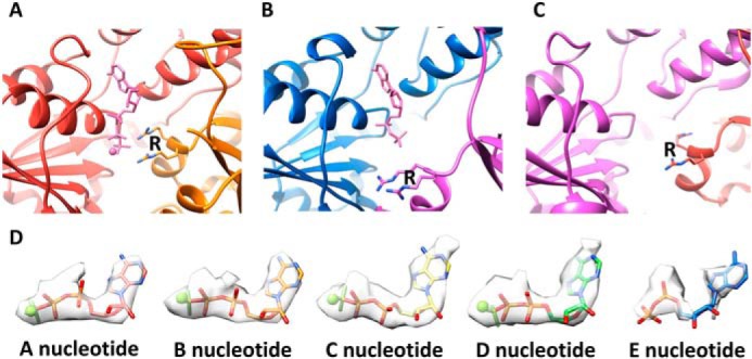
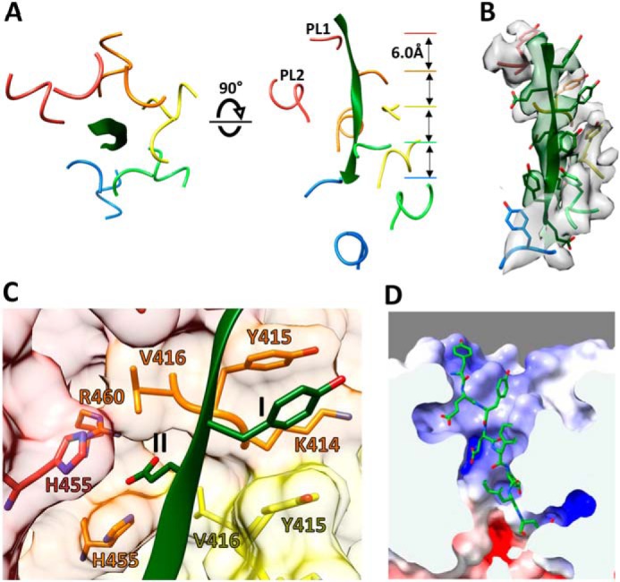
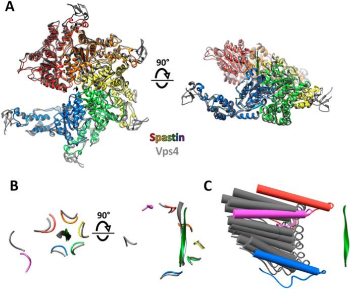
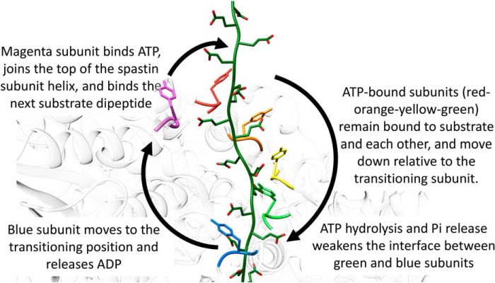
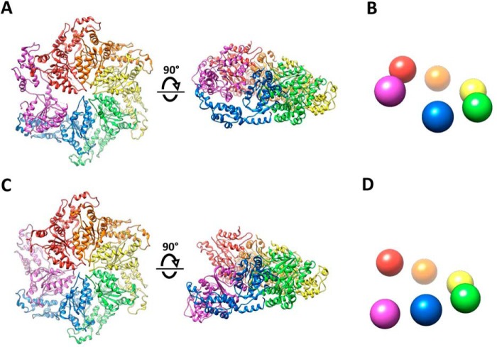
Many members of the AAA+ ATPase family function as hexamers that unfold their protein substrates. These AAA unfoldases include spastin, which plays a critical role in the architecture of eukaryotic cells by driving the remodeling and severing of microtubules, which are cytoskeletal polymers of tubulin subunits. Here, we demonstrate that a human spastin binds weakly to unmodified peptides from the C-terminal segment of human tubulin α1A/B. A peptide comprising alternating glutamate and tyrosine residues binds more tightly, which is consistent with the known importance of glutamylation for spastin microtubule severing activity. A cryo-EM structure of the spastin-peptide complex at 4.2 Å resolution revealed an asymmetric hexamer in which five spastin subunits adopt a helical, spiral staircase configuration that binds the peptide within the central pore, whereas the sixth subunit of the hexamer is displaced from the peptide/substrate, as if transitioning from one end of the helix to the other. This configuration differs from a recently published structure of spastin from Drosophila melanogaster, which forms a six-subunit spiral without a transitioning subunit. Our structure resembles other recently reported AAA unfoldases, including the meiotic clade relative Vps4, and supports a model in which spastin utilizes a hand-over-hand mechanism of tubulin translocation and microtubule remodeling.

Keywords: ATPases associated with diverse cellular activities (AAA); cryo-electron microscopy; microtubule severing mechanism; molecular machine; peptide interaction; protein structure; structure-function.


Conflict of interest statement

The authors declare that they have no conflicts of interest with the contents of this article

Figures

Figure 1.
Figure 1.
Binding of spastin to substrate peptides. Fluorescence polarization anisotropy isotherms for spastin binding to the indicated peptides. Error bars are shown for all data points, but are often smaller than the data point symbol. E8 is an 8-residue glutamylated peptide. (EY)5 is a 10-residue peptide comprised of alternating glutamate and tyrosine residues.
Figure 2.
Figure 2.
Cryo-EM structure of the spastin-peptide complex. A, top view of the cryo-EM density map, color-segmented for the six spastin subunits and the peptide. The contour shown is 6.8x the map root mean square deviation. B, side view of the density map with spastin shown as a transparent surface to reveal the peptide density (green) in the central pore. C, top view of the model. The large and small ATPase domains of subunit B are indicated with dashed lines. ATP (ADP·BeFx) and ADP (pink) are indicated at the subunit interfaces. D, side view of the model with subunit F removed to expose the binding groove and the peptide (green). E, sequence alignment of meiotic clade AAA+ ATPases of known structure. Human spastin secondary structures as defined by DSSP (49) are indicated above, and are numbered according to the scheme used for D. melanogaster spastin (25). Walker A, Walker B, pore loop 1 (PL1), pore loop 2 (PL2), and the arginine fingers are indicated.
Figure 3.
Figure 3.
Binding poses of ATP and ADP. A, coordination of Mg2+ ADP−BeFx (ATP) at the active site of subunit A. Coordination of the β phosphate and BeFx is completed by the two arginine finger (R) residues of subunit B. The BC, CD, and DE interfaces are very similar. B, ADP at the subunit E active site, where the subunit F finger arginines are displaced ∼14 Å relative to the AB, BC, CD, and DE interfaces. C, the subunit F active site lacks sufficient density to determine whether or not nucleotide is bound. D, modeling of nucleotides in the density for nucleotide at the A–E active sites.
Figure 4.
Figure 4.
Binding of substrate peptide as an extended strand. A, top and side views of peptide (green) and residues from pore loop 1 (PL1; 414–416) and pore loop 2 (PL2; 455–460) of subunits A–E. The pore loops of these subunits form a double helix that binds the peptide. Pore loops of subunit F (not shown) are displaced away from the helix axis and the peptide. B, density for the bound peptide and pore loop 1 residues, contoured at 7.0 × σ. All side chains of the peptide are resolved except for the two terminal residues. C, close-up views of the class I and class II pockets that bind the alternating side chains of the peptide. This view shows pockets at the interface of subunits B and C (and His-455 of subunit A). The equivalent binding pockets at the AB, CD, and DE interfaces are very similar. D, electrostatic potential surface within the pore computed for subunits A–E. The scale was −10 (red) to 10 (blue) kb·T·ec−1. The calculation was performed at pH 7.2.
Figure 5.
Figure 5.
Close similarity between spastin and Vps4 and mechanistic implications. A, top and side views of spastin subunits A–E overlapped with Vps4 (12) using PyMol (Schrödinger, LLC). B, top and side views of spastin (colors) and Vps4 (gray) peptide and pore loop 1 residues after overlap on the large ATPase domains. Subunit F (magenta for spastin) is distant from the bound peptide. C, spastin subunit F (magenta) lies on the spectrum of subunit F positions seen from focused classification of multiple Vps4 structures (gray) (13). Pore loop 1 is shown with the preceding strand and following helix. Spastin subunits A (red) and E (blue) are indicated.
Figure 6.
Figure 6.
Proposed mechanism of substrate translocation. Spastin is colored white with the pore loop 1 Tyr-415 of all six subunits in color. Substrate (green) is modeled on the bound peptide, extended at either end by continuing the β-strand conformation, and shown with modeled glutamate side chains.
Figure 7.
Figure 7.
Comparison of ring and spiral configurations of katanin. A, top and side views of the katanin ring structure (PDB 5WCB). The human spastin structure described herein, and Vps4 (12) are essentially identical. B, schematic representation of the side view of the ring structure. C, top and side views of the katanin spiral structure (PDB 5WC0) structure (24). The D. melanogaster spastin structure (25) is essentially identical. D, schematic representation of the side view of the spiral structures. The ring and spiral configurations are essentially identical to each other over the red-blue subunits, and vary only in the position of the magenta subunit.

References

    1. Erzberger J. P., and Berger J. M. (2006) Evolutionary relationships and structural mechanisms of AAA+ proteins. Annu. Rev. Biophys. Biomol. Struct. 35, 93–114 10.1146/annurev.biophys.35.040405.101933 - DOI - PubMed
    1. Nyquist K., and Martin A. (2014) Marching to the beat of the ring: polypeptide translocation by AAA+ proteases. Trends Biochem. Sci. 39, 53–60 10.1016/j.tibs.2013.11.003 - DOI - PMC - PubMed
    1. Monroe N., and Hill C. P. (2016) Meiotic Clade AAA ATPases: protein polymer disassembly machines. J. Mol. Biol. 428, 1897–1911 10.1016/j.jmb.2015.11.004 - DOI - PMC - PubMed
    1. Plaud C., Joshi V., Kajevu N., Pous C., Curmi P. A., and Burgo A. (2018) Functional differences of short and long isoforms of spastin harboring missense mutation. Dis. Model Mech. 11, pii: dmm033704 - PMC - PubMed
    1. Nogales E., and Wang H. W. (2006) Structural mechanisms underlying nucleotide-dependent self-assembly of tubulin and its relatives. Curr. Opin. Struct. Biol. 16, 221–229 10.1016/j.sbi.2006.03.005 - DOI - PubMed

Publication types