

Global skin gene expression analysis of early diffuse cutaneous systemic sclerosis shows a prominent innate and adaptive inflammatory profile
- PMID: 31767698
- PMCID: PMC7386329
- DOI: 10.1136/annrheumdis-2019-215894
Global skin gene expression analysis of early diffuse cutaneous systemic sclerosis shows a prominent innate and adaptive inflammatory profile
Abstract
Objectives: Determine global skin transcriptome patterns of early diffuse systemic sclerosis (SSc) and how they differ from later disease.
Methods: Skin biopsy RNA from 48 patients in the Prospective Registry for Early Systemic Sclerosis (PRESS) cohort (mean disease duration 1.3 years) and 33 matched healthy controls was examined by next-generation RNA sequencing. Data were analysed for cell type-specific signatures and compared with similarly obtained data from 55 previously biopsied patients in Genetics versus Environment in Scleroderma Outcomes Study cohort with longer disease duration (mean 7.4 years) and their matched controls. Correlations with histological features and clinical course were also evaluated.
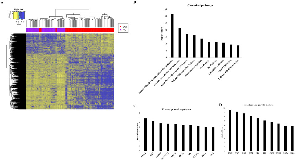
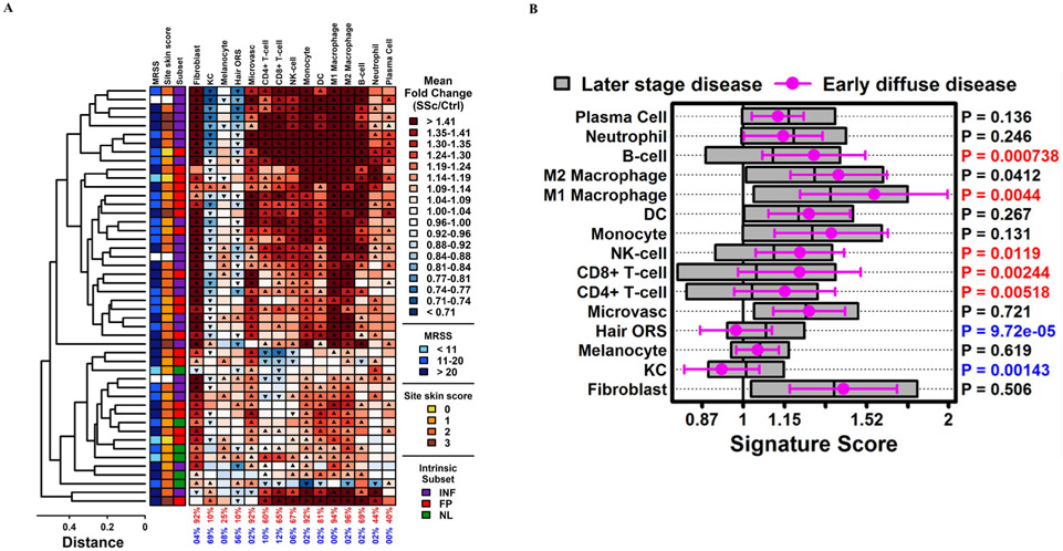
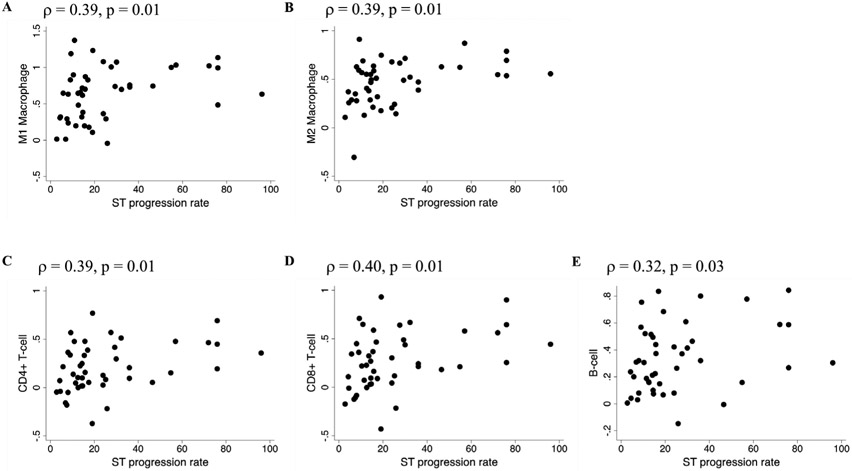
Results: SSc patients in PRESS had a high prevalence of M2 (96%) and M1 (94%) macrophage and CD8 T cell (65%), CD4 T cell (60%) and B cell (69%) signatures. Immunohistochemical staining of immune cell markers correlated with the gene expression-based immune cell signatures. The prevalence of immune cell signatures in early diffuse SSc patients was higher than in patients with longer disease duration. In the multivariable model, adaptive immune cell signatures were significantly associated with shorter disease duration, while fibroblast and macrophage cell type signatures were associated with higher modified Rodnan Skin Score (mRSS). Immune cell signatures also correlated with skin thickness progression rate prior to biopsy, but did not predict subsequent mRSS progression.
Conclusions: Skin in early diffuse SSc has prominent innate and adaptive immune cell signatures. As a prominently affected end organ, these signatures reflect the preceding rate of disease progression. These findings could have implications in understanding SSc pathogenesis and clinical trial design.
Keywords: autoimmunity; inflammation; systemic Sclerosis.
© Author(s) (or their employer(s)) 2020. No commercial re-use. See rights and permissions. Published by BMJ.
Conflict of interest statement
Competing interests: DK reports consultancy fees from Acceleron, Actelion, Bayer, Blade Therapeutics, BMS, Galapagos, Genentech/Roche, GSK, Mitsubishi Tanabi, Sanofi-A ventis/Genzyme, and UCB Pharma, and reports grants from Bayer, BMS, and Genentech/Roche outside the submitted work, and reports ownership interest in Eicos Sciences, Inc. WRS reports consulting fees from UT Health Science Center at Houston. JKG reports grants from Corbus Pharmaceuticals, Cumberland Pharmaceuticals and Eicos Pharmaceuticals outside the submitted work. AAS reports support from Sanofi outside the submitted work. MLW reports grants and personal fees from Celdara Medical, LLC and personal fees from Abbvie, Acceleron, BMS, Corbus Pharmaceuticals and Boehringer Ingelheim outside the submitted work. JLB reports grants from Hoffman La Roche and the Scleroderma Foundation outside the submitted work. SB and PB are employees of Boehringer Ingelheim. FVC reports personal fees from Boehringer Ingelheim outside the submitted work. EJB reports a grant from Pfizer and support from Bohringer Ingelheim, Corbus Pharmaceuticals and Eicos outside the submitted work. MDM reports personal fees from Medtelligence, Actelion Pharma, Astellas, Mitsubishi-Tanabe, and Boehringer Ingelheim and grants from Bayer, Reata, Sanofi, Corbus, Eicos and Boehringer Ingelheim outside the submitted work. SA reports personal fees from Boehringer Ingelheim and grants from Boehringer Ingelheim, Bayer and Momenta outside the submitted work.
Figures
References
-
- Denton CP, Khanna D. Systemic sclerosis. Lancet 2017;390:1685–99. - PubMed
-
- Varga J, Trojanowska M, Kuwana M. Pathogenesis of systemic sclerosis: recent insights of molecular and cellular mechanisms and therapeutic opportunities. J Scleroderma Relat Disord 2017;2:137–52.
Publication types
MeSH terms
Substances
Grants and funding
LinkOut - more resources
Full Text Sources
Other Literature Sources
Molecular Biology Databases
Research Materials
