. 2019 Dec;26(12):1132-1140.
doi: 10.1038/s41594-019-0331-x. Epub 2019 Nov 25.

Mechanism of ribosome stalling during translation of a poly(A) tail

Affiliations

Mechanism of ribosome stalling during translation of a poly(A) tail

Viswanathan Chandrasekaran et al. Nat Struct Mol Biol. 2019 Dec.

Abstract

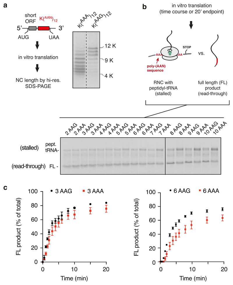
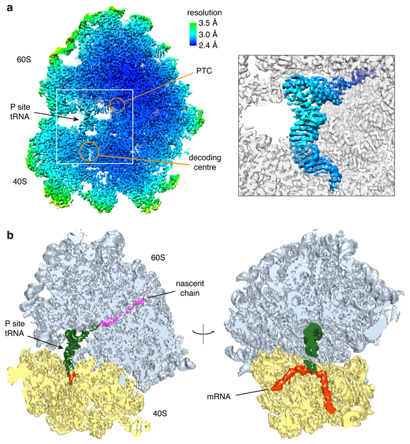
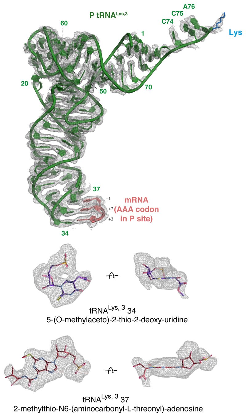
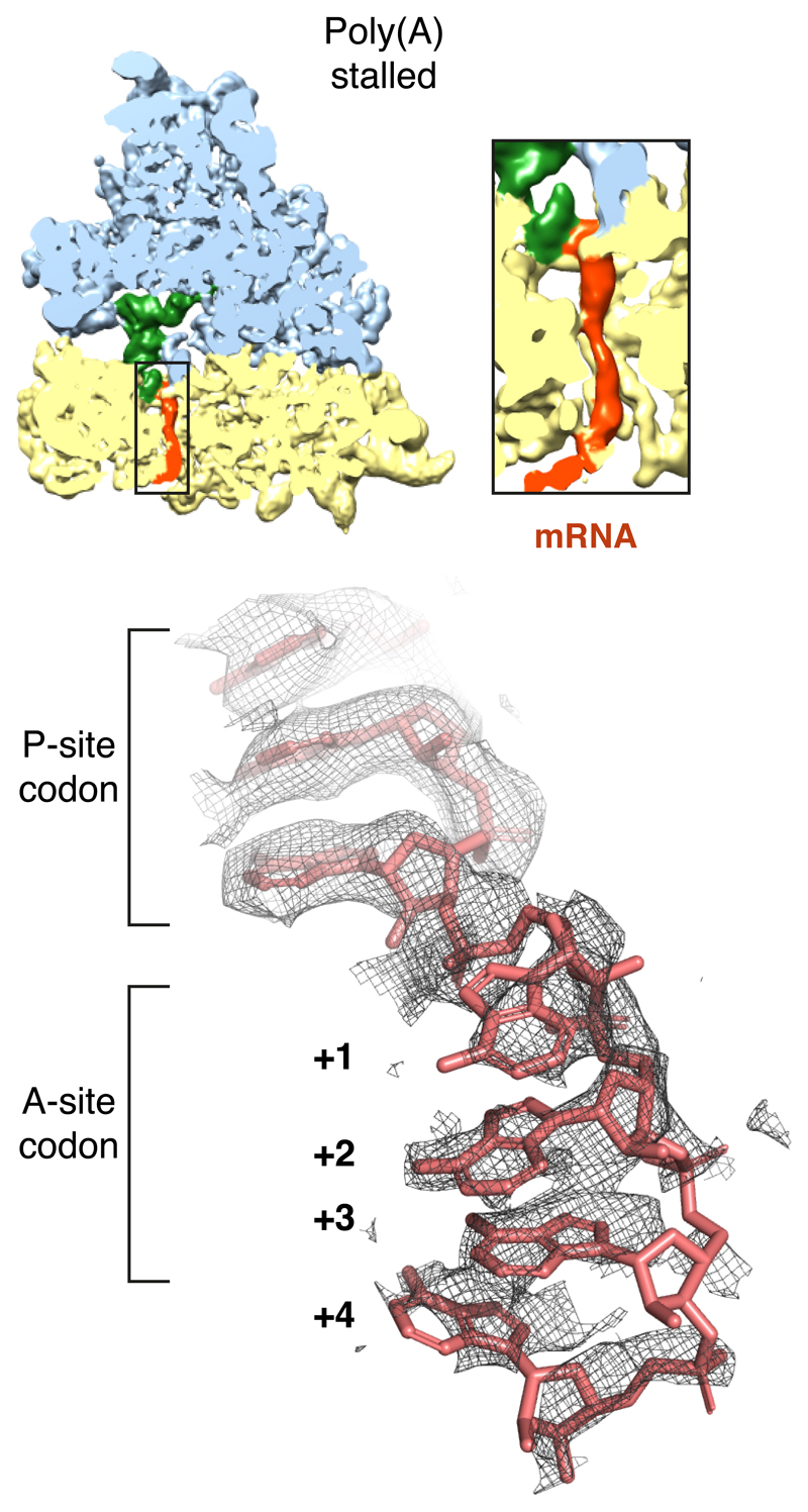
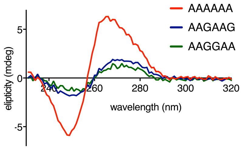
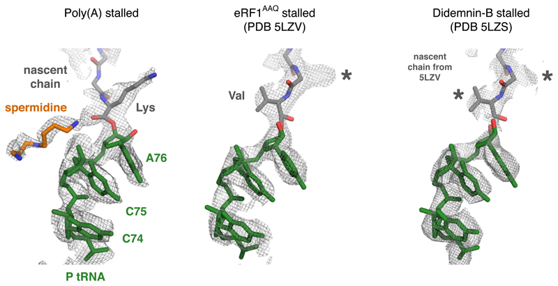
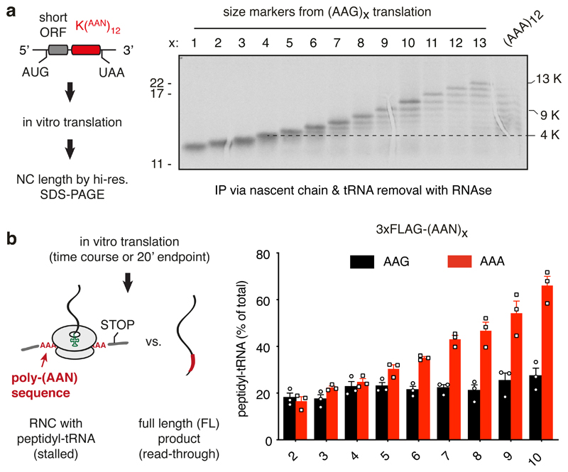
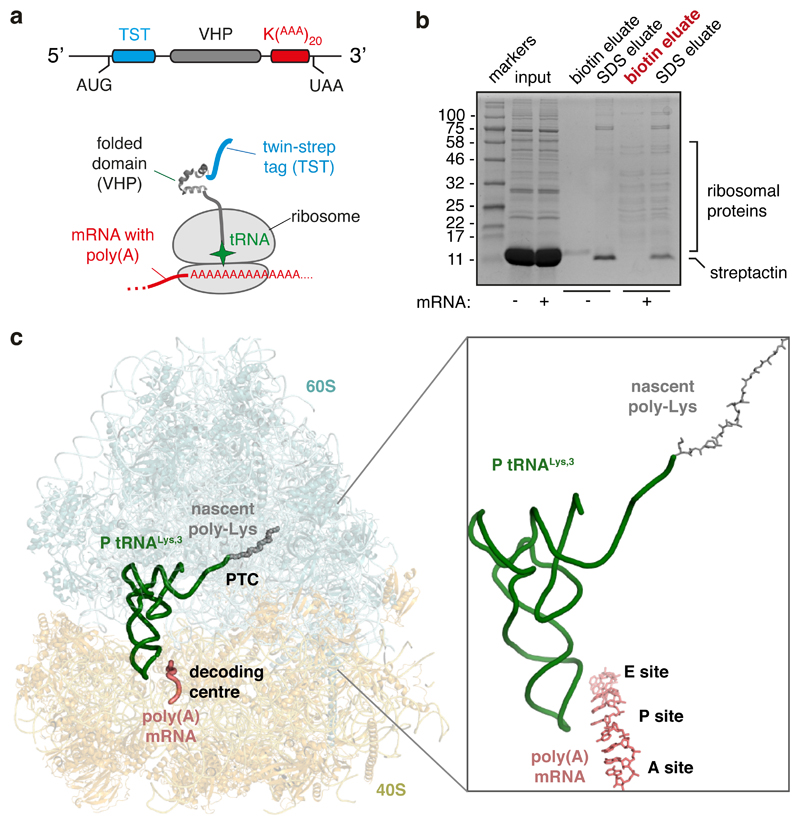
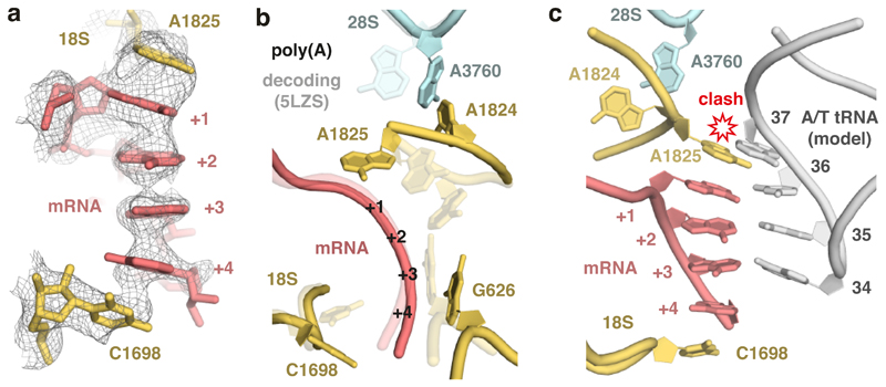
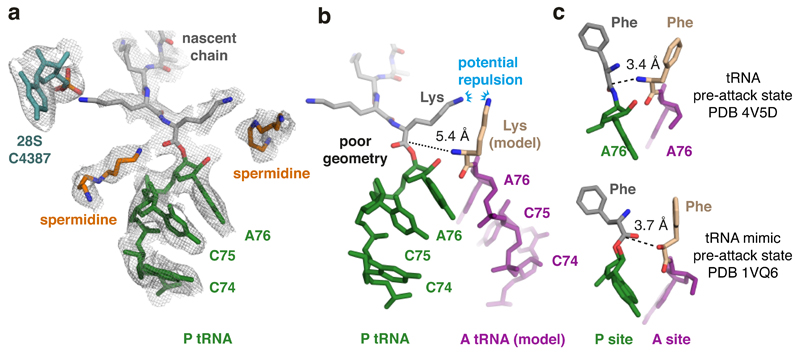
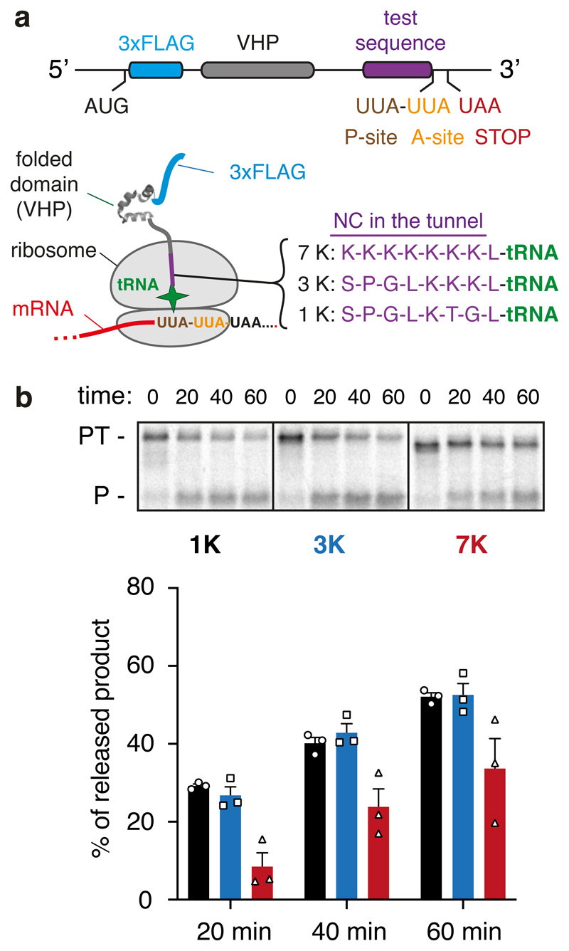
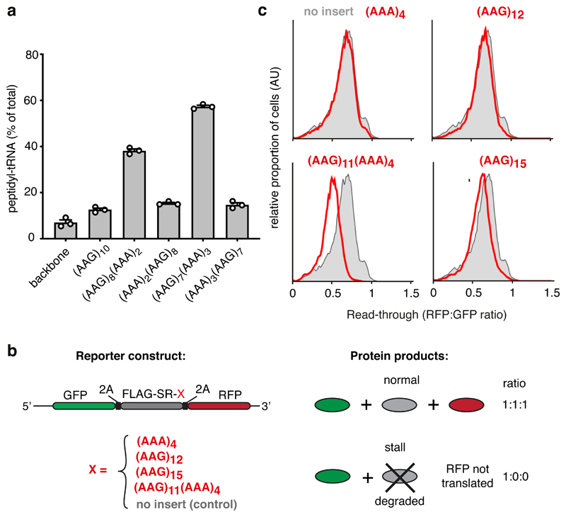
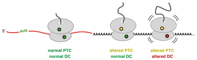
Faulty or damaged messenger RNAs are detected by the cell when translating ribosomes stall during elongation and trigger pathways of mRNA decay, nascent protein degradation and ribosome recycling. The most common mRNA defect in eukaryotes is probably inappropriate polyadenylation at near-cognate sites within the coding region. How ribosomes stall selectively when they encounter poly(A) is unclear. Here, we use biochemical and structural approaches in mammalian systems to show that poly-lysine, encoded by poly(A), favors a peptidyl-transfer RNA conformation suboptimal for peptide bond formation. This conformation partially slows elongation, permitting poly(A) mRNA in the ribosome's decoding center to adopt a ribosomal RNA-stabilized single-stranded helix. The reconfigured decoding center clashes with incoming aminoacyl-tRNA, thereby precluding elongation. Thus, coincidence detection of poly-lysine in the exit tunnel and poly(A) in the decoding center allows ribosomes to detect aberrant mRNAs selectively, stall elongation and trigger downstream quality control pathways essential for cellular homeostasis.


Conflict of interest statement

Competing Interests Statement: The authors declare no competing interests.

Figures

Extended Data Fig. 1
Extended Data Fig. 1. Additional characterization of ribosome stalling in vitro.
(A) A second example of nascent chain products resulting from in vitro translation of iterated AAG or AAA lysine codons in human cell lysate, as in Fig. 1A. The positions of nascent chain products containing 4, 9, or 12 lysines are indicated. (B) Analysis of iterated AAG versus AAA codons for stalling in rabbit reticulocyte lysate. The translation reaction was performed for 20 min after which the proportion of stalled products was assessed by the relative amounts of peptidyl-tRNA versus full length polypeptide. The ‘background’ of ~20% peptidyl-tRNA even in the absence of stalling is due to failed termination at the stop codon, which is located within a few nucleotides of the 3’ end of the mRNA. Later in vitro stalling experiments with a longer 3’UTR that protrudes outside the mRNA channel showed improved termination efficiency (~95%). An overly short 3′UTR presumably makes the mRNA more flexible in the mRNA channel and less able to recruit eRF1. Multiple experiments such as this one were quantified to produce the graph shown in Fig. 1B. (C) Time course of the appearance of full length (FL) product for constructs containing the indicated number of iterated AAG or AAA codons. Translation was synchronised by first pausing the ribosome at a run of rare leucine codons just preceding the poly-basic encoding sequence, then restarting translation at time 0 by addition of tRNA. The mean ± SEM for each time point calculated from two experiments are plotted.
Extended Data Fig. 2
Extended Data Fig. 2. Cryo-EM analysis of ribosomes stalled on poly(A).
(A) Representative micrograph of poly(A)-stalled ribosomes used for single particle analysis. (B) Data processing scheme used for structure determination in Relion 3.0. 3D classification reveals that ~90% of active ribosomes are in the canonical state with P/P tRNA while ~10% are seen in the rotated state with A/P and P/E hybrid state tRNAs. The majority of the rotated state ribosomes also contain density for a preceding ribosome and therefore represent ribosomes that have collided with a poly(A)-stalled ribosome. (C) Fourier shell correlation (FSC) curve of the final map illustrating an overall resolution of 2.8 Å.
Extended Data Fig. 3
Extended Data Fig. 3. Characterization of cryo-EM map.
(A) Local resolution of the poly(A)-stalled ribosome sliced through the centre. The positions of key elements are indicated. PTC: peptidyl-transferase centre. Inset (right) highlights the high local resolution at the PTC and decoding centre (B) Slices through the density map at the plane of the polypeptide exit tunnel (left) and mRNA channel (right). Continuous nascent chain density corresponding to a mixture of poly-Lys lengths and Cα positions is contoured at a different level to the rest of the map and is shown in magenta, and mRNA density is shown in red. The P site tRNA is green, 40S subunit in yellow, and 60S subunit in light blue.
Extended Data Fig. 4
Extended Data Fig. 4. Experimental EM density for P-site Lys-tRNALys,3.
Map-to-model fits for the P-site Lys-tRNALys,3 with the AAA codon of the mRNA in the P-site and the first nascent chain side chain (Lysine). Base modifications at positions 34 and 37 of the tRNA are shown within the EM density.
Extended Data Fig. 5
Extended Data Fig. 5. Views of the mRNA density in the EM map of the poly(A)-stalled ribosome.
The density map is sliced through the ribosome in a plane that reveals the decoding centre and shows the mRNA within the small subunit. The large and small subunits (blue and yellow, respectively), P-site tRNA (green) and mRNA (red) are colored. The inset shows a zoomed in region of the mRNA channel, illustrating that the poly(A) mRNA is ordered through most of the channel. The bottom panel shows the mRNA density in the P- and A-sites in the final refined and sharpened map. The mRNA is well ordered in the P-site due to base-pairing with the P-site tRNA, and ordered in the A-site due to stabilizing interactions with rRNA as shown in Fig. 3.
Extended Data Fig. 6
Extended Data Fig. 6. Guanosine interrupts the intrinsic helical propensity of poly(A).
Circular dichroism (CD) spectra of AAAAAA (red), AAGAAG (blue) and AAGGAA (green) RNA oligonucleotides are plotted. These spectra are averaged from 9 independent measurements performed on the same samples. The AAAAAA oligo displays a CD signature characteristic for the helical conformation of poly(A), as described previously . Introduction of guanosines significantly disrupts this helical structure.
Extended Data Fig. 7
Extended Data Fig. 7. Comparison of peptidyl-tRNA geometry in different mammalian RNC structures.
Shown are the EM density maps for the peptidyl-tRNA region at the PTC for the indicated structures. The fitted models are shown for the poly(A)-stalled ribosome and the RNC stalled at the stop codon with a dominant-negative eRF1AAQ mutant (PDB code 5LZV). The 5LZV RNC is in a geometry competent for peptidyl-transfer (or in this case, peptide release by eRF1). The structure from the didemnin-B stalled RNCs contains a mixture of nascent chains stalled at different positions. Thus, the nascent chain density represents an average of a variety of peptidyl-tRNAs. Note that the nascent chain model from 5LZV fits well into the density map, indicating that the majority of peptidyl-tRNAs assume this configuration during active elongation. The geometry for the poly(A) peptidyl-tRNA is unambiguously different from this optimal geometry. Lys and Val refer to the lysine and valine side chains of modeled nascent chains. The asterisks indicate density for side chains that are not shown.
Fig. 1
Fig. 1. Reconstitution of ribosome stalling on poly(A) mRNA in vitro.
(A) Analysis of nascent chain products resulting from in vitro translation of iterated AAG or AAA lysine codons in human cell lysate. Uncropped gel image is available online (Source Data). (B) Analysis of iterated AAG versus AAA codons for stalling in rabbit reticulocyte lysate. The translation reaction was performed for 20 min after which the proportion of stalled products was assessed by the relative amounts of peptidyl-tRNA versus full length polypeptide. The mean ± SEM (n=3) is plotted together with individual data points from independent experiments. The ‘background’ of ~20% peptidyl-tRNA even in the absence of stalling is due to failed termination at the stop codon, which is located within a few nucleotides of the 3’ end of the mRNA. Later in vitro stalling experiments with a longer 3’UTR that protrudes outside the mRNA channel (e.g., Fig. 6a) showed improved termination efficiency (~95%). An overly short 3’UTR presumably makes the mRNA more flexible in the mRNA channel and less able to recruit eRF1.
Fig. 2
Fig. 2. Structure of the ribosome stalled during poly(A) translation.
(A) Schematic of the construct and ribosome-nascent chain complex (RNC) used for structure determination. (B) Characterization of the RNC purification strategy for structure determination. Rabbit reticulocyte lysate translation reactions containing or lacking the mRNA depicted in panel A were subjected to affinity purification via the N-terminal Twin-Strep-tag and eluted sequentially with biotin and SDS. Aliquots of the total translation reaction (input) and 32-fold excess of each eluate were analyzed by SDS-PAGE and staining with Coomassie blue. The sample subjected to cryo-EM analysis is indicated in red. (C) Overview of the poly(A)-stalled RNC structure with key elements indicated in the inset.
Fig. 3
Fig. 3. Poly(A)-induced decoding center rearrangement.
(A) The ribosome A-site containing the poly(A) single-stranded helix (pink), capped by stacking interactions with 18S rRNA bases (yellow), is shown fitted within the EM density map (mesh). (B) Comparison of the decoding center configurations in the poly(A)-stalled ribosome (solid model) and the ribosome trapped during decoding (PDB 5LZS; transparent model). The mRNA bases and tRNA (in the case of decoding) are omitted for clarity. (C) Superimposed models of the poly(A)-stalled ribosome and the A/T tRNA (grey) positioned as it would be during decoding (PDB 5LZS). Base 37 of the tRNA clashes with 18S rRNA base A1825, which cannot move to its ‘flipped in’ position because A3760 of 28S rRNA occupies this space. Base modifications would be present at position 37 (and 34) when the incoming tRNA is the cognate Lys-tRNALys,3, but this is not shown for clarity.
Fig. 4
Fig. 4. Peptidyl-tRNA is mis-positioned in the poly(A) stalled ribosome.
(A) The P-site tRNA (green), attached nascent chain with the first three lysines (grey), and 28S rRNA residue C4387, which interacts with the penultimate Lys side chain, are shown fitted within the EM density map (mesh). Putative spermidine molecules (orange), hundreds of which are thought to bind ribosomes and facilitate translation,, were modeled into otherwise unaccounted density. (B) Superimposed models of the peptidyl-tRNA in the poly(A)-stalled ribosome with the A-site tRNA positioned for peptidyl transfer (PDB 4V5D). The amino acid in this structure (phenylalanine) was replaced with lysine to model the situation during poly(A) translation. The proximal lysine of the nascent chain faces the lysine of the accommodating A-site tRNA, resulting in potential charge repulsion between their respective epsilon amines. The backbone geometry of the nascent chain attachment to A76 is suboptimal for peptidyl transfer, as highlighted by the 5.4 Å distance between the α-amino group of the aminoacyl tRNA and the incorrectly oriented backbone carbonyl of the peptidyl tRNA. (C) Shown are models depicting the P- and A-site amino acids attached to the P- and A-site nucleotide (A76) for the indicated structures. Note that in both pre-attack structures, the attacking atom is within 4 Å of the P-site target bond, unlike the 5.4 Å distance in the poly(A)-stalled ribosome.
Fig. 5
Fig. 5. Analysis of PTC geometry by puromycin reactivity.
(A) Experimental strategy. A construct was designed to cause ribosome stalling at the second of two rare UUA leucine codons (see Methods). This RNC is known to be functional because translation resumes when liver tRNA is added. RNCs were produced using this strategy in which one of three test sequences (7K, 3K, and 1K, as indicated) are positioned inside the ribosome tunnel. The lysine residues were encoded with AAG codons to minimize stalling before the desired UUA codon. After the translation reaction to synchronise ribosomes at the rare codon stall, the sample was moved to ice and the salt concentration was increased to 500 mM to prevent splitting of ribosomal subunits by rescue factors. Puromycin was then added to 2 μM final concentration and polypeptide release from tRNA was judged by SDS-PAGE. (B) Stalled RNCs containing the 1K, 3K, and 7K test sequences were evaluated for their reactivity to puromycin over a 60 min time course as a measure of peptidyl-transfer capacity. A representative experiment out of three is shown in the top panel, with the bottom panel showing a graph depicting the mean ± SEM (n=3) and individual data points from independent experiments. The positions of peptidyl-tRNA (PT) and puromycin-released peptide (P) are indicated to the left of the gel. Uncropped gel image is available online (Source Data).
Fig. 6
Fig. 6. Coincidence detection of the nascent chain and mRNA mediate stalling.
(A) Iterated combinations of AAG and AAA codons inserted near the end of an open reading frame were analyzed for stalling by in vitro translation. The proportion of stalled product was assessed by the relative amount of peptidyl-tRNA versus full length terminated polypeptide and plotted as % peptidyl-tRNA. A construct lacking the AAG/AAA insert is shown for comparison (‘backbone’). The mean ± SEM (n=3) together with individual data points from independent experiments are plotted. (B) Schematic diagram of the dual-color reporter construct for detection of terminal stalling by flow cytometry. “2A” indicates the viral P2A sequence which causes skipping of peptide bond formation without interrupting elongation. If a test sequence inserted downstream of the FLAG-SR region causes terminal stalling, production of GFP is unaffected while RFP is not produced. Thus, the RFP:GFP ratio will be less than 1 and serves as a quantitative measure of terminal stalling. (C) Iterated combinations of AAG and AAA codons were analyzed for stalling in HEK293T cells using a dual color reporter in which the test sequence of interest (red text) is inserted between an N-terminal GFP and C-terminal RFP. Shown are histograms from flow cytometry analysis of the RFP:GFP ratio as a measure of ribosome read-through of the test sequence (red traces) relative to that seen in the absence of an insert (grey trace). Gating strategy for the histogram is shown in Source Data online.
Fig. 7
Fig. 7. Coincidence detection model for ribosome stalling on poly(A).
Translation of the first few AAA codons progressively slows elongation due to lysine interactions within the exit tunnel favoring a peptidyl-tRNA geometry that is suboptimal for peptide bond formation. In the context of slow peptide bond formation, the decoding center has greater opportunities for rearrangement, which further slows elongation and can cause the ribosome to stall. PTC and DC refer to peptidyl-transferase center and decoding center, respectively.

References

    1. Wolff S, Weissman JS, Dillin A. Differential scales of protein quality control. Cell. 2014;157:52–64. - PubMed
    1. Balchin D, Hayer-Hartl M, Hartl FU. In vivo aspects of protein folding and quality control. Science (80-. ) 2016;353 - PubMed
    1. Labbadia J, Morimoto RI. The Biology of Proteostasis in Aging and Disease. Annu Rev Biochem. 2015;84:435–464. - PMC - PubMed
    1. Kurosaki T, Popp MW, Maquat LE. Quality and quantity control of gene expression by nonsense-mediated mRNA decay. Nat Rev Mol Cell Biol. 2019;20:406–420. - PMC - PubMed
    1. van Hoof A, Wagner EJ. A brief survey of mRNA surveillance. Trends Biochem Sci. 2011;36:585–592. - PMC - PubMed

Publication types

MeSH terms