. 2020 May;41(5):638-649.
doi: 10.1038/s41401-019-0320-y. Epub 2019 Nov 25.

Zinc protects against cadmium-induced toxicity in neonatal murine engineered cardiac tissues via metallothionein-dependent and independent mechanisms

Affiliations

Zinc protects against cadmium-induced toxicity in neonatal murine engineered cardiac tissues via metallothionein-dependent and independent mechanisms

Hai-Tao Yu et al. Acta Pharmacol Sin. 2020 May.

Abstract

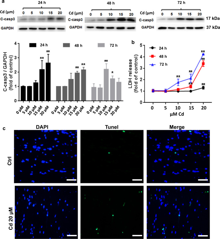
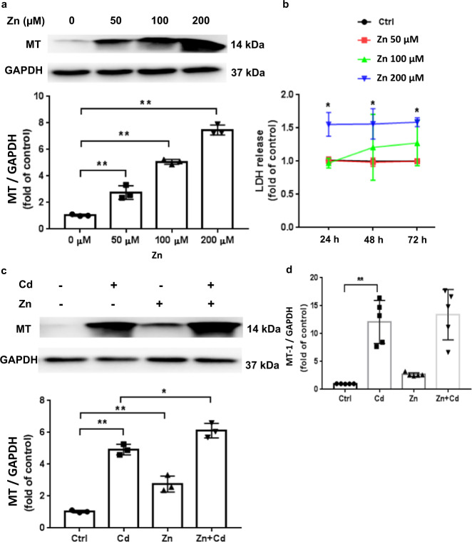
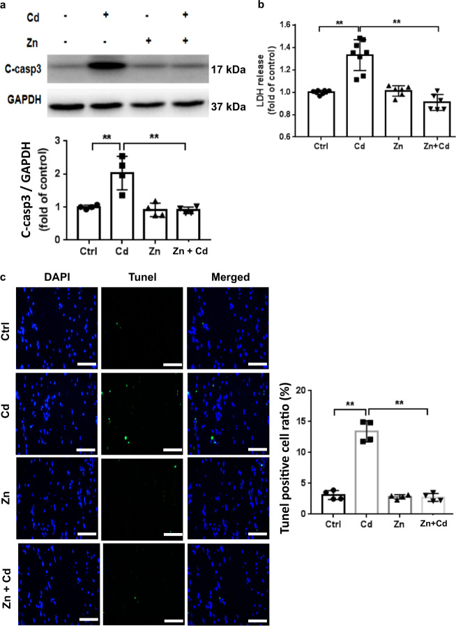
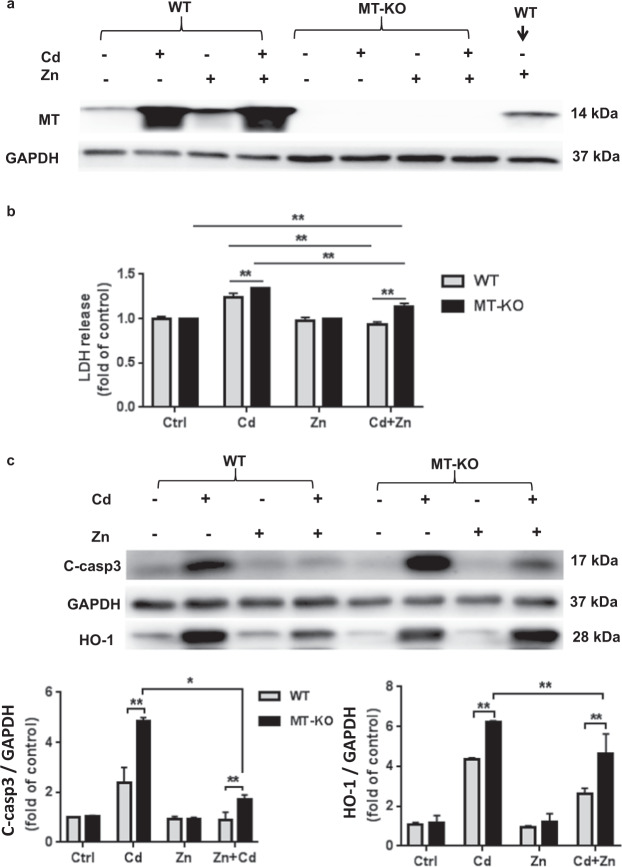
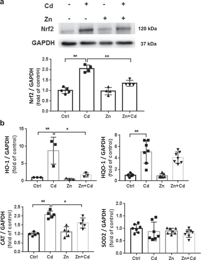
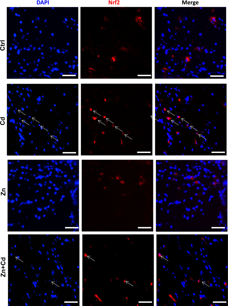
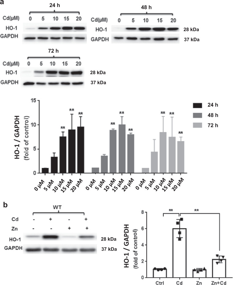
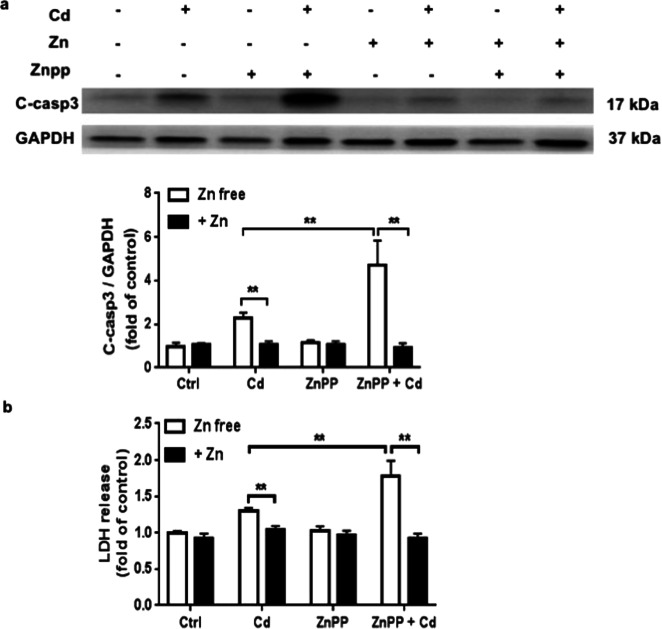
Cadmium (Cd) is a nonessential heavy metal and a prevalent environmental toxin that has been shown to induce significant cardiomyocyte apoptosis in neonatal murine engineered cardiac tissues (ECTs). In contrast, zinc (Zn) is a potent metallothionein (MT) inducer, which plays an important role in protection against Cd toxicity. In this study, we investigated the protective effects of Zn against Cd toxicity in ECTs and explore the underlying mechanisms. ECTs were constructed from neonatal ventricular cells of wild-type (WT) mice and mice with global MT gene deletion (MT-KO). In WT-ECTs, Cd (5-20 μM) caused a dose-dependent toxicity that was detected within 8 h evidenced by suppressed beating, apoptosis, and LDH release; Zn (50-200 μM) dose-dependently induced MT expression in ECTs without causing ECT toxicity; co-treatment of ECT with Zn (50 µM) prevented Cd-induced toxicity. In MT-KO ECTs, Cd toxicity was enhanced; but unexpectedly, cotreatment with Zn provided partial protection against Cd toxicity. Furthermore, Cd, but not Zn, significantly activated Nrf2 and its downstream targets, including HO-1; inhibition of HO-1 by a specific HO-1 inhibitor, ZnPP (10 µM), significantly increased Cd-induced toxicity, but did not inhibit Zn protection against Cd injury, suggesting that Nrf2-mediated HO-1 activation was not required for Zn protective effect. Finally, the ability of Zn to reduce Cd uptake provided an additional MT-independent mechanism for reducing Cd toxicity. Thus, Zn exerts protective effects against Cd toxicity for murine ECTs that are partially MT-mediated. Further studies are required to translate these findings towards clinical trials.

Keywords: Nrf2; Zinc; ZnPP; cadmium toxicity; engineered cardiac tissue; heme oxygenase-1; metallothionein.


Figures

Fig. 1
Fig. 1. Dose and time response of toxic Cd effect on murine ECTs.
a Expression of cleaved caspase 3 after increasing Cd concentration (0, 5, 10, 15, 20 µM) for increasing duration (24, 48, 72 h). Group sizes ranged from n = 3–8 per time point. b LDH release after increasing Cd concentration (0, 5,10, 15, 20 µM) for increasing duration (24, 48, 72 h). Group sizes ranged from n = 3–8 per time point. c Representative ECT immunofluorescent staining for terminal transferase dUTP nick end labeling assay (Tunel, green) and nuclei (DAPI, blue) following 24 h 20 μM Cd exposure (×40 magnification, scale bar = 50 μm). *P < 0.05 and **P < 0.01 vs. corresponding Ctrl ECT for the corresponding treatment duration.
Fig. 2
Fig. 2. Zn and Cd effects on ECT MT expression.
a MT expression after increasing Zn dose (50, 100, 200 µM) for 24 h. Group size is n = 3 per dose. b LDH release after increasing Zn dose (50, 100, 200 µM) for different durations (24; 48; 72 h). Group size is n = 3 per time point. c Effect of Zn (50 µM) and/or Cd (20 µM) on ECT MT protein content. Group size is n = 3 per group. d Effect of Zn (50 µM) and/or Cd (20 µM) on MT-1 gene expression (qPCR). Group size is n = 5 per group. *P < 0.05 and **P < 0.01 vs. corresponding Ctrl ECT.
Fig. 3
Fig. 3. Zn reduces ECT Cd toxicity.
a Cleaved caspase 3 expression following Zn (50 µM) and/or Cd (20 µM) for 24 h. Group size is n = 4 per group. b LDH release following Zn (50 µM) and/or Cd (20 µM) for 24 h. Group size ranged from n = 6–8 per group. c Representative ECT immunofluorescent staining for terminal transferase dUTP nick end labeling assay (Tunel, green) and nuclei (DAPI, blue) and summary Tunel data for each group. Group size is n = 4 per group. ×40 magnification, Scale bar = 50 μm, **P < 0.01 vs. corresponding groups.
Fig. 4
Fig. 4. MT plays a role in Zn protection from ECT Cd toxicity.
a MT expression in WT and in MT-KO ECTs following Zn (50 µM) and/or Cd (20 µM) for 24 h. b LDH release into media following Zn (50 µM) and/or Cd (20 µM) for 24 h. Group size ranged from n = 6–8 per group. c Cleaved caspase 3 and HO-1 expression in WT and MT-KO ECT in each treatment group (Ctrl; Cd; Zn; Zn + Cd). Group size is n = 4 per group. *P < 0.05 and **P < 0.01 vs. corresponding Ctrl ECT.
Fig. 5
Fig. 5. Cd and Zn induce ECT Nrf2 and downstream genes.
a ECT Nrf2 following Zn (50 µM) and/or Cd (20 µM) for 24 h. Group size is n = 4 per group. b qPCR results of Nrf2 downstream genes (HO-1, NQO-1, CAT, SOD2) following Zn (50 µM) and/or Cd (20 µM) for 24 h. Group size ranged from n = 3–7 per group. *P < 0.05 vs. corresponding groups. **P < 0.01 vs. corresponding groups.
Fig. 6
Fig. 6. Evidence of ECT Nrf2 activation after Cd and/or Zn treatment.
Representative ECT stained for Nrf2 (Red) and nuclei (DAPI, blue) following Zn (50 µM) and/or Cd (20 µM) for 24 h. Arrows indicate representative positive cells in which Nrf2 translocated into nucleus, ×40 magnification, scale bar = 50 μm.
Fig. 7
Fig. 7. Cd and/or Zn alter ECT HO-1 expression.
a ECT HO-1 expression after increasing Cd concentration (0, 5, 10, 15, 20 µM) for increasing duration (24, 48, 72 h). Group size ranged from n = 3–8 per time point. **P < 0.01 vs. corresponding Ctrl group. b ECT HO-1 expression following Zn (50 µM) and/or Cd (20 µM) for 24 h. (Ctrl, Cd, Zn, Zn + Cd). Group size is n = 4 per group. **P < 0.01 vs. corresponding group.
Fig. 8
Fig. 8. The HO-1 inhibitor ZnPP increases ECT Cd toxicity.
a Cleaved caspase 3 following treatment with Zn (50 µM) and/or Cd (20 µM) and/or ZnPP (10 µM) for 24 h by WB. Group sizes ranged from n = 3–4 per group. **P < 0.01 vs. corresponding group. b LDH release following treatment with Zn (50 µM) and/or Cd (20 µM) and/or ZnPP (10 µM) for 24 h. Group sizes ranged from n = 3–4 per group. **P < 0.01 vs. corresponding group.

References

    1. Thevenod F, Lee WK. Cadmium and cellular signaling cascades: interactions between cell death and survival pathways. Arch Toxicol. 2013;87:1743–86. - PubMed
    1. Kawakami T, Nishiyama K, Kadota Y, Sato M, Inoue M, Suzuki S. Cadmium modulates adipocyte functions in metallothionein-null mice. Toxicol Appl Pharmacol. 2013;272:625–36. - PubMed
    1. Shen J, Wang X, Zhou D, Li T, Tang L, Gong T, et al. Modelling cadmium-induced cardiotoxicity using human pluripotent stem cell-derived cardiomyocytes. J Cell Mol Med. 2018;22:4221–35. - PMC - PubMed
    1. Yu H, Ye F, Yuan F, Cai L, Ji H, Keller BB. Neonatal murine engineered cardiac tissue toxicology model: Impact of metallothionein overexpression on cadmium-Induced Injury. Toxicol Sci. 2018;165:499–511. - PubMed
    1. Caciari T, Sancini A, Fioravanti M, Capozzella A, Casale T, Montuori L, et al. Cadmium and hypertension in exposed workers: a meta-analysis. Int J Occup Med Environ Health. 2013;26:440–56. - PubMed