. 2019 Dec;25(12):1839-1842.
doi: 10.1038/s41591-019-0653-6. Epub 2019 Nov 25.

Activating mutations in CSF1R and additional receptor tyrosine kinases in histiocytic neoplasms

Affiliations

Activating mutations in CSF1R and additional receptor tyrosine kinases in histiocytic neoplasms

Benjamin H Durham et al. Nat Med. 2019 Dec.

Abstract

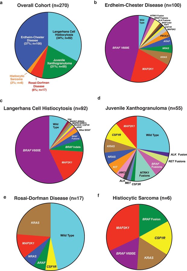
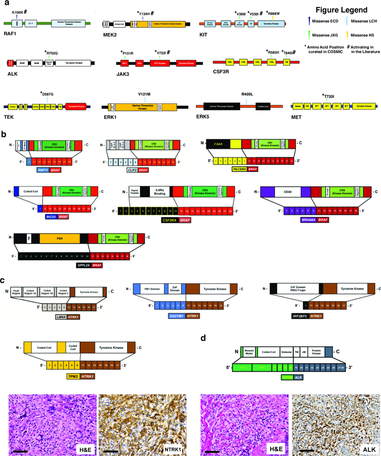
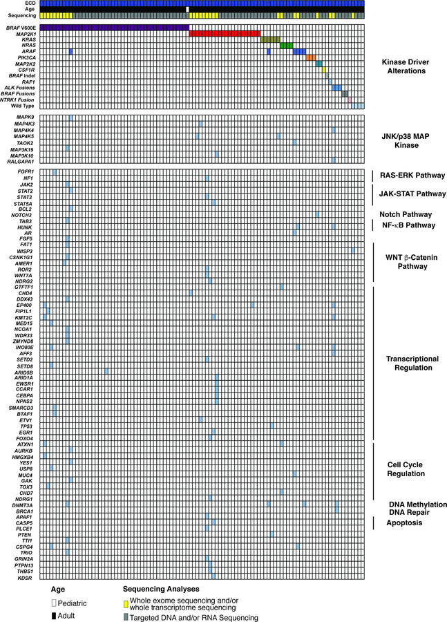
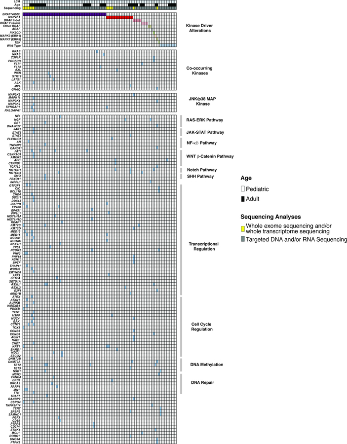
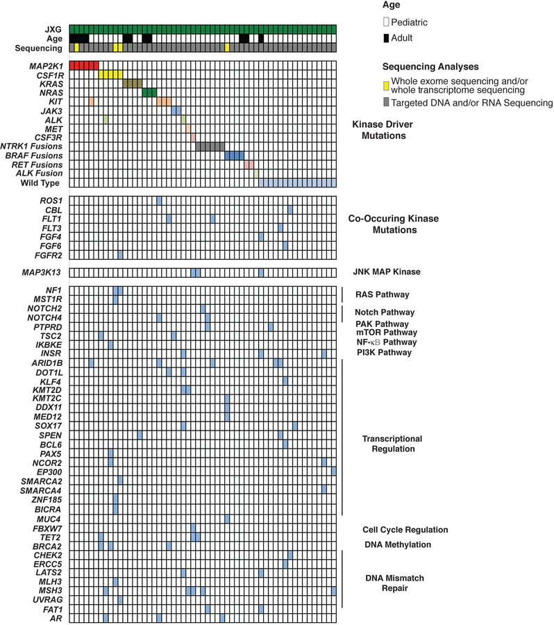
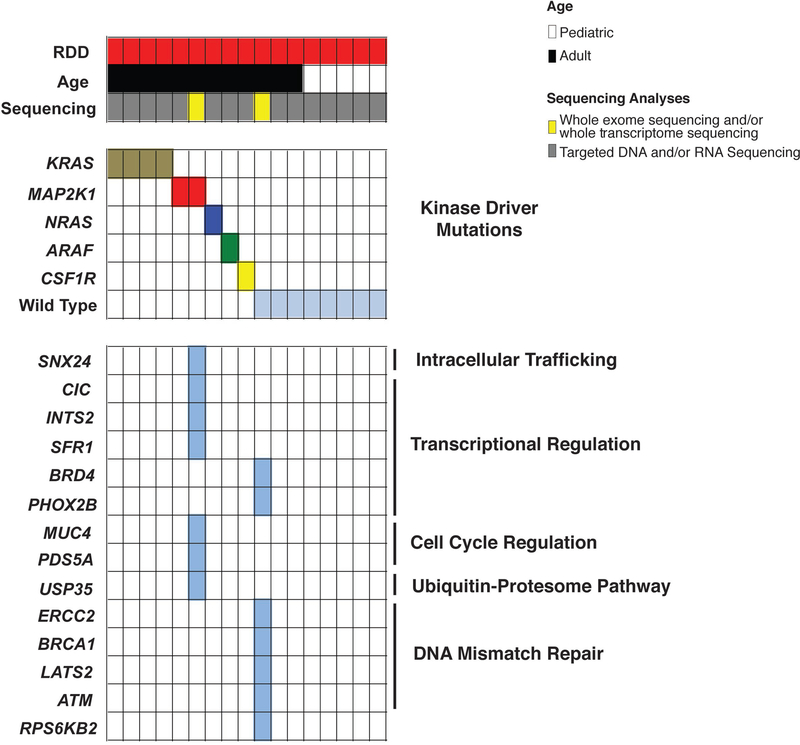
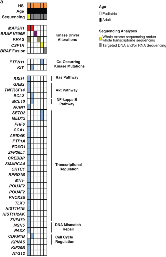
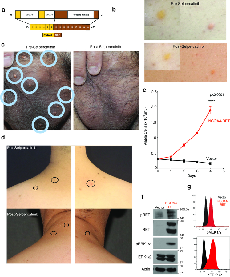
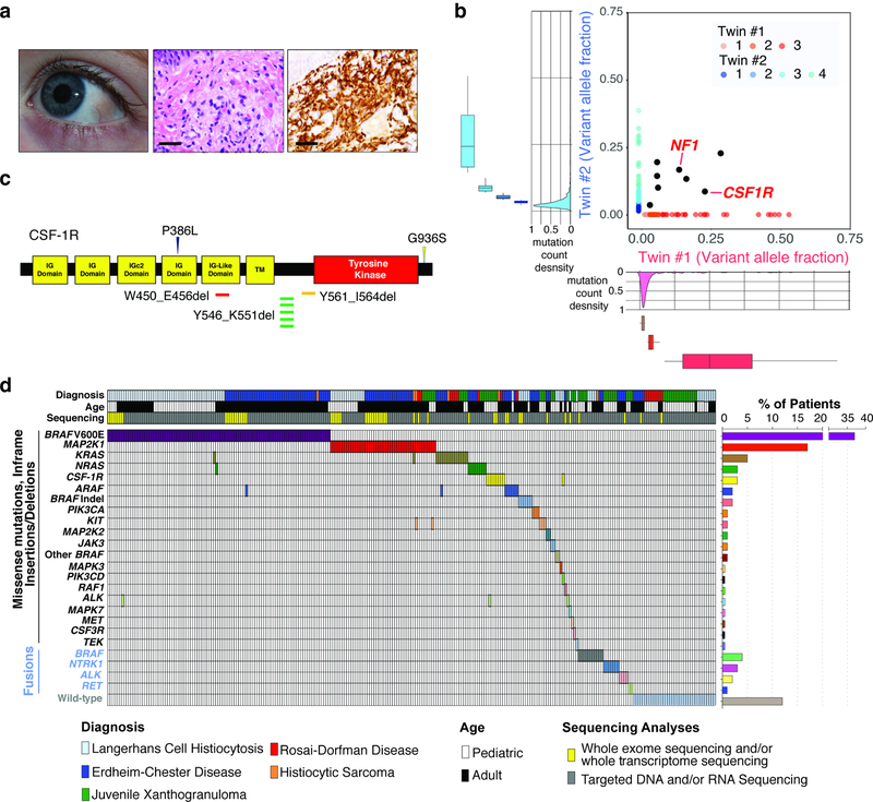
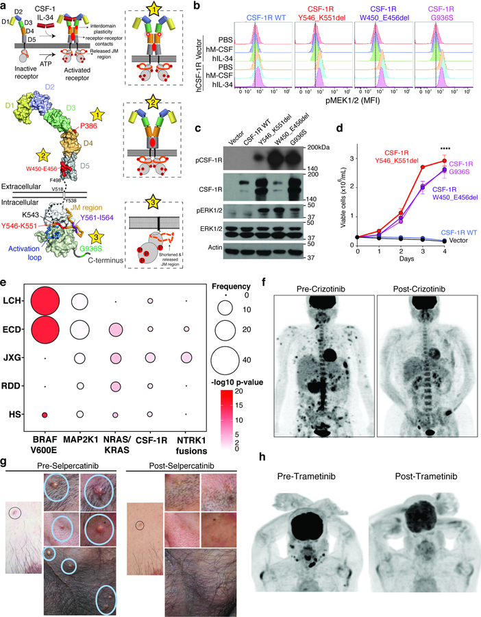
Histiocytoses are clonal hematopoietic disorders frequently driven by mutations mapping to the BRAF and MEK1 and MEK2 kinases. Currently, however, the developmental origins of histiocytoses in patients are not well understood, and clinically meaningful therapeutic targets outside of BRAF and MEK are undefined. In this study, we uncovered activating mutations in CSF1R and rearrangements in RET and ALK that conferred dramatic responses to selective inhibition of RET (selpercatinib) and crizotinib, respectively, in patients with histiocytosis.


Conflict of interest statement

Competing financial interests

The authors declare the following competing interests: B.H.D.: grants from National Cancer Institute, American Society of Hematology, and Erdheim-Chester Disease Global Alliance Foundation. A.Y.: grants from the Leukemia & Lymphoma Society, Aplastic Anemia & MDS International Foundation, and the Lauri Strauss Leukemia Foundation. G.A.U.: personal fees from Sanofi and grants from Sanofi, Novartis, Genentech. M. Ladanyi: advisory board compensation from Merck, AstraZeneca, BristolMyers Squibb, Takeda, and Bayer, and research support from LOXO Oncology and Helsinn Healthcare. D.B.S.: served as a consultant and received honoraria from Pfizer, Loxo Oncology, Lilly Oncology, Vivideon Therapeutics and Illumina. Stock options from Loxo Oncology. M.F.B.: personal fees from Roche and research support from Illumina. D.M.H.: personal fees from Atara Biotherapeutics, Chugai Pharma, Boehringer Ingelheim, AstraZeneca, Pfizer, Bayer, Debiopharm Group, and Genentech, and grants from National Cancer Institute, AstraZeneca, Puma Biotechnology, and Loxo Oncology. I.D.: served as a consultant for Apexigen, Bayer, and Celgene and research support from BMS and Novartis. S.N.S.: program grants from the Flanders Institute for Biotechnology, Belgium (VIB) and the special research fund of Ghent University, and grants for Research Foundation Flanders (FWO). M.E.L.: personal fees from LOXO, AstraZeneca, Roche/Genentech, Novartis. AD: Research fuding from Foundation Medicine and personal fees from Ignyta/Genentech/Roche, Loxo/Bayer/Lilly, Takeda/Ariad/Millenium, TP Therapeutics, AstraZeneca, Pfizer, Blueprint Medicines, Helsinn, Beigene, BergenBio, Hengrui Therapeutics, Exelixis, Tyra Biosciences, Verastem, and MORE Health. E.L.D.: grants from Erdheim-Chester Disease Global Alliance, The Histiocytosis Association, the Society of Memorial Sloan Kettering, the Frame Fund, the Joy Family West Foundation, and the American Society for Clinical Oncology. O.A.-W.: grants from National Cancer Institute, National Heart Lung and Blood Institute, Pershing Square Sohn Foundation, the Histiocyte Society, and H3B Biomedicine, and personal fees from H3B Biomedicine, Foundation Medicine Inc., Merck, and Jansen unrelated to this manuscript. The remaining authors have nothing to disclose.

Figures

Extended Data Fig. 1.
Extended Data Fig. 1.. Pedigree and genomic analyses of monozygotic twins with CSF-1R mutant Juvenile Xanthogranuloma (JXG).
(a) Pedigree of twins with JXG. Twin 1 and 2 are monochorionic, diamniotic identical twins born to an otherwise healthy 35-year-old mother. At one year of age, both twins developed small skin lesions on their forehead, which over the course of four months grew in number and diameter extending to the scalp and upper chest. Scalp biopsies were consistent with a diagnosis of JXG. Ophthalmic examination of both twins also identified multiple, bilateral subconjunctival lesions compatible with JXG in twin 1 only. Both twins were monitored in our institution until 2.5 years-of-age with stable, persistent JXG with no visual compromise or neurologic signs or symptoms, and therefore no indication for therapy. Complete blood counts and comprehensive metabolic profiles of both twins have been within normal limits since birth. (b) Mutational profile of the JXG lesion from each twin displaying the fraction of mutations found in each trinucleotide context (performed using deconstructSigs). (c) Piechart showing the relative contribution of each mutational signature in the JXG lesion from (b) in each twin (based on the 30 mutational signatures detected by Sanger/COSMIC30). Signatures 6 and 15 are associated with defective DNA mismatch repair. (d) Histogram illustrating the fraction of indels among all mutations identified in the JXG lesions of twins 1 and 2 (red dashed lines) along with the distribution of indel fractions for TCGA samples (colorectal, endometrial, ovarian, stomach) with known microsatellite instability (MSI) status assayed from mononucleotide markers (as described previously29; MSI-H: high microsatellite instability; MSS: microsatellite stable). (e) Bar graph depicting proportion of histiocytosis samples that are MSS (90%; n=28/31) versus MSI-H (10%; n=3/31) based on WES.
Extended Data Fig. 2.
Extended Data Fig. 2.. Pie charts of histologic subsets of histiocytoses analyzed with kinase mutations identified in each subset.
(a) Numbers and percentage of patients with each histological subtype of histiocytosis sequenced in this study. Frequency of kinase mutations in the subset of patients with (b) Erdheim-Chester Disease, (c) Langerhans Cell Histiocytosis, (d) Juvenile Xanthogranuloma, (e) Rosai-Dorfman Disease, and (f) Histiocytic Sarcoma.
Extended Data Fig. 3.
Extended Data Fig. 3.. CSF-1R mutants are expressed on the cell surface and result in phosphorylation of CSF-1R.
(a) Representative anti-CSF-1R and GFP flow cytometry analysis of Ba/F3 cell expressions of human CSF-1R constructs from an MSCV-IRES-GFP construct. Experiments were performed with n=3 independent biological replicates. (b) Representative histograms of the median fluorescent intensity (MFI) of phospho-CSF-1R Tyrosine 723 intracellular flow cytometry in cells from (a) in cytokine depletion conditions following 60 minutes of stimulation with PBS, human M-CSF (hM-CSF; 100ng/mL), or hIL-34 (100ng/mL). Cells expressing empty vector (“vector”) are shown in the top three rows of each plot) and those expressing wild-type (WT) or mutant CSF-1R constructs are shown in the bottom three rows of each plot. Experiments were performed with n=3 independent biological experiments. (c) Bar graphs of IC50 values of Ba/F3 cells expressing an empty vector control or CSF-1R mutations in response to Pexidartinib (left) or BLZ945 (right). Mean values of n=3 biologically independent experiments ± standard deviation is shown. Calculation of p-values performed using ordinary one-way ANOVA; ****p<0.0001.
Extended Data Fig. 4.
Novel kinase mutations and fusions identified in patients with histiocytic neoplasms in this study. (a) Protein diagrams of novel, somatic point mutations uncovered in kinases in histiocytoses in this study, as well as whether they have been previously described as somatic in cancer (noted by an asterisk, “*”) and/or functionally characterized (noted by pound sign, “#”). Of the 14 mutations illustrated, the RAF1 K106N, MEK2 Y134H, JAK3 V722I31, KIT V530I32, and CSF3R T64033 mutations have been shown to be activating previously. Also, four co-existed with other kinase alterations in the following combinations: CSF3R R583H mutation co-occurred with CSF3R T640I in a JXG patient, KIT R888W mutation co-occurred with MEK1 F53L in a histiocytic sarcoma patient, KIT V530I mutations co-occurred with a MEK1 V93I mutation in a JXG patient. (b) BRAF fusions, (c) NTRK1 fusions, (d) and ALK fusions identified. Hematoxylin and eosin (H&E) and anti-NTRK1 and anti-ALK immunohistochemical staining shown in TPM3-NTRK1-rearranged juvenile xanthogranuloma and KIF5B-ALK-rearranged Erdheim-Chester Disease tumor biopsies, respectively.
Extended Data Fig. 5.
Extended Data Fig. 5.. Oncoprint of mutations identified in the Erdheim-Chester Disease cohort (n=100 patients).
Results of whole exome and targeted DNA and RNA sequencing of non-LCH neoplasms. Each patient is represented in one column. Diagnosis (ECD), age category, and sequencing method are in the first 3 rows. Somatic mutations identified are in the lower rows and subdivided based on mutations known to activate kinases, affect the JNK/p38 MAP kinase pathway, or involve a diverse array of co-occurring pathways (shown on the right).
Extended Data Fig. 6.
Extended Data Fig. 6.. Oncoprint of mutations identified in the Langerhans Cell Histiocytosis cohort (n=92 patients).
Results of whole exome and targeted DNA and RNA sequencing of LCH neoplasms. Each patient is represented in one column. Diagnosis (LCH), age category, and sequencing method are in the first 3 rows. Somatic mutations identified are in the lower rows and subdivided based on mutations known to activate kinases, affect the JNK/p38 MAP kinase pathway, or involve a diverse array of co-occurring pathways (shown on the right).
Extended Data Fig. 7.
Extended Data Fig. 7.. Oncoprint of mutations identified in the Juvenile Xanthogranuloma cohort (n=55 patients).
Results of whole exome and targeted DNA and RNA sequencing of non-LCH neoplasms. Each patient is represented in one column. Diagnosis (JXG), age category, and sequencing method are in the first 3 rows. Somatic mutations identified are in the lower rows and subdivided based on mutations known to activate kinases, affect the JNK/p38 MAP kinase pathway, or involve a diverse array of co-occurring pathways (shown on the right).
Extended Data Fig. 8.
Extended Data Fig. 8.. Oncoprint of mutations identified in the Rosai Dorfman Disease cohort (n=17 patients).
Results of whole exome and targeted DNA and RNA sequencing of non-LCH neoplasms. Each patient is represented in one column. Diagnosis (RDD), age category, and sequencing method are in the first 3 rows. Somatic mutations identified are in the lower rows and subdivided based on mutations known to activate kinases or involve a diverse array of co-occurring pathways (shown on the right).
Extended Data Fig. 9.
Extended Data Fig. 9.. Mutations identified in histiocytic sarcoma (HS) (n=6 patients).
Results of whole exome and targeted DNA and RNA sequencing of non-LCH neoplasms. Each patient is represented in one column. Diagnosis (HS), age category, and sequencing method are in the first 3 rows. Somatic mutations identified are in the lower rows and subdivided based on mutations known to activate kinases or involve a diverse array of co-occurring pathways (shown on the right).
Extended Data Fig. 10.
Extended Data Fig. 10.. Characteristics of RET fusion-driven histiocytosis and response to selpercatinib.
(a) Protein diagram of NCOA4-RET fusion identified in a cutaneous xanthogranuoma patient. Photographs of NCOA4-RET JXG skin lesions pre- and 12-weeks post selpercatinib on back (b), scrotum (c), and neck (d). (e) Number of Ba/F3 cells expressing an empty vector or the NCOA4-RET fusion following IL-3 withdrawal (mean of n=3 independent biological experiments ± standard deviation). Calculation of p-values performed using two-way ANOVA; ****p<0.0001. Representative western blotting (f) and phospho-protein flow cytometric analysis (g) of phospho-MEK1/2 and phosho-ERK1/2 in the cells from (e). Experiments performed in three independent biological experiments with similar results.
Figure 1.
Figure 1.. Genomic analysis of 270 patients with sporadic and familial histiocytoses.
(a) Subconjunctival and skin lesions from one-year-old, monozygotic twins with JXG (middle: hematoxylin and eosin stain; right: CD68 immunohistochemistry; bars: 50μM). (b) Whole exome sequencing of histiocytosis lesions compared with fingernails from the twins in (a) reveal concordant somatic mutations (black) in both twins including shared CSF-1RY546_K551del and NF1E19X mutations. In addition, each child’s tumor harbors a set of unique genetic alterations (in blue and red, respectively; n=195 and n=816 mutations detected in twin 1 and 2 respectively). The density distribution of the variant allele frequency (VAF) of mutations is depicted outside of the axes with number of clones estimated in the inset (median VAF is shown within box, box edges represent 25th and 75th percentile values, and errors bars depict minimum and maximum values). (c) Location of somatic mutations in CSF-1R in histiocytoses. (d) Oncoprint of mutated kinases and their frequencies across the 270 patient cohort.
Figure 2.
Figure 2.. Activating mutations in CSF-1R and benefit of ALK and RET inhibition in histiocytosis.
(a) Structural mapping of CSF-1R mutations and proposed impact on CSF-1R activation. Top left: Binding of CSF-1 or IL-34 to autoinhibited CSF-1R leads to receptor dimerization and activation hallmarked by conformational switching in the extracellular domains and receptor-receptor contacts to elicit phosphorylation of intracellular domains. Expulsion of the juxtamembrane (JM) region is a prerequisite to activate CSF-1R. Lower left: Structural mapping of mutations to crystal structures of the extracellular and intracellular segments of human CSF-1R (stars 1–3). Proposed mechanistic consquences of these mutations are elaborated via three insets with the corresponding star number. (b) Representative histograms of the median fluorescent intensity (MFI) of pMEK1/2 in BaF/3 cells expressing empty vector (shown in top three rows of each plot) or WT or mutant CSF-1R (each shown in the bottom three rows of each plot). Cells were grown in absence of cytokines and then analyzed following 60 minutes of stimulation with PBS, human M-CSF (100ng/mL), or hIL-34 (100ng/mL). Experiment performed with three independent biological experiments with similar results. (c) Representative western blot of phospho-CSF-1R (Tyr723), phospho-ERK1/2, and loading controls of cells from (b) in cytokine starvation conditions. Experiment performed with three independent biological exeperiments with similar results. (d) Cell numbers from (c) following IL-3 withdrawal (mean value from n=3 biological, independent experiments ± standard deviation). Calculation of p-values performed using the two-way ANOVA; ****p<0.0001. (e) Frequency and statistical enrichment of kinase mutations (x-axis) across histologic subtypes of histiocytic neoplasms (y-axis; LCH (n=67); ECD (n=80); JXG (n=24); RDD (n=8); HS (n=5)). Two-sided p-values, Fisher’s exact test. (f) Maximum Intensity Projection (MIP) images from 18F-Fluorodeoxyglucose (FDG) positron emission tomography (PET) scans pre- and 64-weeks post-crizotinib in a KIF5B-ALK ECD patient. (g) Photographs of NCOA4-RET xanthogranuloma skin lesions pre- and 12-weeks post selpercatinib. (h) MIP images from FDG PET scan pre- and 12-weeks post-Trametinib in a BICD2-BRAF LCH patient.

References

    1. Badalian-Very G, et al. Blood 116, 1919–1923 (2010). - PMC - PubMed
    1. Chakraborty R, et al. Blood 124, 3007–3015 (2014). - PMC - PubMed
    1. Diamond EL, et al. Cancer discovery 6, 154–165 (2016). - PMC - PubMed
    1. Hyman DM, et al. The New England journal of medicine 373, 726–736 (2015). - PMC - PubMed
    1. Diamond EL, et al. Nature 567, 521–524 (2019). - PMC - PubMed

Publication types

MeSH terms