

Massive Intra-abdominal Germ Cell Tumors: A Case Series and Review of Literature
- PMID: 31768145
- PMCID: PMC6864910
Massive Intra-abdominal Germ Cell Tumors: A Case Series and Review of Literature
Abstract
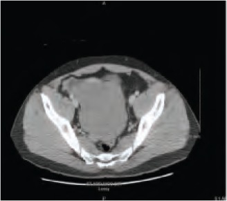
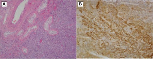
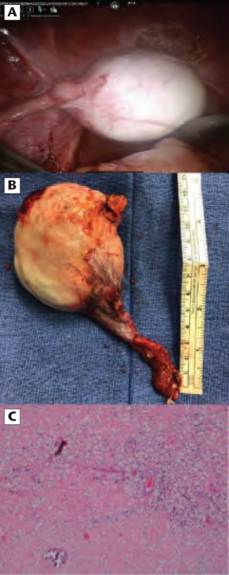
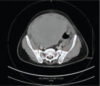
Intra-abdominal testes are at increased risk of malignant transformation and can manifest as large abdominal masses with a wide variation in presenting symptoms. In the setting of cryptorchid or nonpalpable testes, large abdominal masses are highly suspect for germ cell tumors. Without standard guidelines, management can vary extensively. Surgical management may not be trivial and can entail a major abdominal operation in the context of a multimodal approach. The use of biopsy and serum tumor markers may effectively guide sequence of management based upon expected histology. In advanced cases, neoadjuvant chemotherapy may be pursued, and retroperitoneal lymph node dissection may be accomplished at the time of orchiectomy to minimize morbidity. The development of these massive late stage tumors reaffirms current guidelines on the early correction of cryptorchidism.
Keywords: Nonseminomatous; Seminoma; Testicular.
© 2019 MedReviews®, LLC.
Figures
Similar articles
-
Improved management of abdominal undescended testicular tumors with bulky confluent retroperitoneal nodal metastases.J Urol. 1996 Oct;156(4):1341-4. J Urol. 1996. PMID: 8808867 Clinical Trial.
-
Nonpalpable testicular pure seminoma with elevated serum alpha-fetoprotein presenting with retroperitoneal metastasis: a case report.J Med Case Rep. 2016 May 5;10(1):114. doi: 10.1186/s13256-016-0906-7. J Med Case Rep. 2016. PMID: 27150447 Free PMC article.
-
Surgical management of the nonpalpable testis: the Children's Hospital of Philadelphia experience.J Urol. 1998 Apr;159(4):1340-3. J Urol. 1998. PMID: 9507881
-
Retroperitoneal lymph node dissection in the treatment of low-stage nonseminomatous germ cell tumors of the testicle.Expert Rev Anticancer Ther. 2005 Feb;5(1):75-85. doi: 10.1586/14737140.5.1.75. Expert Rev Anticancer Ther. 2005. PMID: 15757440 Review.
-
Current controversies on the role of retroperitoneal lymphadenectomy for testicular cancer.Urol Oncol. 2019 Mar;37(3):209-218. doi: 10.1016/j.urolonc.2018.09.009. Epub 2018 Nov 13. Urol Oncol. 2019. PMID: 30446455 Free PMC article. Review.
Cited by
-
Unusual presentation of an intra-abdominal testicular seminoma in an adult.Urol Case Rep. 2022 May 20;43:102119. doi: 10.1016/j.eucr.2022.102119. eCollection 2022 Jul. Urol Case Rep. 2022. PMID: 35646599 Free PMC article.
-
Unusual Location of Residual Mass in an Uncorrected Undescended Testis with Germ Cell Tumor.Case Rep Urol. 2023 Oct 3;2023:6626799. doi: 10.1155/2023/6626799. eCollection 2023. Case Rep Urol. 2023. PMID: 37822958 Free PMC article.
-
A giant intra-abdominal right testicular seminoma in a bilateral undescended testicle: a case report.Pan Afr Med J. 2023 Jan 3;44:3. doi: 10.11604/pamj.2023.44.3.37512. eCollection 2023. Pan Afr Med J. 2023. PMID: 36818032 Free PMC article.
-
Is Human Chorionic Gonadotropin a Reliable Marker for Testicular Germ Cell Tumor? New Perspectives for a More Accurate Diagnosis.Cancers (Basel). 2025 Jul 21;17(14):2409. doi: 10.3390/cancers17142409. Cancers (Basel). 2025. PMID: 40723290 Free PMC article. Review.
References
-
- AbouZeid AA, Mousa MH, Soliman HA, et al. Intraabdominal testis: histological alterations and significance of biopsy. J Urol. 2011;185:269–274. - PubMed
-
- Pettersson A, Richiardi L, Nordenskjold A, et al. Age at surgery for undescended testis and risk of testicular cancer. N Engl J Med. 2007;356:1835–1841. - PubMed
-
- Wood HM, Elder JS. Cryptorchidism and testicular cancer: separating fact from fiction. J Urol. 2009;181:452–461. - PubMed
-
- Ford TF, Parkinson MC, Pryor JP. The undescended testis in adult life. Br J Urol. 1985;57:181–184. - PubMed
LinkOut - more resources
Full Text Sources