

Orbital Cellular Epithelioid Hemangioma
- PMID: 31768366
- PMCID: PMC6873054
- DOI: 10.1159/000496207
Orbital Cellular Epithelioid Hemangioma
Abstract
Purpose: To report a case of orbital cellular epithelioid hemangioma (EH) in which FOSB and CAMTA1 immunostains were used to detect a cytogenetic rearrangement as an adjunctive tool in diagnosis.
Methods: Case report.
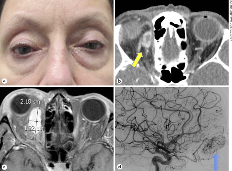
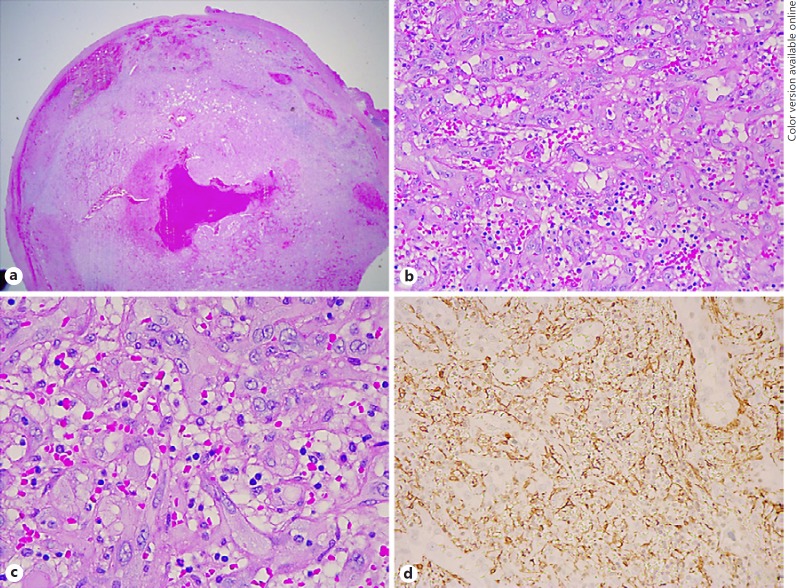
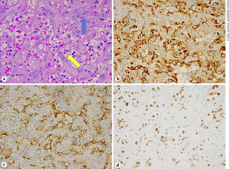
Results: A patient with a history of prior ligation of a presumed orbital varix presented with recurrent proptosis. Imaging revealed a highly vascular right orbital mass. Microscopic examination revealed a circumscribed neoplasm composed of plump epithelioid endothelial cells with copious mildly eosinophilic cytoplasm and relatively uniform vesicular nuclei. To aid in diagnosis, immunostains for FOSB and CAMTA1 were performed to detect corresponding cytogenetic rearrangements. The presence of multifocal nuclear positivity for FOSB, indicating FOSB genetic rearrangement, and negativity for CAMTA1 were considered reassuring features against a diagnosis of a malignant epithelioid hemangioendothelioma (EHE), supporting a diagnosis of benign cellular EH.
Conclusions: This case report demonstrates that the use of immunohistochemical stains to detect cytogenetic rearrangements may aid in the distinction between benign EH and malignant EHE. It also reminds providers of the clinical and histopathologic features of this lesion, which occurs rarely in the orbit, and helps clarify the evolving nomenclature surrounding epithelioid hemangioma.
Keywords: Angiolymphoid hyperplasia with eosinophilia; Cytogenetics; Epithelioid hemangioma.
Copyright © 2019 by S. Karger AG, Basel.
Conflict of interest statement
The authors declare that they have no conflicts of interest or financial interests to disclose.
Figures
References
-
- Fletcher CD, Bridge JA, Hogendoorn PC, Mertens F. WHO Classification of Tumours of Soft Tissue and Bone. 4th ed. Volume 5. Lyon, France: IARC Press; 2013.
-
- Wells GC, Whimster IW. Subcutaneous angiolymphoid hyperplasia with eosinophilia. Br J Dermatol. 1969 Jan;81((1)):1–14. - PubMed
-
- Rosai J, Gold J, Landy R. The histiocytoid hemangiomas. A unifying concept embracing several previously described entities of skin, soft tissue, large vessels, bone, and heart. Hum Pathol. 1979 Nov;10((6)):707–30. - PubMed
-
- Weiss SW, Enzinger FM. Epithelioid hemangioendothelioma: a vascular tumor often mistaken for a carcinoma. Cancer. 1982 Sep;50((5)):970–81. - PubMed
Grants and funding
LinkOut - more resources
Full Text Sources
Miscellaneous
