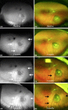
Fundus Autofluorescence Change as an Early Indicator of Treatment Effect of Brachytherapy for Choroidal Melanomas
- PMID: 31768369
- PMCID: PMC6873041
- DOI: 10.1159/000499403
Fundus Autofluorescence Change as an Early Indicator of Treatment Effect of Brachytherapy for Choroidal Melanomas
Abstract
Background: Early confirmation of the effect of brachytherapy for choroidal melanoma showing that tumour coverage is valuable. The irradiated retinal pigment epithelium (RPE) commonly develops atrophy. This study compares the fundus autofluorescence (AF) changes to the development of RPE atrophy following brachytherapy.
Methods: Retrospective study of 19 patients treated with 106Ru and 2 with 125I plaques with either a 3- or 6-month follow-up period. Ultra-widefield (UW) composite colour and AF images were obtained with Optomap 200Tx and interpreted as complete, partial, or no RPE changes and complete or partial hyperautofluorescence, hypoautofluorescence, or isoautofluorescence.
Results: At the 3-month follow-up, 9 of 13 patients (69%) (95% confidence interval [CI], 0.389-0.896) treated with 106Ru plaques developed complete homogenous hyperautofluorescence surrounding the tumour, but only 1 of 13 (8%) (95% CI, 0.004-0.379) developed complete RPE atrophy at the same time point. Six patients in the 106Ru plaque group had their first follow-up with UW imaging at 6 months. Four of them developed homogenous hyperautofluorescence and none developed complete RPE atrophy around the tumour. The 2 patients treated with 125I did not demonstrate any clear RPE or AF changes.
Conclusion: The effect of 106Ru plaque treatment on fundus UW imaging is detected as homogenous and well-demarcated hyperautofluorescence before visible RPE atrophy.
Keywords: 106Ru plaque; Brachytherapy; Choroidal melanoma; Fundus autofluorescence; Ultra-widefieldfundus imaging.
Copyright © 2019 by S. Karger AG, Basel.
Conflict of interest statement
The authors have no conflicts of interest or sources of funding to declare.
Figures

Similar articles
-
Fundus Autofluorescence Imaging in Patients with Choroidal Melanoma.Cancers (Basel). 2022 Apr 2;14(7):1809. doi: 10.3390/cancers14071809. Cancers (Basel). 2022. PMID: 35406581 Free PMC article. Review.
-
Autofluorescence of intraocular tumours.Curr Opin Ophthalmol. 2013 May;24(3):222-32. doi: 10.1097/ICU.0b013e32835f8ba1. Curr Opin Ophthalmol. 2013. PMID: 23429597 Review.
-
Autofluorescence of choroidal melanoma in 51 cases.Br J Ophthalmol. 2008 May;92(5):617-22. doi: 10.1136/bjo.2007.130286. Br J Ophthalmol. 2008. PMID: 18441171
-
Stages of Drusen-Associated Atrophy in Age-Related Macular Degeneration Visible via Histologically Validated Fundus Autofluorescence.Ophthalmol Retina. 2021 Aug;5(8):730-742. doi: 10.1016/j.oret.2020.11.006. Epub 2020 Nov 18. Ophthalmol Retina. 2021. PMID: 33217617 Free PMC article.
-
Ultra-widefield autofluorescence imaging findings in retinoschisis, rhegmatogenous retinal detachment and combined retinoschisis retinal detachment.Acta Ophthalmol. 2021 Mar;99(2):195-200. doi: 10.1111/aos.14521. Epub 2020 Jun 29. Acta Ophthalmol. 2021. PMID: 32602221
Cited by
-
Fundus Autofluorescence Imaging in Patients with Choroidal Melanoma.Cancers (Basel). 2022 Apr 2;14(7):1809. doi: 10.3390/cancers14071809. Cancers (Basel). 2022. PMID: 35406581 Free PMC article. Review.
-
Edge Creep: Increased Pigmentation at the Border of Choroidal Melanomas Treated with Plaque Brachytherapy.Ocul Oncol Pathol. 2023 Aug;9(1-2):56-61. doi: 10.1159/000531006. Epub 2023 May 11. Ocul Oncol Pathol. 2023. PMID: 38376093 Free PMC article.
References
-
- Damato BE, Foulds WS. Tumour-associated retinal pigment epitheliopathy. Eye (Lond) 1990;4((Pt 2)):382–7. - PubMed
-
- Hashmi F, Rojanaporn D, Kaliki S, Shields CL. Orange pigment sediment overlying small choroidal melanoma. Arch Ophthalmol. 2012 Jul;130((7)):937–9. - PubMed
-
- Shields CL, Furuta M, Mashayekhi A, Berman EL, Zahler JD, Hoberman DM, et al. Clinical spectrum of choroidal nevi based on age at presentation in 3422 consecutive eyes. Ophthalmology. 2008 Mar;115((3)):546–552.e2. - PubMed
-
- Mashayekhi A, Siu S, Shields CL, Shields JA. Slow enlargement of choroidal nevi: a long-term follow-up study. Ophthalmology. 2011 Feb;118((2)):382–8. - PubMed
-
- Simpson ER, Gallie B, Laperrierre N, Beiki-Ardakani A, Kivelä T, Raivio V, et al. American Brachytherapy Society - Ophthalmic Oncology Task Force. Electronic address ABS – OOTF Committee The American Brachytherapy Society consensus guidelines for plaque brachytherapy of uveal melanoma and retinoblastoma. Brachytherapy. 2014 Jan-Feb;13((1)):1–14. paulfinger@eyecancer.com. - PubMed
Grants and funding
LinkOut - more resources
Full Text Sources

