Review
. 2019 Nov 22;3(4):041503.
doi: 10.1063/1.5111549. eCollection 2019 Dec.

Microphysiological systems modeling acute respiratory distress syndrome that capture mechanical force-induced injury-inflammation-repair

Affiliations
Review

Microphysiological systems modeling acute respiratory distress syndrome that capture mechanical force-induced injury-inflammation-repair

Hannah Viola et al. APL Bioeng. .

Abstract

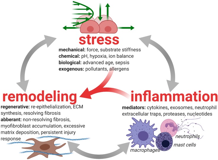
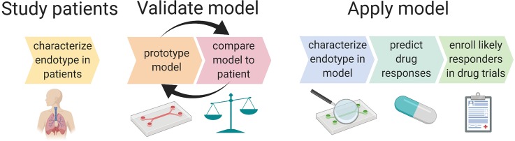
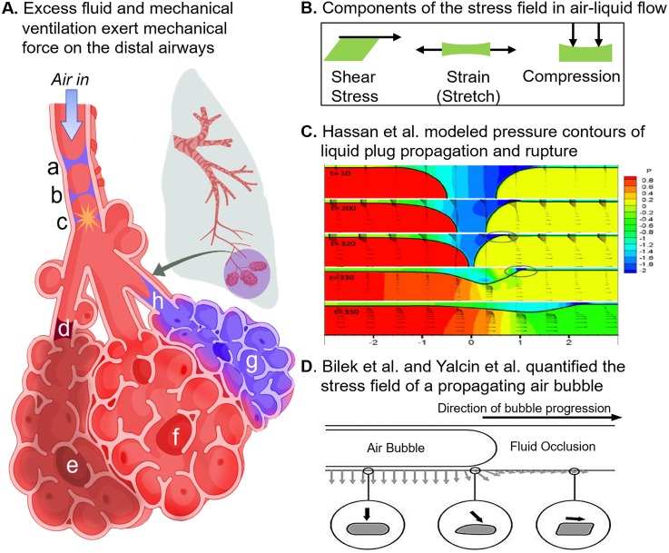
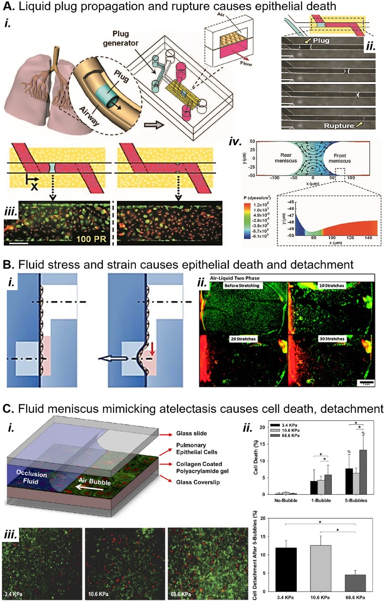
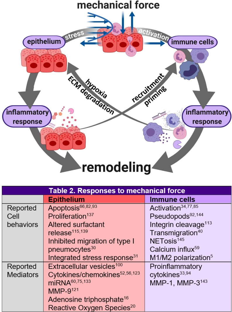
Complex in vitro models of the tissue microenvironment, termed microphysiological systems, have enormous potential to transform the process of discovering drugs and disease mechanisms. Such a paradigm shift is urgently needed in acute respiratory distress syndrome (ARDS), an acute lung condition with no successful therapies and a 40% mortality rate. Here, we consider how microphysiological systems could improve understanding of biological mechanisms driving ARDS and ultimately improve the success of therapies in clinical trials. We first discuss how microphysiological systems could explain the biological mechanisms underlying the segregation of ARDS patients into two clinically distinct phenotypes. Then, we contend that ARDS-mimetic microphysiological systems should recapitulate three critical aspects of the distal airway microenvironment, namely, mechanical force, inflammation, and fibrosis, and we review models that incorporate each of these aspects. Finally, we recognize the substantial challenges associated with combining inflammation, fibrosis, and/or mechanical force in microphysiological systems. Nevertheless, complex in vitro models are a novel paradigm for studying ARDS, and they could ultimately improve patient care.


Figures

FIG. 1.
FIG. 1.
Injury-inflammation-repair in lung disease. Inflammation and fibrosis (resolving or nonresolving) cooperate to remodel lung tissue after injury. Both processes are heavily influenced by stressors in the tissue microenvironment. In ARDS, mechanical forces arising from surfactant dysfunction and mechanical ventilation interfere with the tissue repair process by reinjuring the tissue, thereby inducing inflammation and promoting fibrosis. The interactions between tissue stress, inflammation, and remodeling can direct the tissue toward successful tissue repair or toward aberrant remodeling that consists of progressive fibrosis and systemically dysregulated immunity.
FIG. 2.
FIG. 2.
Iterative model design with validation against patient phenotype will lead to an endotype-specific model of ARDS that can be used for predictive enrichment of ARDS clinical trials. Endotype-specific metrics such as cytokine ratios, immune cell functions (e.g., bacterial killing, metabolism, and NETosis), and degree of surfactant production can be compared between patients and in vitro models. Iterative adjustments to model parameters, such as genetics (e.g., MUC5A upregulated epithelium), physical forces, degree of initial injury, degree of fibrosis, and the type and ratio of inflammatory mediators, could enable the development of a model that produces biomarkers or functional characteristics (e.g., response to therapeutics, response to mechanical strain, and immune cell phenotype changes such as enhanced NETosis), mimicking those of a specific endotype. The model should also be validated by testing functional outputs such as barrier function of the epithelium and tissue healing (scratch wound assay). The final model provides the opportunity for pathophysiological mechanisms of disease to be clarified and for drug candidates to be tested in vitro. Both pathophysiology and drug testing will help predict whether a certain endotype is likely to respond to novel treatments.
FIG. 3.
FIG. 3.
Physiologic mechanical forces in the bronchoalveolar region and their computational models. (A) The acinus consists of alveoli sacs that branch off of common terminal bronchiole (d) or (h); sacs (e), (f), and (g) are depicted in this figure. Sac (e) is cut off from air flow by the stagnant plug at (d); sac (f) is overinflated, and sac (g) is flooded with proteinaceous fluid. (B) Shear, strain, and compression are the main components of force present in the lungs, either independently or in concert. In the above depiction, strain results from overinflation of sac (f) due to obstruction of sac (d) and collapse of sac (g). Compression of adjacent sac results from the overinflation of (f). Shear stress is a component of the stress field produced during airway reopening at (h). Interfacial flow damages the small airways when liquid plugs propagate and rupture during inspiration (a)–(c) Transient liquid plugs form when the small airways collapse slightly and liquid on either side of the airways meets, forming the plug depicted in (a). Upon inspiration, the plug is pushed by pressure-driven flow, becoming thinner and thinner (b) until it loses integrity and pops (c), creating the crackle sounds that are observed upon auscultation of the lungs. (C) Hassan et al. modeled liquid plug propagation and rupture and found that the leading edge of the plug creates a narrow capillary wave (circled). The wave's extreme pressure gradient imparts severe stress on the airway wall. (D) The first in vitro model of airway reopening was introduced by Bilek et al. Using this model, Yalcin et al. found that smaller airway diameters experience greater stress. Reproduced with permission from Hassan et al., Int. J. Numer. Methods Fluids 67, 1373 (2011). Copyright 2011, John Wiley and Sons. Reproduced with permission from Bilek et al. J. Appl. Physiol. 94, 770 (2003). Copyright 2003, American Physiological Society.
FIG. 4.
FIG. 4.
MPS models of mechanical force in lung disease. (a) (i) A microfluidic device replicates the generation of crackle sounds frequently heard in the distal airways of patients with pulmonary edema. (ii) The device generates liquid plugs that propagate and rupture in channels over alveolar type I pneumocytes. (iii) Plugs result in epithelial cell death (red) during propagation (left) and especially at the rupture site (right). Scale bar, 150 μM. (iv) Fluid dynamic simulations show that the leading edge of the plug applies severe shear stress to the epithelium. Reproduced with permission from Huh et al. Proc. Natl. Acad. Sci. U. S. A. 104(48), 18886–18891 (2007). Copyright 2007 National Academy of Sciences. (b) (i) Douville et al. report a device that applies simultaneous fluid shear stress and mechanical strain to alveolar epithelium; vacuum stretches the membrane and simultaneously lowers the fluid level. (ii) Fluid stress and strain result in death (red cells) and detachment of alveolar epithelium in the device. Scale bar, 1 mm. Reproduced with permission from Lab on a Chip 11, 609–619 (2011). Copyright 2011 Royal Society of Chemistry. (c) (i) Higuita-Castro et al. mimic small airway or alveolar reopening by propagating an air bubble over the epithelium. The device design, first conceived by Bilek et al., has been extensively used to characterize the damage of liquid stress during atelectasis and airway reopening. (ii) and (iii) Higuita-Castro et al. show that the fluid meniscus causes increasing cell death and detachment with increasing substrate stiffness. Reproduced with permission from Higuita-Castro et al., J. Appl. Physiol. 117, 1231 (2014). Copyright 2014 American Physiological Society.
FIG. 5.
FIG. 5.
Reported mechanical stress-induced cell behaviors and inflammatory mediators that enable cross talk between pulmonary epithelium and immune cells. Mechanical stresses due to surfactant dysfunction, edema, and mechanical ventilation cause the epithelium to produce inflammatory and fibrotic mediators and may induce the integrated stress response, a key mediator of mechanical stress-induced epithelial injury. Mechanical force-induced cell behaviors and mediators of cross talk between the epithelium and immune cells are listed in Fig. 5. Such cross talk drives remodeling pathways; for example, hyperactivated neutrophils deplete oxygen in their microenvironment through the excessive production of reactive oxygen species (ROSs). This hypoxia stresses the epithelium and leads to apoptosis or inflammatory signaling that promotes fibroblast activation and epithelial proliferation. Neutrophils also contribute directly to remodeling during epithelial transmigration; massive neutrophil influx compromises tight junction integrity and stimulates repair of the lamina propria. Mechanically activated neutrophils and alveolar macrophages also secrete proteases that degrade the extracellular matrix (ECM), such as neutrophil elastase, and promote continued ECM destruction and activation of fibroblasts to repair the ECM damage. Because neutrophils are significant drivers of this destructive remodeling cycle, they are a target of therapeutic research.
FIG. 6.
FIG. 6.
Models of pulmonary inflammation in vitro. (a) (i) A microfluidic small airway-on-a-chip replicates the endothelium, interstitial fibroblasts, and epithelium. (ii) Fungal infection is simulated by inoculating the epithelium with wild type Aspergillus fumigatus, a model fungal pathogen. (iii) Neutrophils are added to the endothelial channels after the hyphae have extended into the interstitial space. Scale bar, 200 μM. (iv) Neutrophils chemotax from the endothelium through the interstitium toward fungal hyphae, attracted by volatile compounds produced by the fungus. Scale bars, 100 μM. Reproduced with permission from Barkal et al. Nat. Commun. 8, 1770 (2017). Copyright 2017 Authors, licensed under a CC BY 4.0 (https://creativecommons.org/licenses/by/4.0/). (b) (i) and (ii) A microfluidic alveolus-on-a-chip incorporates strain and neutrophil transmigration in a bilayer epithelium-endothelium coculture model. (iii) E. coli on the epithelium attracts neutrophils to transmigrate from the basal channel through endothelium and epithelium. (iv) Two E. coli bacteria (green) on the epithelium are chased and phagocytosed by a neutrophil (red) on the epithelium of the device. Reproduced with permission from Huh et al., Science 328, 1662 (2010). Copyright 2010 AAAS.
FIG. 7.
FIG. 7.
In vitro models of the lung microenvironment could be applied to study fibroproliferative disease in ARDS. (i) A lung-on-a-chip that replicates vascular leakage, leading to pulmonary edema and fibrin clotting. (ii) Strain is applied, by pulling vacuum on either side of the chamber, to a membrane (iii) with alveolar epithelium on the apical side and endothelium on the basal side. Scale bar, 200 μM. (iv) IL-2 induces endothelial and epithelial permeability allowing basal media loaded with prothrombin and fibrin to pass through the membrane and flood the apical channel, simulating pulmonary edema. Scale bar, 200 μM. (v) and (vi) Fibrin clots form on the apical channel after it becomes flooded with basal media containing fibrin and prothrombin. Scale bar (v), 50 μM. Scale bar (vi), 5 μM. Reproduced with permission from Huh et al. Sci. Transl. Med. 4, 159ra147 (2012). Copyright 2012 AAAS.

References

    1. Albelda S. M., “ Endothelial and epithelial cell adhesion molecules,” Am. J. Respir. Cell Mol. Biol. 4(3), 195–203 (1991). 10.1165/ajrcmb/4.3.195 - DOI - PubMed
    1. Albert R. K., “ The role of ventilation-induced surfactant dysfunction and atelectasis in causing acute respiratory distress syndrome,” Am. J. Respir. Crit. Care Med. 185(7), 702–708 (2012). 10.1164/rccm.201109-1667PP - DOI - PubMed
    1. Aogáin M. M., Tiew P. Y., Lim A. Y. H. et al. , “ Distinct “immunoallertypes” of disease and high frequencies of sensitization in non-cystic fibrosis bronchiectasis,” Am. J. Respir. Crit. Care Med. 199(7), 842–853 (2019). 10.1164/rccm.201807-1355OC - DOI - PubMed
    1. Ariel A. and Timor O., “ Hanging in the balance: Endogenous anti-inflammatory mechanisms in tissue repair and fibrosis,” J. Pathol. 229(2), 250–263 (2013). 10.1002/path.4108 - DOI - PubMed
    1. Ballotta V., Driessen-Mol A., Bouten C. V. C., and Baaijens F., “ Strain-dependent modulation of macrophage polarization within scaffolds,” Biomaterials 35(18), 4919–4928 (2014). 10.1016/j.biomaterials.2014.03.002 - DOI - PubMed