Review
. 2019 Dec;593(24):3583-3608.
doi: 10.1002/1873-3468.13693. Epub 2019 Dec 11.

Genomic foundations of evolution and ocular pathogenesis in human adenovirus species D

Affiliations
Review

Genomic foundations of evolution and ocular pathogenesis in human adenovirus species D

Ashrafali Mohamed Ismail et al. FEBS Lett. 2019 Dec.

Abstract

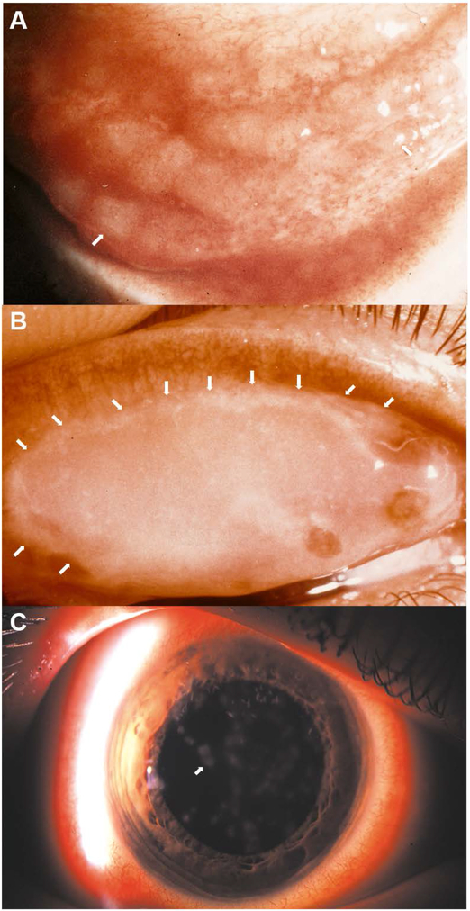
Human adenovirus commonly causes infections of respiratory, gastrointestinal, genitourinary, and ocular surface mucosae. Although most adenovirus eye infections are mild and self-limited, specific viruses within human adenovirus species D are associated with epidemic keratoconjunctivitis (EKC), a severe and highly contagious ocular surface infection, which can lead to chronic and/or recurrent, visually disabling keratitis. In this review, we discuss the links between adenovirus ontogeny, genomics, immune responses, and corneal pathogenesis, for those viruses that cause EKC.

Keywords: conjunctivitis; epidemic keratoconjunctivitis; genomics; homologous recombination; human adenovirus; inflammation; ocular infection; pink eye; tropism; virus evolution.


Figures

Fig. 1.
Fig. 1.
Photomicrographs of common clinical manifestations of ocular surface in epidemic keratoconjunctivitis (EKC). (A) Inferior conjunctival fornix of a patient with acute EKC shows conjunctival lymphoid hyperplasia presenting as jelly bean-shaped milky elevations of the mucosa (arrows point to two out of many follicles). (B) Superior eyelid tarsal surface with a conjunctival membrane (with visible margins of the membrane delineated by white arrows). (C) Corneal subepithelial infiltrates (arrow points to one of many such infiltrates). Image in C reproduced with permission.[264]
Fig. 2.
Fig. 2.
Phylogenetic analysis of human adenovirus (HAdV) DNA polymerase gene, including 103 genotypes, for HAdV species A-G. The tree was constructed in MEGA7 using the Maximum parsimony analysis with a bootstrap test of 500 replicates. HAdV-D is the largest species. The tree was obtained using the Tree-Bisection-Regrafting (TBR) algorithm with search level 1 in which the initial trees were obtained by the random addition of sequences (10 replicates). The tree is drawn to scale with branch lengths calculated using the average pathway method and is in the units of the number of changes over the whole sequence. All positions with less than 95% site coverage were eliminated and there were a total of 3270 positions in the final dataset.
Fig. 3.
Fig. 3.
Nucleotide diversity Pi (π) plots showing the average number of nucleotide differences per site for the known hypervariable regions in HAdV-Ds, including penton base, hexon, fiber, and E3 CR1 genes, and performed with all 73 HAdV-D genotypes. The plot was constructed using DnaSP v6 with 100 nt window and 25 nt step size (gaps are excluded).
Fig. 4.
Fig. 4.
Molecular HAdV-D types known to cause epidemic keratoconjunctivitis (EKC) and those predicted to be possible causes. (A) The known EKC viruses all have a fiber gene from either type HAdV-D8, D9, or D37, with otherwise disparate hexon genes. (B) Putative EKC-causing viruses based on the aforementioned fiber gene association. It is proposed that any virus with the fiber knob of HAdV-D8, D9, or D37 is a potential cause of EKC.
Fig. 5.
Fig. 5.
Cornea facsimile model of adenovirus keratitis. (A). Facsimiles shown in the sketch were generated in 12-ml transwell plates with a 3um pore size using primary human keratocytes mixed with collagen type 1, and layered with reduced growth factor containing Matrigel®. Infection with HAdV-D37 was performed overnight, and leukocytes then added to the lower chamber for one hour prior to harvest for confocal microscopy and flow cytometry for myeloperoxidase and CD45, respectively. (B) By myeloperoxidase staining (red/orange), as shown both en face (left column) and in cross section (stacked 3-D view, middle column), infected (V) corneal facsimiles developed neutrophil infiltration, whereas mock (M) infected facsimiles did not. DAPI staining (blue) to show cell nuclei (right column) showed similar pockets of cellular infiltration only in virus infected wells. Pretreatment with inhibitors of p38 (SB203580) and Src (PP2) blocked infiltration. (C) Flow cytometry for CD45 positive cells (leukocytes) in HAdV-D37 infected facsimiles showed CD45 events only in the presence of both virus and keratocytes. Therefore, virus alone was insufficient to induce chemotaxis. Figure modified with permission from Rajaiya et al.[215]
Fig. 6.
Fig. 6.
Cotton rat and rabbit models of adenovirus ocular infection. (A) The cotton rat model, figure adapted with permission from Tsai et al.[217] The image shown is of an adult cotton rat cornea 18 days after inoculation with HAdV-D8 (1×10⁵ plaque-forming units). Arrows indicate subepithelial opacities observed two weeks after inoculation. (B). New Zealand White (NZW) rabbit ocular model, figure adapted with permission from Clement et al.[221] Image of Ad5-infected NZW rabbit eye at 7 days post-infection. a. Intact cornea. b. Discharge and exudates on the cornea (arrow). c. Lower eyelid inflammation/vascular dilation. d. Upper eyelid inflammation/vascular dilation. e. Prominent blood vessel (arrow) close to the caruncle of the eye. f. Corneal neovascularization. (C) Hollander rabbit corneal model, figure adapted with permission from Hauwere et al.[222] Rabbits were infected with HAdV-C5 by intracorneal and subconjunctival injections and topical instillation. The infected eye developed subepithelial opacities by day 56 post infection.
Fig. 7.
Fig. 7.
Mouse model of adenovirus keratitis. (A) Induction of the mouse model of adenovirus keratitis. a. BALB/c mouse cornea retroilluminated to show size of heat-pulled, glass micropipette needle (arrow) as compared to a 33 gauge metal needle. b. Diffuse illumination view of mouse cornea prior to, and c. during injection with HAdV-D37 or virus-free dialysis buffer using the glass needle (arrow) and a gas-powered microinjection system. The injection causes transient whitening of the corneal stroma (arrow points to tip of glass needle within the corneal stroma. d. Composite confocal image of a mouse cornea taken immediately after injection of Cy3 dye demonstrates successful intrastromal injection. (B) Thin-section electron microscopy of C57Bl/6j mouse corneal stroma at intermediate time points after intrastromal injection of HAdV-D37. Micrographs show the corneal stroma at a, 4 hours, and b-d, 8 hours after injection. Intracellular structures are labeled as follows: Cy, cytoplasm; Nu, nucleus. The inset in a shows a higher magnification of intracellular virus. All micrographs show densely packed intracellular viral arrays. Scale bars: 2 μm in a and b, and 0.5 μm in c and d, and the inset in a. Adapted with permission from Mukherjee et al.[233] (C) Representative clinical photographs of C57BL/6j mouse corneas after mock infection with dialysis buffer or 105 TCID HAdV-D37 at days shown post infection. Buffer-injected corneas remained clear at all times. Opacities in HAdV-D37 injected corneas were seen as early as 1 day after infection, and appeared to peak at 4 days. The opacities then regressed slowly but in approximately one-third of mice recurred as characteristic subepithelial infiltrates at about 6 weeks after infection (day 42, right panel, arrows). Figure adapted with permission from Chintakuntlawar et al.[232]

References

    1. Harrach B, Benkö M, Both GW, Brown M, Davison AJ, Echavarría M, Hess M, Jones MS, Kajon A, Lehmkuhl HD, Mautner V, Mittal SK and Wadell G (2012) Family - Adenoviridae In Virus taxonomy : classification and nomenclature of viruses : ninth report of the International Committee on Taxonomy of Viruses (Andrew MQ King MJA, Carstens Eric B., and Lefkowitz Elliot J., ed.êds), pp. 125–141. Elsevier, Oxford United Kingdom, Amsterdam.
    1. Trentin JJ, Yabe Y and Taylor G (1962). The quest for human cancer viruses. Science 137, 835–41. - PubMed
    1. Huebner RJ, Rowe WP and Lane WT (1962). Oncogenic effects in hamsters of human adenovirus types 12 and 18. Proc Natl Acad Sci U S A 48, 2051–8. - PMC - PubMed
    1. Berget SM, Moore C and Sharp PA (1977). Spliced segments at the 5’ terminus of adenovirus 2 late mRNA. Proc Natl Acad Sci U S A 74, 3171–5. - PMC - PubMed
    1. Chow LT, Gelinas RE, Broker TR and Roberts RJ (1977). An amazing sequence arrangement at the 5’ ends of adenovirus 2 messenger RNA. Cell 12, 1–8. - PubMed

Publication types

MeSH terms