. 2019 Nov 26:8:e47533.
doi: 10.7554/eLife.47533.

Nucleolar dynamics and interactions with nucleoplasm in living cells

Affiliations

Nucleolar dynamics and interactions with nucleoplasm in living cells

Christina M Caragine et al. Elife. .

Abstract

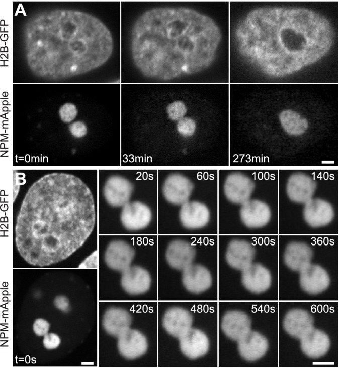
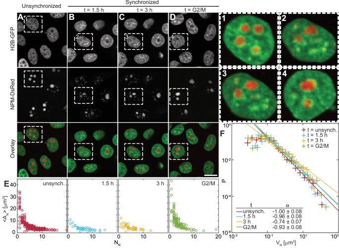
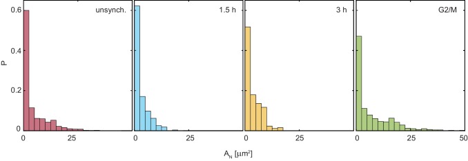
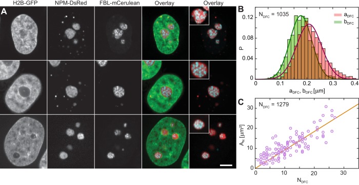
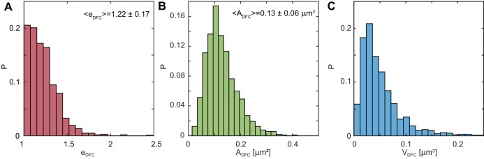
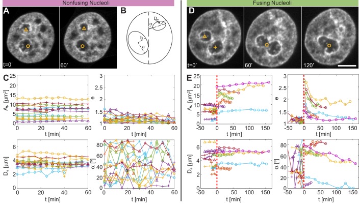
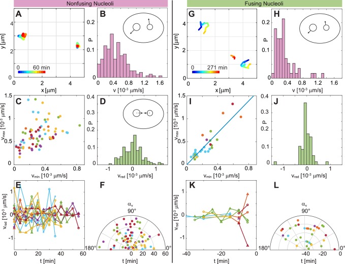
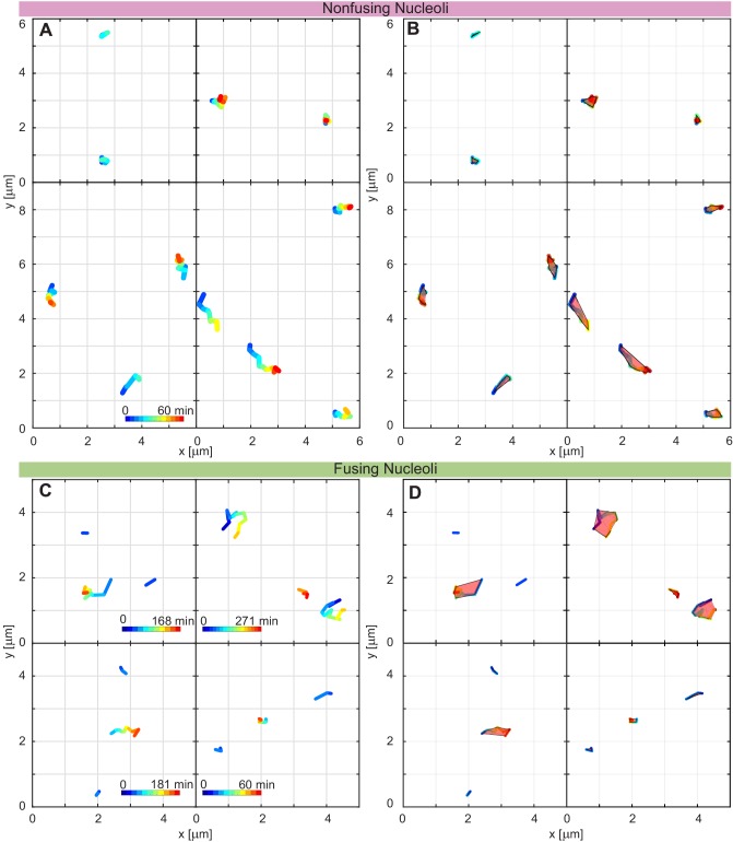
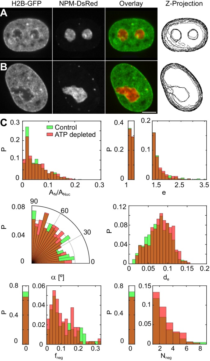
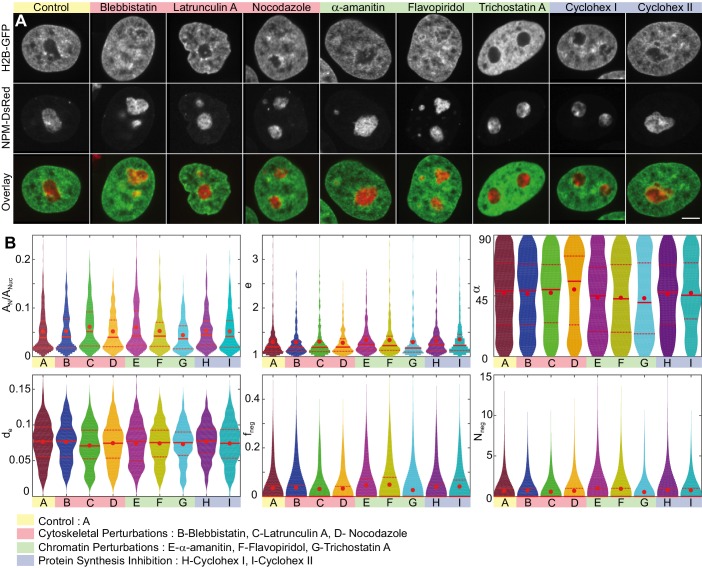
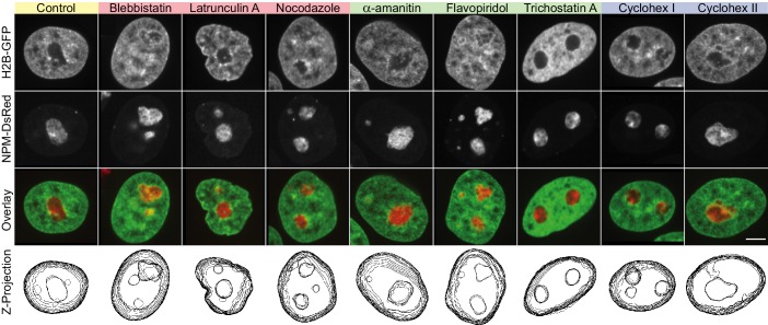
Liquid-liquid phase separation (LLPS) has been recognized as one of the key cellular organizing principles and was shown to be responsible for formation of membrane-less organelles such as nucleoli. Although nucleoli were found to behave like liquid droplets, many ramifications of LLPS including nucleolar dynamics and interactions with the surrounding liquid remain to be revealed. Here, we study the motion of human nucleoli in vivo, while monitoring the shape of the nucleolus-nucleoplasm interface. We reveal two types of nucleolar pair dynamics: an unexpected correlated motion prior to coalescence and an independent motion otherwise. This surprising kinetics leads to a nucleolar volume distribution, [Formula: see text], unaccounted for by any current theory. Moreover, we find that nucleolus-nucleoplasm interface is maintained by ATP-dependent processes and susceptible to changes in chromatin transcription and packing. Our results extend and enrich the LLPS framework by showing the impact of the surrounding nucleoplasm on nucleoli in living cells.

Keywords: human; liquid-liquid phase separation; molecular biophysics; nucleolar dynamics; nucleolus; nucleoplasm; nucleus; physics of living systems; structural biology.


Conflict of interest statement

CC, SH, AZ No competing interests declared

Figures

Figure 1.
Figure 1.. Nucleolar coalescence.
(A) Time lapse of a nucleus with fluorescently labeled chromatin (H2B-GFP) and two fusing nucleoli (NPM-mApple). Time points depict: pre-fusion (t = 0 min), with two distinct nucleoli, fusion in progress (t = 33 min), with a clearly visible neck connecting the two nucleoli, and post-fusion (t = 273 min), where a resultant nucleolus can be seen still rounding up. (B) Time series showing the growth of the neck connecting two coalescing nucleoli. At t = 0 s, both the fluorescently labeled chromatin (H2B-GFP) and coalescing nucleoli (NPM-mApple) are depicted, the later frames, 20–600 s, show the progress of the nucleolar coalescence (NPM-mApple). Parts of (B) adapted from Figure 3b in Caragine et al. (2018). Scale bar, 2 µm.
Figure 2.
Figure 2.. Nucleolar size distribution as a function of cell cycle.
(A–D) Micrographs of HeLa cell nuclei with fluorescently labeled chromatin (H2B-GFP) and nucleoli (NPM-DsRed) under the following conditions: unsynchronized cells (A), synchronized cells 1.5 hr (B) and 3 hr (C) after mitosis, and cells arrested at G2/M checkpoint (D). (1–4) enlarged view of the boxed nuclei from (A–D). (E) Average nucleolar area AN as a function of number of nucleoli per nucleus NN for all conditions from (A–D). For unsynchronized cells, total number of nucleoli analyzed NN = 1331 in 228 nuclei, for t = 1.5 hr, NN = 275 in 42 nuclei, for t = 3 hr, NN = 257 in 51 nuclei, and for t = G2/M, NN = 497 in 124 nuclei. (F) Distributions of nucleolar volume VN and their fit to f(VN)VNα for all conditions from (A–D). For all fits, the goodness-of-fit, R2 > 0.98. Scale bar, 15 µm.
Figure 2—figure supplement 1.
Figure 2—figure supplement 1.. Nucleolar area distributions, p(AN), for all conditions shown in Figure 2E: unsynchronized cells, synchronized cells 1.5 hr after mitosis, synchronized cells 3 hr after mitosis, and cells arrested in G2/M.
For unsynchronized cells, number of analyzed nucleoli is NN = 1331 in 228 nuclei, for t = 1.5 hr, NN = 275 in 42 nuclei, for t = 3 hr, NN = 257 in 51 nuclei, and for t = G2/M, NN = 497 in 124 nuclei.
Figure 3.
Figure 3.. Nucleolar internal structure.
(A) Micrographs of HeLa nuclei with fluorescently labeled chromatin (H2B-GFP, green), nucleolar granular component (NPM-DsRed, red) and nucleolar dense fibrillar component (DFC) (FBL-mCerulean, blue) and overlays of all three signals (green, red, blue) and red and blue signal. The insets in overlay images present an enlarged view of a nucleolus from the image. (B) Distributions measured for semi-major axis aDFC (red) and semi-minor axis bDFC (green) of single DFCs (NDFC = 1035). The solid red and green lines correspond to the Gaussian fits of distributions of aDFC and bDFC, respectively, with aDFC ≈ 210 nm and bDFC ≈ 180 nm. (C) Nucleolar area, AN, as a function of DFC number per nucleolus, NDFC, with a linear fit AN ≈ 0.92NDFC. We evaluated 1279 DFCs over 114 nucleoli in 63 nuclei. Scale bar, 5 µm.
Figure 3—figure supplement 1.
Figure 3—figure supplement 1.. Distributions of DFC eccentricity, area and volume.
(A) Distribution of the measured eccentricities, e=a/b, where a and b are the DFC semi-major and semi-minor axes, respectively (NDFC= 1035). (B) Distribution of the DFC area, A=πab, measured for single DFCs (NDFC= 1035). (C) DFC volume distribution, p(VDFC). DFC volume, VDFC, was calculated as VDFC=4πa3/3, where a is the semi-major axis of the DFC, providing the upper boundary on the volume estimate. p(VDFC) exhibits a sharp maximum at VDFC = 0.030 μm3, suggesting largely a monodisperse population of DFCs (NDFC= 1035).
Figure 4.
Figure 4.. Comparison of size, shape and nuclear positioning between fusing and nonfusing nucleoli.
(A) Micrographs of a nucleus with fluorescently labeled chromatin (H2B-GFP), where two void spaces (labeled by yellow triangle and yellow circle) correspond to two nucleoli that did not fuse between t = 0 and 60 min. (B) Schematics of measured variables. (C) Measured variables for nonfusing nucleoli: nucleolar area, AN, nucleolar eccentricity, e, shortest distance from the nucleolar centroid to the nuclear envelope, De, and the angle between the major nuclear and nucleolar axes, α (NN = 17, Ncell = 6). All characteristics are calculated in the nucleolar focal plane. (D) Micrographs of a nucleus with fluorescently labeled chromatin (H2B-GFP), where two void spaces (labeled by yellow triangle and yellow cross) correspond to two nucleoli before fusion at t = 0 min, while at t = 60 and 120 min they can be seen fusing (yellow circle). (E) Measured variables for fusing nucleoli: AN, e, De and α (NN = 12, Ncell = 7). The dashed red line at t = 0 min indicates fusion. All measurements are carried out in the nucleolar focal plane. Scale bar, 5 µm.
Figure 5.
Figure 5.. Comparison of dynamics between fusing and nonfusing nucleoli.
(A) Trajectories of two nonfusing nucleoli color-coded by their temporal evolution (blue to red). The time step is 5 min. (B) Histogram of the velocity magnitude, v, for the nonfusing nucleoli (NN = 17, Ncell = 6). (C) Velocities for pairs of nonfusing nucleoli, where vmax and vmin is the larger and the smaller nucleolar velocity, respectively. (D) Histogram of radial velocity, vrad, for nonfusing nucleoli, with vrad calculated with respect to the midpoint distance between nucleoli. (E) vrad as a function of time for nonfusing nucleoli. (F) Velocity angle, αv, in polar coordinates as a function of time for nonfusing nucleoli. (G) Trajectories of pair of fusing nucleoli color-coded by their temporal evolution (blue to red). The pre-fusion nucleoli are visible at earlier times (blue to yellow), while the post-fusion nucleolus appears at later times (orange to red). The time step is 15 and 16 min. (H) Histogram of the v for the fusing nucleoli (NN = 12, Ncell = 7). (I) Velocities for pairs of fusing nucleoli, where vmax and vmin is the larger and the smaller nucleolar velocity, respectively, with a linear fit vmax 1.74vmin. (J) Histogram of vrad for fusing nucleoli. (K) vrad as a function of time for fusing nucleoli. (L) αv for fusing nucleoli as a function of time.
Figure 5—figure supplement 1.
Figure 5—figure supplement 1.. Additional nucleolar trajectories and enlarged view of nucleolar trajectories from Figure 5A and Figure 5G.
(A) Trajectories of nucleoli which do not fuse, time increases from blue to red (enlarged view of Figure 5A). (B) The trajectory areas determined as the convex hull of the trajectories of nucleoli from A. (C) Trajectories of nucleoli which fuse, time increases from blue to red (enlarged view of Figure 5G). The post fusion nucleolus is in between the other two trajectories. (D) The trajectory areas determined as the convex hull of the trajectories of nucleoli from C.
Figure 6.
Figure 6.. Nucleoli under physiological conditions and upon ATP-depletion.
(A–B) Micrographs of HeLa nuclei with fluorescently labeled chromatin (H2B-GFP, green) and nucleoli (NPM-DsRed, red), their color overlay and z-projections of nucleolar and nuclear contours: (A) under physiological conditions (control) and (B) after ATP depletion. (C) Distributions of the following nucleolar measurements under physiological conditions (NN= 648, NCell= 208) vs. upon ATP depletion (NN= 345, NCell= 127): nucleolar area normalized by nuclear area, AN/ANuc, nucleolar eccentricity, e, angle between the major nuclear and nucleolar axes, α, the shortest distance from the nucleolar centroid to the nuclear envelope normalized by the nuclear circumference, de, fraction of the nucleolar contour with negative curvature, fneg, and number of continuous regions of negative curvature along the nucleolar contour, Nneg. All measurements are carried out in the nucleolar focal plane. Scale bar, 5 µm.
Figure 7.
Figure 7.. Nucleoli upon biochemical perturbations.
(A) Micrographs of HeLa nuclei with fluorescently labeled chromatin (H2B-GFP, green) and nucleoli (NPM-DsRed, red), and the overlay, under the following conditions: control, upon addition of blebbistatin, latrunculin A, nocodazole, α-amanitin, flavopiridol, trichostatin A, and cycloheximide (at t1= 30 min and t2= 6.5 hr). (B) Histograms of the following measurements for all conditions (width indicates probability): nucleolar area normalized by nuclear area, AN/ANuc, nucleolar eccentricity, e, angle between the major nuclear and nucleolar axes, α, the shortest distance from the nucleolar centroid to the nuclear envelope normalized by the nuclear circumference, de, fraction of the nucleolar contour with negative curvature, fneg, and number of continuous regions of negative curvature along the nucleolar contour, Nneg. All data collected in the nucleolar focal plane. Red dot, solid red line and dotted red lines indicate the mean, median and quartiles, respectively. Table 1 provides p-values for all measured data with respect to the control. The number of nucleoli and cells are as follows: control (NN= 648, NCell= 208), blebbistatin (NN= 399, NCell= 127), latrunculin A (NN= 307, NCell= 104), nocodazole (NN= 310, NCell= 106), α-amanitin (NN= 268, NCell= 95), flavopiridol (NN= 309, NCell= 105), trichostatin A (NN= 278, NCell= 95), and cycloheximide at t1= 30 min (NN= 291, NCell= 91), and cycloheximide at t2= 6.5 hr (NN= 294, NCell= 105). Scale bar, 5 µm.
Figure 7—figure supplement 1.
Figure 7—figure supplement 1.. Nucleoli upon biochemical perturbations, including z-projections.
Micrographs of HeLa nuclei with fluorescently labeled chromatin (H2B-GFP, green) and nucleoli (NPM-DsRed, red), the two signal overlay, and z-projections of nuclear and nucleolar contours, under the following conditions: control, upon addition of blebbistatin, latrunculin A, nocodazole, α-amanitin, flavopiridol, trichostatin A, and cycloheximide (at t1= 30 min and t2= 6.5 hr). Scale bar, 5 µm.

References

    1. Alberts B, Johnson A, Lewis J, Morgan D, Raff M, Roberts K, Walter P. Molecular Biology of the Cell. Garland Science; 2014. - DOI
    1. Amenta PS. Fusion of nucleoli in cells cultured from the heart of Triturus viridescens. The Anatomical Record. 1961;139:155–165. doi: 10.1002/ar.1091390207. - DOI - PubMed
    1. Berry J, Weber SC, Vaidya N, Haataja M, Brangwynne CP. RNA transcription modulates phase transition-driven nuclear body assembly. PNAS. 2015;112:E5237–E5245. doi: 10.1073/pnas.1509317112. - DOI - PMC - PubMed
    1. Berry J, Brangwynne CP, Haataja M. Physical principles of intracellular organization via active and passive phase transitions. Reports on Progress in Physics. 2018;81:046601. doi: 10.1088/1361-6633/aaa61e. - DOI - PubMed
    1. Bickmore WA, van Steensel B. Genome architecture: domain organization of interphase chromosomes. Cell. 2013;152:1270–1284. doi: 10.1016/j.cell.2013.02.001. - DOI - PubMed

Publication types