. 2020 Jan-Dec;12(1):1685349.
doi: 10.1080/19420862.2019.1685349.

In vivo safety profile of a CSPG4-directed IgE antibody in an immunocompetent rat model

Affiliations

In vivo safety profile of a CSPG4-directed IgE antibody in an immunocompetent rat model

Iwan P Williams et al. MAbs. 2020 Jan-Dec.

Abstract

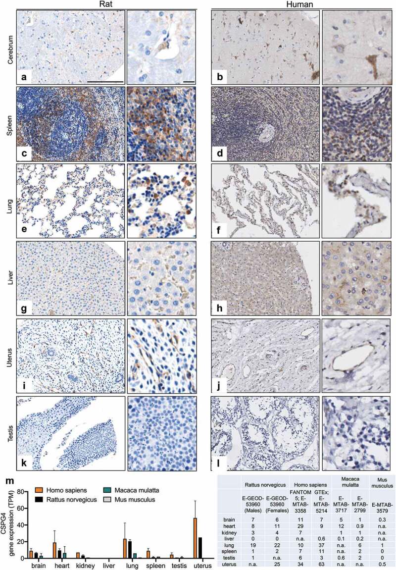
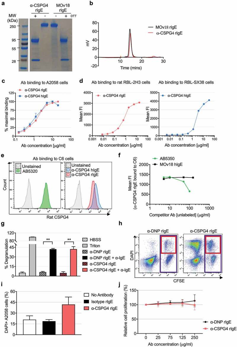
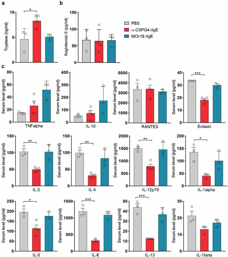
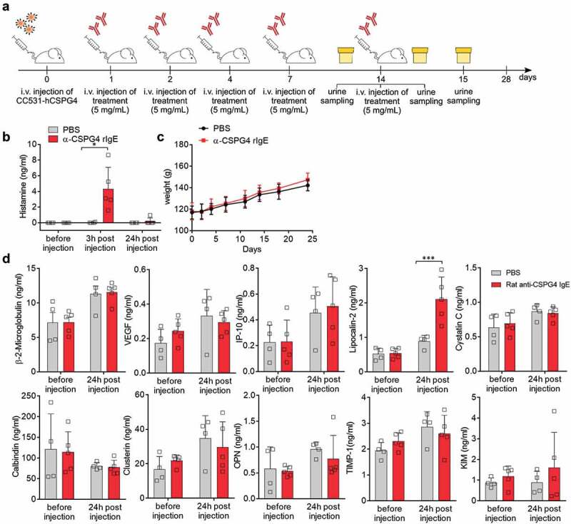
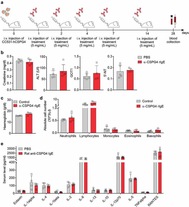
IgE monoclonal antibodies hold great potential for cancer therapy. Preclinical in vivo systems, particularly those in which the antibody recognizes the host species target antigen and binds to cognate Fc receptors, are often the closest approximation to human exposure and represent a key challenge for evaluating the safety of antibody-based therapies. We sought to develop an immunocompetent rat system to assess the safety of a rodent anti-tumor IgE, as a surrogate for the human therapeutic candidate. We generated a rat IgE against the human tumor-associated antigen chondroitin sulfate proteoglycan 4 (CSPG4) and cross-reactive for the rat antigen. We analyzed CSPG4 distribution in normal rat and human tissues and investigated the in vivo safety of the antibody by monitoring clinical signs and molecular biomarkers after systemic administration to immunocompetent rats. Human and rat CSPG4 expression in normal tissues were comparable. Animals receiving antibody exhibited transient mild to moderate adverse events accompanied by mild elevation of serum tryptase, but not of angiotensin II or cytokines implicated in allergic reactions or cytokine storm. In the long term, repeated antibody administration was well tolerated, with no changes in animal body weight, liver and kidney functions or blood cell counts. This model provides preclinical support for the safety profiling of IgE therapeutic antibodies. Due to the comparable antigen tissue distribution in human and rat, this model may also comprise an appropriate tool for proof-of-concept safety evaluations of different treatment approaches targeting CSPG4.

Keywords: CSPG4; IgE; allergooncology; antibody; immunotherapy; rat model; species cross-reactivity.


Figures

Figure 1.
Figure 1.
CSPG4 expression in normal human and rat tissues. (A-L) CSPG4 expression in rat (left) and human (right) tissues was investigated by immunohistochemistry of tissue microarrays using commercial anti-CSPG4 antibodies and developed using DAB chromogen. Hematoxylin was used to counterstain. Scale bars represent 200 μm (lower magnification) and 20 μm (higher magnification). M, Histogram (left) summarizing CSPG4 gene expression (Transcripts Per Million, TPM) in the specified tissues of four different species based on the transcriptomic dataset (EMBL-EBI Expression Atlas, https://www.ebi.ac.uk/gxa/). Dataset and respective mRNA expressions are listed in the table (right); n.a.: data not available.
Figure 2.
Figure 2.
In vitro characterization of α-CSPG4 rIgE antibody. A, SDS-PAGE of reduced (DTT+) and non-reduced (DTT-) α-CSPG4 rIgE and MOv18 rIgE. B, HPLC-SEC profile of α-CSPG4 rIgE and MOv18 rIgE. C, Dose-dependent binding of α-CSPG4 rIgE (left) and α-CSPG4 hIgE (right) to CSPG4-expressing human A2058 cells detected by flow cytometry and expressed as % of maximal binding (maximal Mean Fluorescence Intensity). D, Dose-dependent binding of α-CSPG4 hIgE (left) and α-CSPG4 rIgE (right) to FcϵRI-expressing RBL-SX38 (left) and RBL-2H3 cells (right) detected by flow cytometry. Representative results of one of four (RBL-SX38) and one (RBL-2H3) independent experiments. E, Binding profiles of commercial polyclonal anti-rat CSPG4 (AB5320), α-CSPG4 rIgE and α-CSPG4 hIgE to CSPG4-expressing rat C6 cells. F, Competition between increasing concentrations of rabbit AB5320 antibody (or MOv18 hIgE as control) and α-CSPG4 rIgE (detected with a secondary anti-rat IgE antibody) binding to C6 cells. G, RBL-2H3 cells degranulation in the absence of stimuli (HBSS), in the presence of α-DNP rIgE or α-CSPG4 rIgE alone, or upon crosslinking with α-IgE secondary antibody. Cells treated with Triton-X100 were set as 100%. Bars indicate mean ± SD from 3 independent experiments. **p < .01 by one-way ANOVA. H and I, RBL-2H3 cells-mediated ADCC against CFSE-labeled A2058 cells pre-coated with α-CSPG4 rIgE or α-DNP rIgE. H, Representative dot plot obtained in the presence of α-DNP rIgE (left panel) or α-CSPG4 rIgE (right panel). The percentage of dead cells was defined as DAPI+ A2058 cells (red box)*100/total A2058 cells (purple box). I, ADCC data obtained from 3 independent experiments. Bars indicate mean + SD; *p < .05 by one-way ANOVA. J, A2058 cells proliferation after incubation with α-CSPG4 rIgE or α-DNP rIgE (4 days). Incubation with no antibody was set as 100%. Data are the mean of 3 independent replicates.
Figure 3.
Figure 3.
Molecular analysis of immediate effects of in vivo α-CSPG4 rIgE administration. A-B, Levels of tryptase (ng/ml) and angiotensin II (pg/ml) were measured via ELISA in the sera of WAG rats 30 minutes after intravenous administration of 10mg/ml α-CSPG4 rIgE (n = 4), 10mg/ml MOv18 rIgE (n = 3) or equivalent volume of PBS (n = 3). Bars represent average values ± SD; *p < .05 calculated using One-way ANOVA. C, Levels of selected cytokines (pg/ml) were measured using bead-based multiplex assay in the sera of WAG rats 30 minutes after intravenous administration of α-CSPG4 rIgE (n = 4), MOv18 rIgE (n = 3) or PBS (n = 3). Bars represent average values ± SD; *p < .05, **p < .01, ***p < .005 calculated using one-way ANOVA.
Figure 4.
Figure 4.
Effects of repeated α-CSPG4 rIgE administrations. A, Schematic representation of scheduling of α-CSPG4 rIgE administrations and urine sampling. Urine was collected immediately before, 3 hours and 24 hours after the last administration of 5mg/ml α-CSPG4 rIgE (n = 5) or PBS (n = 4). B, Urine levels of histamine (ng/ml) were measured in the urine at the three time points indicated in A. Bars represent average values ± SD; *p < .05 was calculated using two-way ANOVA. C, Body weight of rats during α-CSPG4 rIgE treatment indicated in A. Bars represent average values ± SD. D, Urine levels of selected markers for kidney function were measured using bead-based multiplex assay at the time points indicated. Bars represent average values ± SD; ***p < .005 calculated using two-way ANOVA.
Figure 5.
Figure 5.
Long-term α-CSPG4 rIgE does not affect blood hematological or biochemical parameters. A, Blood of WAG rats treated with repeated α-CSPG4 rIgE (n = 5) or PBS (n = 4) administrations was collected before necropsy on experimental day 28. B, Serum levels of creatinine, alanine aminotransferase (ALT), Glutamic-Oxaloacetic Transaminase 1 (GOT1), 5ʹ-nucleotidase (5ʹ-NT). Bars represent average ± SD. Each symbol represents one rat. C, Hemoglobin concentration in the blood of WAG rats treated with α-CSPG4 rIgE or untreated (control). Bars represent average + SD. Each symbol represents one rat. D, Absolute blood cells numbers detected in α-CSPG4 rIgE or untreated animals. Bars represent average values ± SD. Each symbol represents one rat. E, Serum levels of selected cytokines (pg/ml) in α-CSPG4 rIgE or PBS treated animals. Bars represent average values ± SD.

Similar articles

Cited by

References

    1. Josephs DH, Spicer JF, Karagiannis P, Gould HJ, Karagiannis SN.. IgE immunotherapy: a novel concept with promise for the treatment of cancer. MAbs. 2014;6:54–13. doi:10.4161/mabs.27029. - DOI - PMC - PubMed
    1. Sutton BJ, Davies AM, Bax HJ, Karagiannis SN. IgE antibodies: from structure to function and clinical translation. Antibodies (Basel). 2019;8(1):19. - PMC - PubMed
    1. Leoh LS, Daniels-Wells TR, Penichet ML. IgE immunotherapy against cancer. Curr Top Microbiol Immunol. 2015;388:109–49. doi:10.1007/978-3-319-13725-4_6. - DOI - PMC - PubMed
    1. Burgess M, Mapp S, Mazzieri R, Cheung C, Chambers L, Mattarollo SR, Mollee P, Gill D, Saunders NA. Increased FcgammaRIIB dominance contributes to the emergence of resistance to therapeutic antibodies in chronic lymphocytic leukaemia patients. Oncogene. 2017;36:2366–76. doi:10.1038/onc.2016.387. - DOI - PubMed
    1. Tada T, Okumura K, Platteau B, Beckers A, Bazin H. Half-lives of two types of rat homocytotropic antibodies in circulation and in the skin. Int Arch Allergy Appl Immunol. 1975;48:116–31. doi:10.1159/000231297. - DOI - PubMed

Publication types

MeSH terms

LinkOut - more resources